Review
. 2021 Aug 24:13:709301.
doi: 10.3389/fnsyn.2021.709301. eCollection 2021.

Current Therapies for Neonatal Hypoxic-Ischaemic and Infection-Sensitised Hypoxic-Ischaemic Brain Damage

Affiliations
Review

Current Therapies for Neonatal Hypoxic-Ischaemic and Infection-Sensitised Hypoxic-Ischaemic Brain Damage

Konstantina Tetorou et al. Front Synaptic Neurosci. .

Abstract

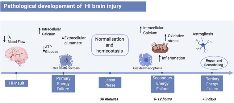
Neonatal hypoxic-ischaemic brain damage is a leading cause of child mortality and morbidity, including cerebral palsy, epilepsy, and cognitive disabilities. The majority of neonatal hypoxic-ischaemic cases arise as a result of impaired cerebral perfusion to the foetus attributed to uterine, placental, or umbilical cord compromise prior to or during delivery. Bacterial infection is a factor contributing to the damage and is recorded in more than half of preterm births. Exposure to infection exacerbates neuronal hypoxic-ischaemic damage thus leading to a phenomenon called infection-sensitised hypoxic-ischaemic brain injury. Models of neonatal hypoxia-ischaemia (HI) have been developed in different animals. Both human and animal studies show that the developmental stage and the severity of the HI insult affect the selective regional vulnerability of the brain to damage, as well as the subsequent clinical manifestations. Therapeutic hypothermia (TH) is the only clinically approved treatment for neonatal HI. However, the number of HI infants needed to treat with TH for one to be saved from death or disability at age of 18-22 months, is approximately 6-7, which highlights the need for additional or alternative treatments to replace TH or increase its efficiency. In this review we discuss the mechanisms of HI injury to the immature brain and the new experimental treatments studied for neonatal HI and infection-sensitised neonatal HI.

Keywords: hypoxia; infection; ischaemia; neonatal brain damage; neonatal encephalopathy.


Conflict of interest statement

The authors declare that the research was conducted in the absence of any commercial or financial relationships that could be construed as a potential conflict of interest.

Figures

FIGURE 1
FIGURE 1
Pathological development of neonatal HI brain injury. The HI insult is initiated by reduction of blood flow and oxygen to the foetal brain, leading to primary energy failure. The main events of this phase include reduction of ATP and glucose, increase of intracellular calcium, and therefore increase of extracellular glutamate. This leads to cell death mainly via necrosis. Following re-oxygenation, a latent phase begins, where the body resumes a “normal” state. A secondary energy failure may take place after 6–12 h post-HI insult, where a subsequent and stronger wave of cell death hits the brain, and events like inflammation, oxidative stress, and mitochondrial damage occur. Depending on the severity of the insult, a tertiary energy failure can occur and persist for months, characterised by brain remodelling and repair, as well as astrogliosis. Figure created with BioRender.com.
FIGURE 2
FIGURE 2
Lipopolysaccharide sensitisation. A bacterial infection sensitises the brain to HI insult via the interaction of LPS and TLR4. This leads to internalisation of NF-κB, mediated by MyD88. NF-κB activates the transcription of pro-inflammatory cytokine genes. Simultaneously, the interaction of LPS with TLR4 activates the NLRP3 inflammasome, which also promotes increase in pro-inflammatory cytokine levels and apoptosis. Figure created with BioRender.com.

References

    1. Adcock I. M. (2007). HDAC inhibitors as anti-inflammatory agents. Br. J. Pharmacol. 150 829–831. 10.1038/sj.bjp.0707166 - DOI - PMC - PubMed
    1. Adrian M., Jeandet P., Douillet-Breuil A. C., Tesson L., Bessis R. (2000). Stilbene content of mature Vitis vinifera berries in response to UV-C elicitation. J. Agric. Food Chem. 48 6103–6105. 10.1021/jf0009910 - DOI - PubMed
    1. Ahmad M. Z., Alkahtani S. A., Akhter S., Ahmad F. J., Ahmad J., Akhtar M. S., et al. (2016). Progress in nanotechnology-based drug carrier in designing of curcumin nanomedicines for cancer therapy: current state-of-the-art. J. Drug Target 24 273–293. 10.3109/1061186X.2015.1055570 - DOI - PubMed
    1. Ahn S. Y., Chang Y. S., Park W. S. (2016). Stem cells for neonatal brain disorders. Neonatology 109 377–383. 10.1159/000444905 - DOI - PubMed
    1. Ahn S. Y., Chang Y. S., Sung D. K., Sung S. I., Park W. S. (2018). Hypothermia broadens the therapeutic time window of mesenchymal stem cell transplantation for severe neonatal hypoxic ischemic encephalopathy. Sci. Rep. 8:7665. 10.1038/s41598-018-25902-x - DOI - PMC - PubMed

LinkOut - more resources