. 2021 Aug 24:12:718477.
doi: 10.3389/fmicb.2021.718477. eCollection 2021.

A Murine Model of Mycobacterium kansasii Infection Reproducing Necrotic Lung Pathology Reveals Considerable Heterogeneity in Virulence of Clinical Isolates

Affiliations

A Murine Model of Mycobacterium kansasii Infection Reproducing Necrotic Lung Pathology Reveals Considerable Heterogeneity in Virulence of Clinical Isolates

Vinicius O Mussi et al. Front Microbiol. .

Abstract

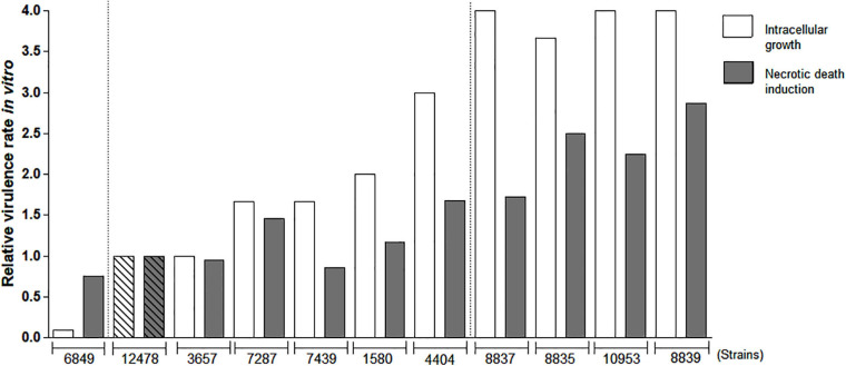
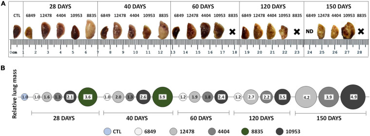
Among non-tuberculous mycobacteria, Mycobacterium kansasii is one of the most pathogenic, able to cause pulmonary disease indistinguishable from tuberculosis in immunocompetent susceptible adults. The lack of animal models that reproduce human-like lung disease, associated with the necrotic lung pathology, impairs studies of M. kansasii virulence and pathogenicity. In this study, we examined the ability of the C57BL/6 mice, intratracheally infected with highly virulent M. kansasii strains, to produce a chronic infection and necrotic lung pathology. As a first approach, we evaluated ten M. kansasii strains isolated from Brazilian patients with pulmonary disease and the reference strain M. kansasii ATCC 12478 for virulence-associated features in macrophages infected in vitro; five of these strains differing in virulence were selected for in vivo analysis. Highly virulent isolates induced progressive lung disease in mice, forming large encapsulated caseous granulomas in later stages (120-150 days post-infection), while the low-virulent strain was cleared from the lungs by day 40. Two strains demonstrated increased virulence, causing premature death in the infected animals. These data demonstrate that C57BL/6 mice are an excellent candidate to investigate the virulence of M. kansasii isolates. We observed considerable heterogeneity in the virulence profile of these strains, in which the presence of highly virulent strains allowed us to establish a clinically relevant animal model. Comparing public genomic data between Brazilian isolates and isolates from other geographic regions worldwide demonstrated that at least some of the highly pathogenic strains isolated in Brazil display remarkable genomic similarities with the ATCC strain 12478 isolated in the United States 70 years ago (less than 100 SNPs of difference), as well as with some recent European clinical isolates. These data suggest that few pathogenic clones have been widely spread within M. kansasii population around the world.

Keywords: Mycobacterium kansasii; animal models; clinical isolates; nontuberculous mycobacteria; pulmonary disease; virulence; virulence factor genes.


Conflict of interest statement

The authors declare that the research was conducted in the absence of any commercial or financial relationships that could be construed as a potential conflict of interest.

Figures

FIGURE 1
FIGURE 1
Colony morphotype variants of Mycobacterium kansasii strains. Mycobacterial strains were grown on Middlebrook 7H10 agar for 21 days (M. kansasii) and 28 days (Mycobacterium tuberculosis), and the macrocolony images were captured. (A) M. kansasii clinical isolates (images are demonstrated for strains 8835, 4404, and 10953) exhibited a rough colony shape (R) similar to that of the reference M. kansasii strain 12478 or the M. tuberculosis strain H37Rv, with exception of the isolate 6849, which showed smooth morphotype (S). (B) All M. kansasii strains grown on the agar exhibited yellow colonies under exposure to light.
FIGURE 2
FIGURE 2
Evaluation of relative virulence of Mycobacterium kansasii strains as measured by in vitro macrophage infection. Mycobacterial ability to intracellular growth in macrophages and induction of macrophage death was evaluated to calculate relative virulence of each M. kansasii isolate compared with that of the reference virulent M. kansasii strain 12478. RAW264.7 macrophage-like cells were infected with the studied strains at a multiplicity of infection (MOI) of 1:1 and cultured for 4 days. Intracellular bacterial growth was evaluated by CFU test. The mean growth value of reference strain 12478 was established as 1.0 and relative growth rate of each clinical isolate was calculated proportionally. To evaluate macrophage death, the cells were infected at a MOI of 10:1, cultured for 3 days, and necrotic cell death was evaluated by quantification of lactate dehydrogenase (LDH) release (LDH test). Relative cytotoxicity of each isolate in relation to the cytotoxicity of reference strain 12478 (established as 1.0) was calculated proportionally.
FIGURE 3
FIGURE 3
Morbidity and mortality of mice after challenge with Mycobacterium kansasii strains. C57BL/6 mice were infected intratracheally with 5 × 10⁴ bacilli of each strain and the disease progression was studied within a 150-day period. (A) Body weight loss was used as an indicator of morbidity. Data are presented as the percentage of initial body weight of each animal prior to infection. Values were reported as mean ± standard deviation, SD, and the differences were considered significant according to ***P < 0.001; **P < 0.01; and *P < 0.05. (B) Survival of the infected animals. The data were obtained in three independent experiments with 10 mice in each group. Kaplan-Meier curves and log rank test were used to evaluate statistical significance. Statistically significant differences between each group infected with the individual clinical isolate and the group infected with reference strain 12478 are presented by symbol ###P < 0.001. (C) Bacterial loads in the lungs were assessed by CFU assay. The data were obtained in three independent experiments with 12–15 mice in each group: 2–3 animals per point. Values were reported as mean ± SD. Mean values that were significantly different from the mean value of the group infected by reference strain 12478 are indicated by asterisks as follows: ***P < 0.001 and **P < 0.01. Significant differences between values obtained for each group at different time points are indicated by symbol ###P < 0.001.
FIGURE 4
FIGURE 4
Macropathological changes in the lungs of mice infected with Mycobacterium kansasii strains. C57BL/6 mice were infected as indicated in the Figure 3 legend, and lungs were examined on day 0, 28, 40, 60, 120, and 150 p.i. (A) Representative images of the upper lobe of the right lung demonstrating the gross pathology in animals of each infected group, observed as numerous giant inflammatory lesions (white nodes of different size). (B) Relative lung mass in different infected groups was determined by the ratio of the mean lung weight of animals in each group to the mean lung weight of control (CTL) animals (1.0). ND, not demonstrated; X-premature animal death.
FIGURE 5
FIGURE 5
Histopathological changes in the lungs of mice infected with low-virulence strain 6849 and high-virulence strain 8835. C57BL/6 mice were infected with strain 6849 (A,B) or strain 8835 (C–F), and lungs were examined on day 28 (A,C,E) and day 40 p. i. (B,D,F). Representative hematoxylin-and-eosin (H&E) -stained lung sections. Few small-size granulomas (A,B). Extensive pneumonia, leading to consolidation of lung tissue (C–F). Note accumulation of exudates in the respiratory bronchioles on day 28 p.i. (E), and blood coagulation in the vessels and diffuse necrotic cell death in the lung parenchima on day 40 p.i. (D,F). Bars, 1,000 μm (A–D), 200 μm (E,F).
FIGURE 6
FIGURE 6
Histopathological changes in the lungs of mice infected with highly virulent Mycobacterium kansasii strain 10953. C57BL/6 mice were infected, and lung pathology was examined during a 150-day period, on day 0 (A), 28 (B), 40 (C), 60 (D–F), 120 (G–I) and 150 (J–L). Representative lung sections stained by H&E (A–E,G,H,J), Ziehl-Neelsen (F) or Masson’s Trichrome methods (I,K,L) are shown. Small granulomas seen on day 28 (B), increased by day 40 (C), and progressed to extensive granulomatous pneumonia by day 60 (D). Area of initial intragranulomatose necrosis, marked by black square in panel (D), is enlarged in panels (E,F), demonstrating large numbers of dying cells (E) and AFB (F). In panel (D), a liver section (l) eventually is localized by side of the lung section; white arrows indicate several granulomas in the liver. By day 120, necrotic lesions progressed to large caseous granulomas marked by black squares (G). The upper and the lower squares refer to areas of higher magnification in panel (H) (peripheral area composed by neutrophil-predominant cell infiltrates) and (I) (central area occupied by acellular caseum), respectively. Note a fibrotic capsule surrounding the necrotic lesion (I). By day 150 (J), necrosis leads to destruction of normal lung structure (symbol *) with easy dislocation of the amorphous necrotic material [lower black square enlarged in panel (L)] and its presence in occluded bronchial airways [black arrows in panel (J)]. The area marked by upper square in panel (J), enlarged in panel (K), demonstrates significant interstitial fibrosis of the lung. Bars, 1,000 μm (A–D,G,J), 100 μm (E,F,H,I,K).
FIGURE 7
FIGURE 7
Morphometric analysis of lung pathology in animals infected with Mycobacterium kansasii strains. For morphometric determination of the inflammatory area occupied in the lungs by cellular or liquid exudates (pneumonia area), images of ten lung sections of each group of animals were captured and analyzed using the Image J program. Statistically significant differences between each group infected with the individual clinical isolate and the respective group infected with reference strain 12478 are presented by asterisks as follows: ***P < 0.001 and **P < 0.01.
FIGURE 8
FIGURE 8
Virulence factors shared between Mycobacterium kansasii ATCC 12478 and Mycobacterium tuberculosis H37Rv, according to their functional categories. The diagram shows the distribution of virulence factors encoded in each mycobacterial genome according to their assigned functional category, as defined in Mycobrowser classification (https://mycobrowser.epfl.ch/). Green dots represent M. tuberculosis H37Rv virulence factors, while blue dots account for homologous counterparts in M. kansasii ATCC 12478.

References

    1. Alcaide F., Richter I., Bernasconi C., Springer B., Hagenau C., Telenti A. (1997). Heterogeneity and clonality among isolates of Mycobacterium kansasii: implications for epidemiological and pathogenicity studies. J. Clin. Microbiol. 35 1959–1964. 10.1128/JCM.35.8.1959-1964.1997 - DOI - PMC - PubMed
    1. Almeida F. M., Ventura T. L., Amaral E. P., Ribeiro S. C., Calixto S. D., Manhães M. R., et al. (2017). Hypervirulent Mycobacterium tuberculosis strain triggers necrotic lung pathology associated with enhanced recruitment of neutrophils in resistant C57BL/6 mice. PLoS One 12:e0173715. 10.1371/journal.pone.0173715 - DOI - PMC - PubMed
    1. Altenhoff A. M., Levy J., Zarowiecki M., Tomiczek B., Vesztrocy A. W., Dalquen D. A., et al. (2019). OMA standalone: orthology inference among public and custom genomes and transcriptomes. Genome Res. 29 1152–1163. 10.1101/gr.243212.118 - DOI - PMC - PubMed
    1. Bakuła Z., Kościuch J., Safianowska A., Proboszcz M., Bielecki J., van Ingen J., et al. (2018a). Clinical, radiological and molecular features of Mycobacterium kansasii pulmonary disease. Respir. Med. 139 91–100. 10.1016/j.rmed.2018.05.007 - DOI - PubMed
    1. Bakuła Z., Modrzejewska M., Pennings L., Proboszcz M., Safianowska A., Bielecki J., et al. (2018b). Drug susceptibility profiling and genetic determinants of drug resistance in Mycobacterium kansasii. Antimicrob. Agents Chemother. 62:e01788-17. 10.1128/AAC.01788-17 - DOI - PMC - PubMed