. 2021 Oct;22(4):1148.
doi: 10.3892/etm.2021.10581. Epub 2021 Aug 9.

Long non-coding RNA SNHG1 relieves microglia activation by downregulating miR-329-3p expression in an in vitro model of cerebral infarction

Affiliations

Long non-coding RNA SNHG1 relieves microglia activation by downregulating miR-329-3p expression in an in vitro model of cerebral infarction

Jianli He et al. Exp Ther Med. 2021 Oct.

Abstract

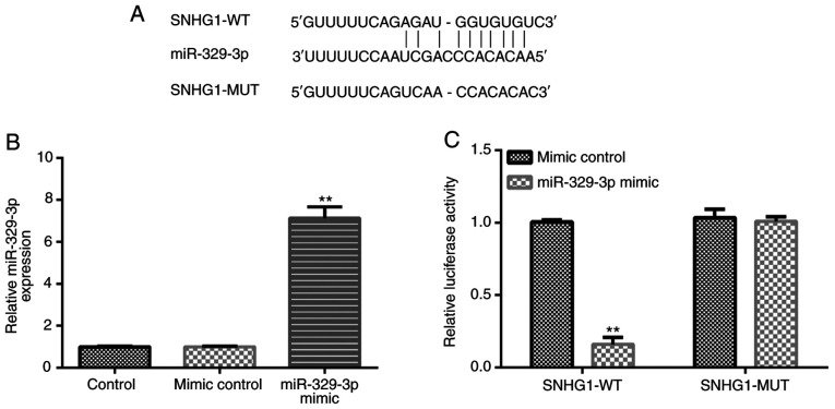
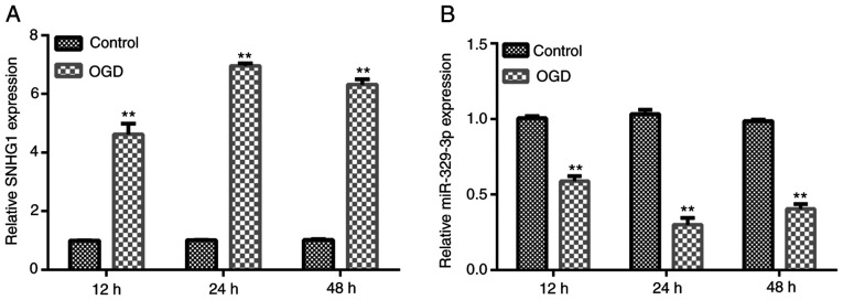
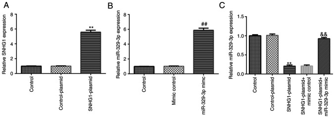
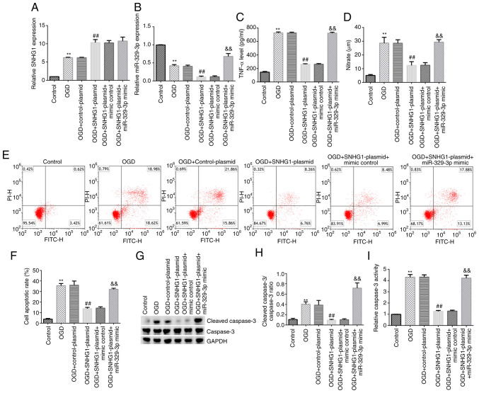
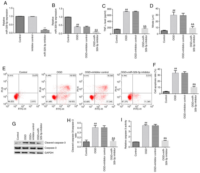
Following cerebral infarction, activated microglia cells can release a large amount of inflammatory cytokines, thereby exacerbating neuronal damage. It has been demonstrated that the long non-coding RNA small nucleolar RNA host gene 1 (SNHG1) exerts a protective effect against cerebral infarction. However, its specific role in cerebral infarction and underlying mechanism have yet to be fully elucidated. The present study aimed to investigate the effects of the SNHG1 and microRNA (miR)-329-3p in cerebral infarction and to determine the underlying molecular mechanisms. An in vitro oxygen-glucose deprivation (OGD) model was established using the BV-2 microglial cell line. The mRNA expression levels of SNHG1 and miR-329-3p were analyzed using reverse transcription-quantitative PCR and the protein expression levels of cleaved caspase-3 and caspase-3 were detected using western blotting. The binding relationship between SNHG1 and miR-329-3p was predicted using starBase and verified using a dual luciferase reporter assay. The release of TNF-α and nitric oxide, as well as caspase-3 activity, were detected using appropriate commercial kits. Flow cytometry analysis was performed to measure cell apoptosis. The results of the present study revealed that the expression levels of SNHG1 were upregulated in the OGD-induced BV-2 cell model. miR-329-3p was discovered to directly target SNHG1, and its mRNA expression levels were downregulated in the OGD-induced BV-2 cell model. The SNHG1-plasmid downregulated miR-329-3p expression levels, while this effect was reversed by transfection with the miR-329-3p mimic. The overexpression of SNHG1 or knockdown of miR-329-3p inhibited OGD-induced BV-2 cell activation. In conclusion, the results of the present study suggested that SNHG1 may reduce microglial cell activity by regulating the expression of miR-329-3p, indicating its potential protective role in cerebral infarction.

Keywords: cerebral infarction; long non-coding RNA small nucleolar RNA host gene 1; microRNA-329-3p; microglia.


Conflict of interest statement

The authors declare that they have no competing interests.

Figures

Figure 1
Figure 1
miR-329-3p directly targets SNHG1. (A) Predicted miR-329-3p binding sites in the 3'-UTR of SNHG1. (B) BV-2 cells were transfected with miR-329-3p mimic or mimic control for 48 h, then the expression levels of miR-329-3p were analyzed using reverse transcription-quantitative PCR. (C) Dual luciferase reporter assay was used to verify the miR-329-3p binding sites in the 3'-UTR of SNHG1. **P<0.01 vs. mimic control. miR, microRNA; SNHG1, small nucleolar RNA host gene 1; UTR, untranslated region; WT, wild-type; MUT, mutant.
Figure 2
Figure 2
Expression levels of SNHG1 and miR-329-3p in OGD-induced BV-2 cells. BV-2 cells were cultured in serum/glucose-free DMEM with 95% N2 and 5% CO2 at 37˚C for 12, 24 or 48 h. Then, relative mRNA expression levels of (A) SNHG1 and (B) miR-329-3p in BV-2 cells were analyzed using reverse transcription-quantitative PCR. **P<0.01 vs. control. SNHG1, small nucleolar RNA host gene 1; miR, microRNA; OGD, oxygen-glucose deprivation.
Figure 3
Figure 3
Transfection efficiency of SNHG1-plasmid and miR-329-3p mimic in BV-2 cells. BV-2 cells were transfected with control-plasmid, SNHG1-plasmid, mimic control, miR-329-3p mimic, SNHG1-plasmid + mimic control, or SNHG1-plasmid + miR-329-3p mimic for 24 h. (A) Reverse transcription-quantitative PCR was performed to analyze the mRNA expression levels of SNHG1 in BV-2 cells following transfection with control-plasmid or SNHG1-plasmid. (B) Reverse transcription-quantitative PCR was performed to analyze the expression levels of miR-329-3p in BV-2 cells following transfection with mimic control or miR-329-3p mimic. (C) Reverse transcription-quantitative PCR was performed to analyze the expression levels of miR-329-3p in BV-2 cells transfected with control-plasmid, SNHG1-plasmid, SNHG1-plasmid + mimic control, or SNHG1-plasmid + miR-329-3p mimic. **P<0.01 vs. control-plasmid; ##P<0.01 vs. mimic control; &&P<0.01 vs. SNHG1-plasmid + mimic control. SNHG1, small nucleolar RNA host gene 1; miR, microRNA.
Figure 4
Figure 4
Overexpression of SNHG1 affects OGD-induced BV-2 cell activation and apoptosis. BV-2 cells were cultured in serum/glucose-free DMEM with 95% N2 and 5% CO2 at 37˚C for 48 h, then the cells were transfected with control-plasmid, SNHG1-plasmid, SNHG1-plasmid + mimic control, or SNHG1-plasmid + miR-329-3p mimic for another 24 h. Reverse transcription-quantitative PCR was performed to analyze the expression levels of (A) SNHG1 and (B) miR-329-3p in BV-2 cells. (C) TNF-α release into the supernatant of the transfected cells was detected using an ELISA. (D) NO production in the supernatant of the transfected cells was detected using a commercial kit. (E and F) Flow cytometric analysis was used to measure the levels of apoptosis of BV-2 cells induced by OGD. (G) Cleaved caspase-3 and caspase-3 protein expression was analyzed using western blotting. (H) Cleaved caspase-3/caspase-3 ratio. (I) Caspase-3 activity was measured using a colorimetric assay kit. **P<0.01 vs. control; ##P<0.01 vs. OGD + control-plasmid; &&P<0.01 vs. OGD + SNHG1-plasmid + mimic control. SNHG1, small nucleolar RNA host gene 1; miR, microRNA; OGD, oxygen-glucose deprivation; NO, nitric oxide.
Figure 5
Figure 5
Knockdown of miR-329-3p expression affects OGD-induced BV-2 cell activation and apoptosis. (A) BV-2 cells were transfected with inhibitor control or miR-329-3p inhibitor for 24 h, and then reverse transcription-quantitative PCR was performed to analyze the expression levels of miR-329-3p. (B) BV-2 cells were cultured in serum/glucose-free DMEM with 95% N2 and 5% CO2 at 37˚C for 48 h; then, the cells were transfected with inhibitor control or miR-329-3p inhibitor for 24 h and reverse transcription-quantitative PCR was performed to analyze the expression levels of miR-329-3p. (C) TNF-α release into the supernatant of the transfected cells was detected using ELISA. (D) NO production in the supernatant of the transfected cells was detected using a commercial kit. (E and F) Flow cytometric analysis was used to measure the levels of apoptosis of BV-2 cells induced by OGD. (G) Cleaved caspase-3 and caspase-3 protein expression was measured using western blotting. (H) Cleaved caspase-3/caspase-3 ratio. (I) Caspase-3 activity was detected using a colorimetric assay kit. **P<0.01 vs. inhibitor control; ##P<0.01 vs. control; &&P<0.01 vs. OGD + inhibitor control. SNHG1, small nucleolar RNA host gene 1; miR, microRNA; OGD, oxygen-glucose deprivation; NO, nitric oxide.

References

    1. Sun W, Li G, Zeng X, Lai Z, Wang M, Ouyang Y, Zeng G, Peng J, Zhong J, Xiao D, et al. Clinical and imaging characteristics of cerebral infarction in patients with nonvalvular atrial fibrillation combined with cerebral artery stenosis. J Atheroscler Thromb. 2018;25:720–732. doi: 10.5551/jat.43240. - DOI - PMC - PubMed
    1. Choi JW, Chong S, Phi JH, Lee JY, Kim HS, Chae JH, Lee J, Kim SK. Postoperative symptomatic cerebral infarction in pediatric moyamoya disease: Risk factors and clinical outcome. World Neurosurg. 2020;136:e158–e164. doi: 10.1016/j.wneu.2019.12.072. - DOI - PubMed
    1. Wang YW, Zhang GM. New silent cerebral infarction in patients with acute non-cerebral amyloid angiopathy intracerebral hemorrhage as a predictor of recurrent cerebrovascular events. Med Sci Monit. 2019;25:418–426. doi: 10.12659/MSM.914423. - DOI - PMC - PubMed
    1. Sveinsson OA, Kjartansson O, Valdimarsson EM. Cerebral ischemia/infarction-epidemiology, causes and symptoms. Laeknabladid. 2014;100:271–279. doi: 10.17992/lbl.2014.05.543. (In Icelandic) - DOI - PubMed
    1. Rojsanga W, Sawanyawisuth K, Chotmongkol V, Tiamkao S, Kongbonkiat K, Kasemsap N. Clinical risk factors predictive of thrombotic stroke with large cerebral infarction. Neurol Int. 2019;11(7941) doi: 10.4081/ni.2019.7941. - DOI - PMC - PubMed