Review
. 2021 Aug 24:8:729786.
doi: 10.3389/fcvm.2021.729786. eCollection 2021.

Imaging of Cardiac Device-Related Infection

Affiliations
Review

Imaging of Cardiac Device-Related Infection

Jose Aguilera et al. Front Cardiovasc Med. .

Abstract

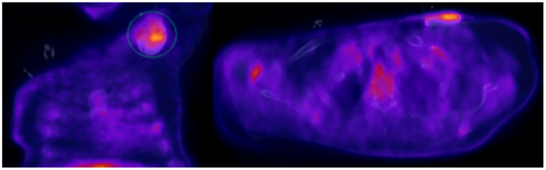
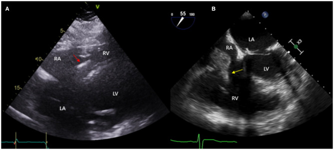
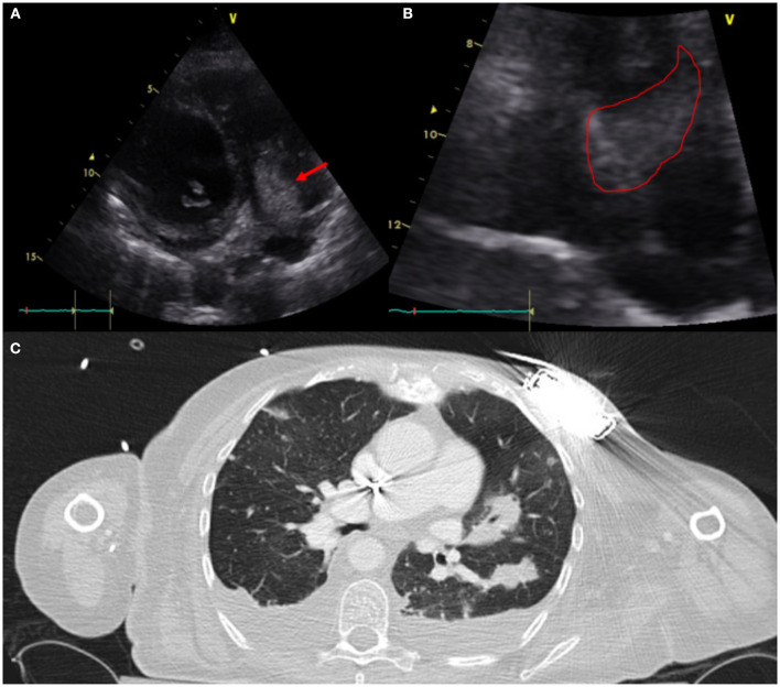
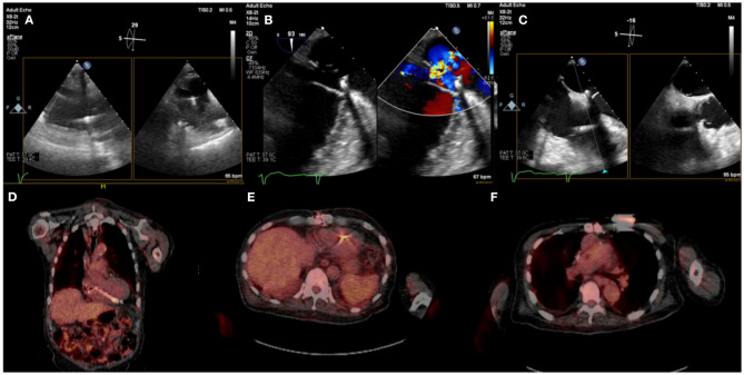
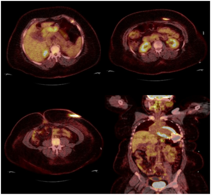
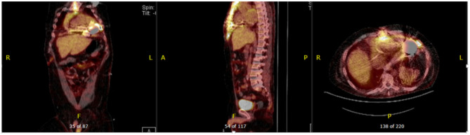
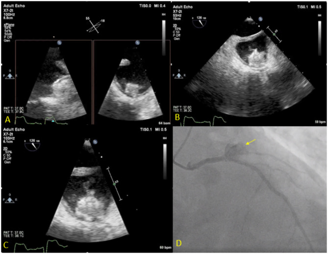
Cardiac devices are frequently used in different cardiovascular conditions for the purpose of morbidity or mortality prevention. These include cardiac implantable electronic devices (CIED) like permanent pacemakers and implantable cardiac defibrillators, ventricular assistance devices (VADs), left atrial appendage occlusion (LAAO) devices like the Watchman™, atrial and ventricular septal occluders like the Amplatzer™, among others. In the past years, there has been an increase in the development of these devices as a result of a rise in the number of indications for implantation, paired with the aging and more medically complex patient population. This has led to an increase in the incidence of cardiac device-related infections, one of the most feared and serious complications which is associated with significant morbidity, mortality and financial burden. Accurate diagnosis of cardiac device-related infections is essential given the management implications which often involve removal of the infected device, removal of other prosthetic material and long-term antimicrobial therapy. Clinical and laboratory data are useful diagnostic tools but multimodality imaging is often necessary. The recently published 2020 European Heart Rhythm Association International Consensus document, which is endorsed by many expert societies, has recommended the use of multimodality imaging for the diagnosis of CIED infections. (1) This allows better disease characterization by identifying abnormal fluid collections and guiding aspiration for both diagnostic and therapeutic purposes (i.e. soft tissue ultrasound and computed tomography), evaluation for local extent of disease (i.e. transesophageal echocardiogram to evaluate for concomitant infective endocarditis), embolic manifestation of disease (i.e. computed tomography and magnetic resonance imaging) and metabolic tissue characterization (positron emission tomography and tagged white blood cell scan). (2) In addition, computed tomography (CT) allows for pre-procedural planning which has shown to be associated with better procedural outcomes.

Keywords: cardiac device; cardiac implantable electronic devices; computed tomography; device infection; endocarditis; left ventricular assist devices; positron emission tomography; transesophageal echocardiogram.


Conflict of interest statement

The authors declare that the research was conducted in the absence of any commercial or financial relationships that could be construed as a potential conflict of interest.

Figures

Figure 1
Figure 1
Seventy year-old male with sick sinus syndrome s/p dual chamber PPM 2 years prior, who presented with fever. PPM pocket site with mild tenderness on palpation but no obvious signs of infection. Infectious work up was unremarkable but given ongoing fever and high clinical suspicion for pocket infection, an 18F-FDG PET-CT was obtained showing increased FDG pocket uptake consistent with pocket infection. The patient underwent complete system removal.
Figure 2
Figure 2
Fifty nine year-old male with a historyof ischemic cardiomyopathy (EF 35%) s/p ICD for primary prevention of sudden cardiac death who presented with fever and MSSE bacteremia. Initial TTE done (A) shows subcostal view with ICD lead in place (red arrow), without evident vegetations. Based on high clinical suspicion for CIED infection, a TEE was obtained (B) which showed a large vegetation attached to the ICD lead prolapsing into the right ventricle (yellow arrow). The patient was treated with IV antibiotics but given persistent bacteremia and large vegetation he underwent sternotomy with ICD system extraction and epicardial defibrillator placement.
Figure 3
Figure 3
Sixty one year-old woman with a history of NICM s/p single lead ICD who presented with fever and malaise. Infectious work up was positive for MSSA bacteremia. TTE showed a very large vegetation attached to the ICD lead in the RA protruding into the RV [(A)-modified apical view and (B)-RV focused view with RA zoom]. A pre-procedural CT (C) was obtained and revealed bilateral, extensive and multifocal septic emboli with cavitary lesions as well as bilateral pleural effusions without evidence of pocket infection. The patient underwent open sternotomy with device extraction and TV repair for septal leaflet perforation for management of CIED systemic infection with IE.
Figure 4
Figure 4
Eighty five year-old male with history ischemic cardiomyopathy s/p dual-chamber ICD for primary prevention, history of CABG and mitral valve repair presented with persistent MRSA bacteremia. TEE showed small linear echodensities attached to right atrial lead (A), multiple small echodensities attached to mitral ring and moderate mitral regurgitation (B). The RV showed no vegetations with normal tricuspid valve (C). 18F-FDG PET-CT was obtained to assess extent of infection and showed focal uptake along the RV lead (D,E). Left chest wall ICD showed focal uptake but no pocket uptake to suggest pocket infection (F). The patient was deemed to have a prohibitive risk for redo cardiac surgery and was treated medically. He passed away 2 months after the diagnosis of CIED systemic infection with MRSA endocarditis.
Figure 5
Figure 5
Thirty eight year-old female with end-stage heart failure s/p LVAD (Heart Mate 3) presented with drainage around driveline exit site and abdominal pain. She was treated with oral antibiotics for several weeks but continued to have symptoms. 18F-FDG PET-CT was obtained to assess extent of infection and showed increase FDG uptake surrounding LVAD driveline in the anterior abdominal wall without collections. She underwent driveline debridement for DLI and the driveline exit was relocated medially.
Figure 6
Figure 6
Forty seven year-old male with ischemic cardiomyopathy s/p LVAD (Heart Mate 3) presented with suspected DLI, with wound cultures positive for Candida Albicans. 18F-FDG PET-CT showed FDG activity in the pericardium, mediastinum, surrounding LVAD pump, driveline and sternum consistent with deep and superficial LVAD infection. He was managed with systemic lifelong antifungal therapy.
Figure 7
Figure 7
Seventy three year-old male with history of CAD s/p multiple PCIs and Afib s/p Watchman implantation 3 weeks prior to presentation, presented with fever, chest pain and confusion. Blood cultures grew Serratiamarcensens. TEE (A–C) shows Watchman device in the left atrial appendage with large mobile vegetations. Course was complicated by distal embolization to the brain an intestine vasculature. Patient developed worsening chest pain and ST depressions, coronary angiogram (D) shows total occlusion of the proximal left circumflex artery secondary to mycotic aneurysm (Arrow). The patient was deemed too high risk for surgery and was treated medically.

Similar articles

Cited by

References

    1. Blomström-Lundqvist C, Traykov V, Erba PA, Burri H, Nielsen JC, Bongiorni MG, et al. . European Heart Rhythm Association (EHRA) international consensus document on how to prevent, diagnose, and treat cardiac implantable electronic device infections-endorsed by the Heart Rhythm Society (HRS), the Asia Pacific Heart Rhythm Society (APHRS), the Latin American Heart Rhythm Society (LAHRS), International Society for Cardiovascular Infectious Diseases (ISCVID), and the European Society of Clinical Microbiology and Infectious Diseases (ESCMID) in collaboration with the European Association for Cardio-Thoracic Surgery (EACTS). Eur Heart J. (2020) 41:2012–32. 10.1093/eurheartj/ehaa010 - DOI - PubMed
    1. De Winter F, Vogelaers D, Gemmel F, Dierckx RA. Promising role of 18-F-fluoro-D-deoxyglucose positron emission tomography in clinical infectious diseases. Eur J Clin Microbiol Infect Dis. (2002) 21:247–57. 10.1007/s10096-002-0708-2 - DOI - PubMed
    1. Sohail MR, Uslan DZ, Khan AH, Friedman PA, Hayes DL, Wilson WR, et al. . Infective endocarditis complicating permanent pacemaker and implantable cardioverter-defibrillator infection. Mayo Clin Proc. (2008) 83:46–53. 10.4065/83.1.46 - DOI - PubMed
    1. Dai M, Cai C, Vaibhav V, Sohail MR, Hayes DL, Hodge DO, et al. . Trends of cardiovascular implantable electronic device infection in 3 decades: a population-based study. JACC Clin Electrophysiol. (2019) 5:1071–80. 10.1016/j.jacep.2019.06.016 - DOI - PubMed
    1. Kusumoto FM, Schoenfeld MH, Wilkoff BL, Berul CI, Birgersdotter-Green UM, Carrillo R, et al. . 2017 HRS expert consensus statement on cardiovascular implantable electronic device lead management and extraction. Heart Rhythm. (2017) 14:e503–51. 10.1016/j.hrthm.2017.09.001 - DOI - PubMed