

Modeling human congenital disorders with neural crest developmental defects using patient-derived induced pluripotent stem cells
- PMID: 34504908
- PMCID: PMC8390449
- DOI: 10.1016/j.reth.2021.08.001
Modeling human congenital disorders with neural crest developmental defects using patient-derived induced pluripotent stem cells
Abstract
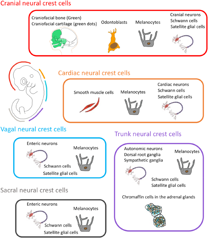
The neural crest is said to be the fourth germ layer in addition to the ectoderm, mesoderm and endoderm because of its ability to differentiate into a variety of cells that contribute to the various tissues of the vertebrate body. Neural crest cells (NCCs) can be divided into three functional groups: cranial NCCs, cardiac NCCs and trunk NCCs. Defects related to NCCs can contribute to a broad spectrum of syndromes known as neurocristopathies. Studies on the neural crest have been carried out using animal models such as Xenopus, chicks, and mice. However, the precise control of human NCC development has not been elucidated in detail due to species differences. Using induced pluripotent stem cell (iPSC) technology, we developed an in vitro disease model of neurocristopathy by inducing the differentiation of patient-derived iPSCs into NCCs and/or neural crest derivatives. It is now possible to address complicated questions regarding the pathogenetic mechanisms of neurocristopathies by characterizing cellular biological features and transcriptomes and by transplanting patient-derived NCCs in vivo. Here, we provide some examples that elucidate the pathophysiology of neurocristopathies using disease modeling via iPSCs.
Keywords: Disease modeling; Neurocristopathy; iPS cells derived neural crest cells.
© 2021 The Japanese Society for Regenerative Medicine. Production and hosting by Elsevier B.V.
Conflict of interest statement
H.O. is a founding scientist and scientific advisor of SanBio Co. Ltd. and K Pharma Inc. Other author indicates no potential conflicts of interest.
Figures
Similar articles
-
Human Pluripotent Stem Cell-Derived Neural Crest Cells for Tissue Regeneration and Disease Modeling.Front Mol Neurosci. 2019 Feb 22;12:39. doi: 10.3389/fnmol.2019.00039. eCollection 2019. Front Mol Neurosci. 2019. PMID: 30853889 Free PMC article. Review.
-
Modeling Early Neural Crest Development via Induction from hiPSC-Derived Neural Plate Border-like Cells.Methods Mol Biol. 2022;2549:281-298. doi: 10.1007/7651_2021_454. Methods Mol Biol. 2022. PMID: 35355234
-
The stemness of neural crest cells and their derivatives.Birth Defects Res C Embryo Today. 2014 Sep;102(3):251-62. doi: 10.1002/bdrc.21079. Epub 2014 Sep 15. Birth Defects Res C Embryo Today. 2014. PMID: 25219876 Review.
-
CHARGE syndrome modeling using patient-iPSCs reveals defective migration of neural crest cells harboring CHD7 mutations.Elife. 2017 Nov 28;6:e21114. doi: 10.7554/eLife.21114. Elife. 2017. PMID: 29179815 Free PMC article.
-
Immunological Properties of Neural Crest Cells Derived from Human Induced Pluripotent Stem Cells.Stem Cells Dev. 2019 Jan 1;28(1):28-43. doi: 10.1089/scd.2018.0058. Epub 2018 Nov 22. Stem Cells Dev. 2019. PMID: 30251915 Free PMC article.
Cited by
-
Clinical and Genetic Correlation in Neurocristopathies: Bridging a Precision Medicine Gap.J Clin Med. 2024 Apr 11;13(8):2223. doi: 10.3390/jcm13082223. J Clin Med. 2024. PMID: 38673496 Free PMC article. Review.
-
Fascial Nomenclature: Update 2022.Cureus. 2022 Jun 13;14(6):e25904. doi: 10.7759/cureus.25904. eCollection 2022 Jun. Cureus. 2022. PMID: 35720786 Free PMC article. Review.
-
Alternative Balance between Transcriptional and Epigenetic Regulation during Developmental Proliferation of Human Cranial Neural Crest Cells.Cells. 2024 Sep 30;13(19):1634. doi: 10.3390/cells13191634. Cells. 2024. PMID: 39404397 Free PMC article.
References
-
- Huang X., Saint-Jeannet J.P. Induction of the neural crest and the opportunities of life on the edge. Dev Biol. 2004;275(1):1–11. - PubMed
-
- Dupin E. The issue of the multipotency of the neural crest cells. Dev Biol. 2018;444(1):S47–S59. - PubMed
-
- Baggiolini A. Premigratory and migratory neural crest cells are multipotent in vivo. Cell Stem Cell. 2015;16(3):314–322. - PubMed
-
- Le Douarin N.M., Dupin E. The “beginnings” of the neural crest. Dev Biol. 2018;444(1):S3–S13. - PubMed
-
- Minoux M., Rijli F.M. Molecular mechanisms of cranial neural crest cell migration and patterning in craniofacial development. Development. 2010;137(16):2605–2621. - PubMed
Publication types
LinkOut - more resources
Full Text Sources
