. 2021 Jun 28:38:100988.
doi: 10.1016/j.eclinm.2021.100988. eCollection 2021 Aug.

High cerebrospinal amyloid-β 42 is associated with normal cognition in individuals with brain amyloidosis

Affiliations

High cerebrospinal amyloid-β 42 is associated with normal cognition in individuals with brain amyloidosis

Andrea Sturchio et al. EClinicalMedicine. .

Abstract

Background: Brain amyloidosis does not invariably predict dementia. We hypothesized that high soluble 42-amino acid β amyloid (Aβ42) peptide levels are associated with normal cognition and hippocampal volume despite increasing brain amyloidosis.

Methods: This cross-sectional study of 598 amyloid-positive participants in the Alzheimer's Disease Neuroimaging Initiative cohort examined whether levels of soluble Aβ42 are higher in amyloid-positive normal cognition (NC) individuals compared to mild cognitive impairment (MCI) and Alzheimer's disease (AD) and whether this relationship applies to neuropsychological assessments and hippocampal volume measured within the same year. All subjects were evaluated between June 2010 and February 2019. Brain amyloid positivity was defined as positron emission tomography-based standard uptake value ratio (SUVR) ≥1.08 for [18] F-florbetaben or 1.11 for [18]F-florbetapir, with higher SUVR indicating more brain amyloidosis. Analyses were adjusted for age, sex, education, APOE4, p-tau, t-tau, and centiloids levels.

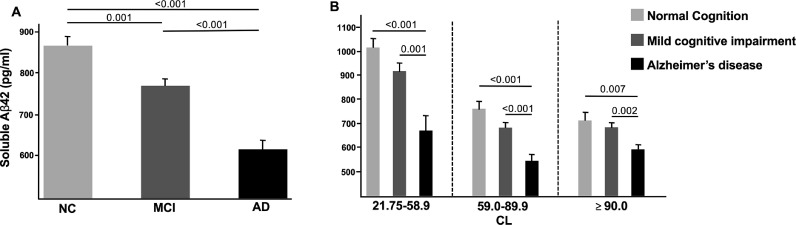
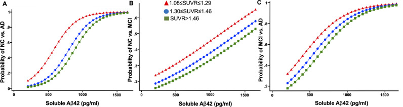
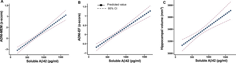
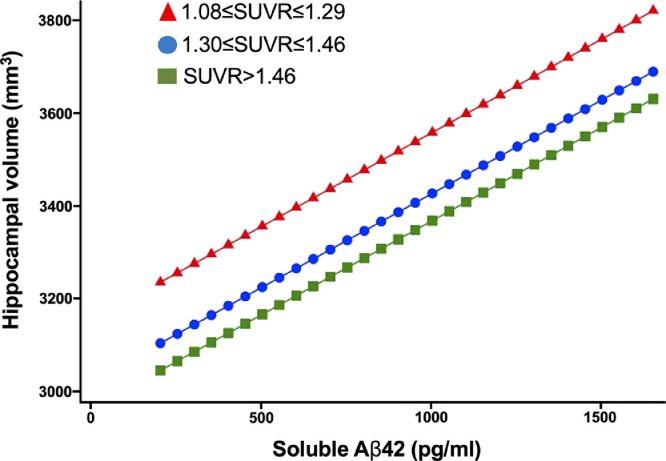
Findings: Higher soluble Aβ42 levels were observed in NC (864.00 pg/ml) than in MCI (768.60 pg/ml) or AD (617.46 pg/ml), with the relationship between NC, MCI, and AD maintained across all amyloid tertiles. In adjusted analysis, there was a larger absolute effect size of soluble Aβ42 than SUVR for NC (0.82 vs. 0.40) and MCI (0.60 vs. 0.26) versus AD. Each standard deviation increase in Aβ42 was associated with greater odds of NC than AD (adjusted odds ratio, 6.26; p < 0.001) or MCI (1.42; p = 0.006). Higher soluble Aβ42 levels were also associated with better neuropsychological function and larger hippocampal volume.

Interpretation: Normal cognition and hippocampal volume are associated with preservation of high soluble Aβ42 levels despite increasing brain amyloidosis.

Funding: Please refer to the Funding section at the end of the article.

Keywords: Alzheimer's disease; Atrophy; Hippocampus; Β-amyloid.


Conflict of interest statement

None.

Figures

Fig. 1
Fig. 1
Adjusted analyses of amyloid-positive normal cognition compared to mild cognitive impairment and Alzheimer's disease subjects. (A) Soluble Aβ42 levels in each diagnostic category; (B) soluble Aβ42 levels in each diagnostic category across CL tertiles. The estimated means of soluble Aβ42 were adjusted for age, sex, education, APOE4, centiloids, p-tau levels, and t-tau levels. The estimated means of soluble Aβ42 by CL tertiles are adjusted for age, sex, education, APOE4, p-tau levels, and t-tau levels. *The corresponding SUVR analysis, which demonstrated similar results, is shown in Supplementary Fig. 2.3. CL: centiloids (higher CL means greater amyloid burden); NC: normal cognition; MCI: mild cognitive impairment, AD: Alzheimer's disease.
Fig. 2
Fig. 2
Adjusted probability of amyloid-positive normal cognition versus Alzheimer's disease (A), normal cognition versus mild cognitive impairment (B), and mild cognitive impairment versus Alzheimer's disease (C) with levels of soluble Aβ42 by SUVR tertiles of brain amyloid. SUVR: standardized uptake value ratio; NC: normal cognition; MCI: mild cognitive impairment; AD: Alzheimer's disease. The results are adjusted for age, sex, education, APOE4, p-tau levels, and t-tau levels.
Fig. 3
Fig. 3
Adjusted association of CSF Aβ42 with cognitive assessments (A, B) and hippocampal volume (average right and left) (C) in the amyloid PET-positive cohorts. SUVR: standardized uptake value ratio; ADNI memory score; ADNI-EF: ADNI executive function; CI: confidence interval. The results are adjusted for age, sex, education, APOE4, centiloids, p-tau levels, and t-tau levels including intracranial volume for hippocampal volume analysis.
Fig. 4
Fig. 4
Adjusted association of CSF Aβ42 with hippocampal volume (average right and left) by SUVR tertiles of brain amyloid. SUVR: standardized uptake value ratio. The results are adjusted for age, sex, education, APOE4, p-tau levels, t-tau levels, and intracranial volume.

References

    1. Selkoe D.J., Hardy J. The amyloid hypothesis of Alzheimer's disease at 25 years. EMBO Mol Med. 2016;8:595–608. - PMC - PubMed
    1. Malmberg M., Malm T., Gustafsson O. Disentangling the amyloid pathways: a mechanistic approach to etiology. Front Neurosci. 2020;14:256. - PMC - PubMed
    1. Nagase T., Iwaya K., Iwaki Y. Insulin-derived amyloidosis and poor glycemic control: a case series. Am J Med. 2014;127:450–454. May. - PubMed
    1. Navalkar A., Ghosh S., Pandey S., Paul A., Datta D., Maji S.K. Prion-like p53 amyloids in cancer. Biochemistry. 2020;59:146–155. - PubMed
    1. Kent S.A., Spires-Jones T.L., Durrant C.S. The physiological roles of tau and Aβ: implications for Alzheimer's disease pathology and therapeutics. Acta Neuropathol. 2020;140:417–447. - PMC - PubMed

LinkOut - more resources