. 2021 Sep 10:10:e72409.
doi: 10.7554/eLife.72409.

A Tad-like apparatus is required for contact-dependent prey killing in predatory social bacteria

Affiliations

A Tad-like apparatus is required for contact-dependent prey killing in predatory social bacteria

Sofiene Seef et al. Elife. .

Abstract

Myxococcus xanthus, a soil bacterium, predates collectively using motility to invade prey colonies. Prey lysis is mostly thought to rely on secreted factors, cocktails of antibiotics and enzymes, and direct contact with Myxococcus cells. In this study, we show that on surfaces the coupling of A-motility and contact-dependent killing is the central predatory mechanism driving effective prey colony invasion and consumption. At the molecular level, contact-dependent killing involves a newly discovered type IV filament-like machinery (Kil) that both promotes motility arrest and prey cell plasmolysis. In this process, Kil proteins assemble at the predator-prey contact site, suggesting that they allow tight contact with prey cells for their intoxication. Kil-like systems form a new class of Tad-like machineries in predatory bacteria, suggesting a conserved function in predator-prey interactions. This study further reveals a novel cell-cell interaction function for bacterial pili-like assemblages.

Keywords: Myxococcus xanthus; infectious disease; microbiology; motility; predation; tad pilus.


Conflict of interest statement

SS, JH, Pd, LM, GB, DR, RJ, RM, EC, BH No competing interests declared, TM Reviewing editor, eLife

Figures

Figure 1.
Figure 1.. A-motility is required for invasion of prey colonies.
Colony plate assays showing invasion of an E. coli prey colony (dotted line) 48 hr after plating by WT (a, Video 1), A+S- (b) and A-S+ (c, Video 2) strains. Scale bar = 2 mm. (a1) Zoom of the invasion front. Myxococcus single cells are labeled with mCherry. Blue arrows show the movement of ‘arrowhead’ cell groups as they invade prey colonies. White arrows point to A-motile single cells that penetrated the prey colony. Scale bar = 10 µm. See associated Video 1 for the full time lapse. (b1) Zoom of the invasion front formed by A+S- cells. The A-motile Myxococcus cells can infiltrate the prey colony and kill prey cells. Scale bar = 10 µm. (c1) Zoom of the invasion front formed by A-S+ cells. Note that the S-motile Myxococcus cells come in contact with the prey colony, but in absence of A-motility, the predatory cells fail to infiltrate the colony and remain stuck at the border. Scale bar = 10 µm. See associated Video 2 for the full time lapse.
Figure 2.
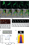
Figure 2.. A-motile cells kill prey cells by contact.
(a) Prey (E. coli) colony invasion by an ‘arrowhead formation’. Activity of the A-motility complex is followed by monitoring Myxococcus cells expressing the bFA-localized AglZ-YFP protein. Upper panel: Cells within the arrowhead (outlined in white) assemble bFAs (white arrowheads). Lower panel: Semantic segmentation (see methods) of the total cell population, E. coli (white) and Myxococcus (green). The numbered and colored E. coli cells (magenta) are the ones that are observed to lyse as the Myxococcus cells penetrate the colony. See associated Video 3 for the full time lapse. Scale bar = 10 µm. (b) bFAs are disassembled when Myxococcus establishes lytic contacts with prey cells. Shown is an AglZ-YFP expressing Myxococcus cell establishing contact with an mCherry-expressing E. coli cell (overlay and phase contrast image). Note that the Myxococcus cell resumes movement and thus re-initiates bFA (white arrowheads) formation immediately after E. coli cell lysis. See associated Video 4 for the full time lapse. Scale bar = 2 µm. (c) Myxococcus (outlined in white) provokes E. coli plasmolysis. Top: shown is a GFP-expressing E. coli cell lysing in contact with a Myxococcus cell. GFP fluorescence remains stable for 5 min after contact and becomes undetectable instantaneously, suggesting plasmolysis of the E. coli cell. Scale bar = 2 µm. Bottom: graphic representation of fluorescence intensity loss upon prey lysis. (d,e) Myxococcus contact provokes local degradation of the E. coli peptidoglycan. (d) E. coli PG was labeled covalently with the fluorescent D-amino acid TADA. Two E. coli cells lyse upon contact. Holes in the PG-labelling are observed at the contact sites (white arrows). Note that evidence for plasmolysis and local IM membrane contraction is visible by phase contrast for the lower E. coli cell (dark arrow). Scale bar = 2 µm. (e) Kymograph of TADA-labeling corresponding to the upper E. coli cell. At time 0, which corresponds to the detection of cell lysis, a hole is detected at the contact site and propagates bi-directionally from the initial site showing that the prey cell wall is degraded in time after cell death. Scale bar = 1 µm.
Figure 2—figure supplement 1.
Figure 2—figure supplement 1.. A-motility appears to be essential for predation.
WT M. xanthus and the different motility mutant strains were mixed with E. coli and spotted on CF 1.5% agar plates (+0.07% glucose). After 24 hr of incubations pictures were taken, showing that WT and ∆t6ss had similar predation efficiencies. A A+S- (∆pilA) strain presented a predation efficiency slightly reduced. However, the A-S+ strain (∆aglQ) was greatly impaired at predating. No predation was observed for the A-S- strain (∆aglQpilA). Therefore, A-motility appears to be essential for predation.
Figure 2—figure supplement 2.
Figure 2—figure supplement 2.. Contact-dependent killing by an A-S- motility mutant (∆aglQpilA).
Growth of E. coli cells leads to contact with non-motile Myxococcus cells and rapid lysis. Example cell reflects events observed for n=20 events. Scale bar = 2 µm.
Figure 2—figure supplement 3.
Figure 2—figure supplement 3.. Contact-dependent killing by a ∆t6ss mutant.
E. coli prey cells are labeled with GFP to monitor contact-dependent lysis. Example cell reflects events observed for n=20 events. Scale bar = 2 µm.
Figure 2—figure supplement 4.
Figure 2—figure supplement 4.. T6SS VipA sheath assembly in Myxococcus cells during predation.
Several assembly patterns are observed as described in other bacteria. Stretched: extended T6SS sheaths. Contracted: retracted T6SS sheath. Scale bars = 2 µm.
Figure 2—figure supplement 5.
Figure 2—figure supplement 5.. Prey contact-dependent lysis is not correlated to T6SS sheath contraction.
Contact-dependent lysis and VipA-GFP dynamics were observed simultaneously. Contraction and lysis at the contacted site were only marginally observed (correlated) suggesting that T6SS intoxication plays a minor role at best in contact-dependent killing.
Figure 2—figure supplement 6.
Figure 2—figure supplement 6.. Contact-dependent lysis in liquid cultures.
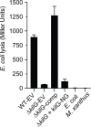
E. coli lysis is detected as extracellular release of the β-galactosidase allows hydrolysis of CPRG which becomes colored after 24 hr. Lysis is not observed when Myxococcus (WT or ∆t6ss) and E. coli are separated by a membrane, showing that it is contact-dependent.
Figure 2—figure supplement 7.
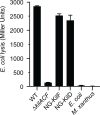
Figure 2—figure supplement 7.. CPRG colorimetric assay performed on motility and T6SS mutant strains.
β-galactosidase activities of the cell lysates (n=4, expressed in Miller Units) were measured for each strain. After 24 hr incubation, only WT, ∆t6ss and A-S+ strains had the ability to lyse E. coli in liquid. PilA appears to be essential for cell-cell contact with E. coli and cell lysis in liquid. This experiment was independently performed four times. Error bars represent the standard deviation to the mean.
Figure 2—figure supplement 8.
Figure 2—figure supplement 8.. Crystal violet assay.
After 24 hr incubation, wells containing the different M. xanthus strains mixed with E. coli were stained with a crystal violet solution to revealed biofilm formation. The ∆pilA strains appeared to be deficient at forming a biofilm in the presence of a prey.
Figure 3.
Figure 3.. A Tad-like apparatus is required for prey recognition and contact-dependent killing.
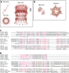
(a) Model structure of the Kil system following bioinformatics predictions. Annotated cluster 1 and cluster 2 genes are shown together with the possible localization of their protein products. Dark triangles indicate the genes that were deleted in this study. (b) kil mutants are impaired in E. coli lysis in liquid. Kinetics of CPRG-hydrolysis by the β-Galactosidase (expressed as Miller Units) observed after co-incubation of Myxococcus wild-type (WT) or the kil mutants with E. coli for 24 hr. M. xanthus and E. coli alone were used as negative controls. This experiment was performed independently four times. (c) The percentage of contacts with E. coli leading to a pause in motility was calculated for M. xanthus wild-type (from five independent predation movies, number of contacts observed n=807) and the kil mutants (number of contacts observed for ∆kilC: n=1780; ∆kilH: n=1219; ∆kilG: n=1141; ∆kilB: n=842; ∆kilK: n=710; ∆kilKLM: n=1446) (d) The percentage of contacts with E. coli leading to cell lysis was also estimated. In panels (b), (c), and (d), error bars represent the standard deviation of the mean. One-way ANOVA statistical analysis followed by Dunnett’s posttest was performed to evaluate if the differences observed, relative to wild-type, were significant (*: p≤0.05, **: p≤0.01, ****: p≤0.0001) or not (ns: p>0.05).
Figure 3—figure supplement 1.
Figure 3—figure supplement 1.. Bioinformatics analyses of Kil proteins.
(a, b) Structural models of the putative KilC secretin (a) and KilF hexameric ATPase (b). KilC Secretin: tertiary and quaternary structural models were based on the structure of E. coli type II secretion system GspD protein D (PDB identifier 5WQ7) and generated with SWISS-MODEL (Materials and methods). KilF ATPase: modeled with Phyre2 and SWISS-MODEL based on the structure of the Sulfolobus acidocaldarius FlaI ATPase (PDB identifier 4II7). (c, d) Analysis of putative pseudo-pilin proteins. For clarity, the multiple alignment is separated in three blocks representing the three different groups, the alpha-proteobacteria, the Myxococcales and the Bdellovibrionales. All sequences, except the one from C. fuscus, were taken from HHPRED matrix alignments. Residues conserved between the pfam domains TadF and the MXAN_4660 family (c), as well as between the TadE and the MXAN_4658 family (d) are highlighted in cyan; residues conserved between the Bd0115 family and the MXAN_4660 family (c), as well as the residues conserved between the Bd0115 family and the MXAN_4658 family (d) are highlighted in brown; residues conserved in all families (TadF, MXAN_4660, Bd0115, TadE, MXAN_4658) are highlighted in red. (c) HHPRED-based multiple sequence alignment of MXAN_4660 (KilM) with the TadF domain and pilus assembly protein Bd0115 from B. bacteriovorus. Myxobacterial sequences correspond to the following NCBI RefSeq IDs: Myxococcus xanthus: WP_011554652; Myxococcus stipitatus: WP_015350653; Myxococcus macrosporus: WP_043711698; Stigmatella aurantiaca: WP_013376800; Cystobacter fuscus: WP_002624349. (d) HHPRED-based multiple sequence alignment of MXAN_4658 with the TadE domain and pilus assembly protein Bd0115 from B. bacteriovorus. Myxobacterial sequences correspond to the following NCBI RefSeq IDs: M. xantus: WP_011554650; M. stipitatus: WP_015350651; M. macrosporus: WP_043711696; S. aurantiaca: WP_013376798; C. fuscus: WP_002624796.
Figure 3—figure supplement 2.
Figure 3—figure supplement 2.. The kil genes are expressed during starvation.
RNA-seq analysis of kil gene expression in rich medium, starvation medium and starvation medium with live prey cells extracted and computed from data by Livingstone et al., 2018. For each gene and condition, the data is compiled from three independent biological replicates. Note addition of prey does not change the expression profile which is significantly induced by starvation alone.
Figure 3—figure supplement 3.
Figure 3—figure supplement 3.. CPRG colorimetric assay.
In a 96-well plate, the different kil strains transformed with pSWU19-EV (Empty Vector) or complemented (comp) with a pSWU19 carrying the different kil genes were incubated with E. coli in liquid. After 24 hr incubation, β-galactosidase activities (expressed as Miller Units) of the different cell lysates were measured. The kil mutants ectopically expressing the different kil genes under the control of the pilA promoter presented a restored predation phenotype similar to WT-EV control. E. coli and M. xanthus alone were used as negative controls. This experiment was independently performed twice with at least two experimental replicates per strain each time. Error bars represent the standard deviation to the mean.
Figure 4.
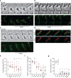
Figure 4.. The Kil Tad-like system assembles upon contact and causes prey cell lysis.
(a) NG-KilF clusters form in contact with the prey and their formation precedes cell lysis. Scale bar = 2 µm. See associated Video 5 for the full time lapse. (b) KilG-NG forms clusters at the contact site with the prey and their formation is followed by the prey cell lysis. Scale bar = 2 µm. See associated Video 6 for the full time lapse. (c) NG-KilD clusters only form in contact with the prey and their formation precedes cell lysis. Scale bar = 2 µm. See associated Video 7 for the full time lapse. (d) PG-holes are formed at the cluster-assembly sites. Representative picture of TADA-labeled E. coli cells in the presence of NG-KilD expressing Myxococcus xanthus cells. PG holes and clusters are indicated with white arrows. Scale bar = 2 µm. (e) The percentage of contacts with E. coli leading to NG-KilD foci formation was calculated for M. xanthus wild-type (from five independent predation movies, number of contacts observed n=807) and the kil mutants (number of contacts observed for ∆kilC: n=1780; ∆kilH: n=1219; ∆kilG: n=1141; ∆kilB: n=842; ∆kilK: n=710; ∆kilKLM: n=1446). (f) The percentage of NG-KilD foci associated with a motility pause was also estimated for M. xanthus WT (from five independent predation movies, number of NG-KilD foci observed n=198) and the kil mutants (number of NG-KilD foci observed for ∆kilC: n=320; ∆kilH: n=270; ∆kilG: n=251; ∆kilB: n=215; ∆kilK: n=94; ∆kilKLM: n=355). (g) The percentage of NG-KilD foci leading to E. coli lysis was estimated as well. In panels (d), (e), and (f), error bars represent the standard deviation to the mean. One-way ANOVA statistical analysis followed by Dunnett’s posttest was performed to evaluate if the differences observed, relative to wild-type, were significant (*: p≤0.05, **: p≤0.01, ****: p≤0.0001) or not (ns: p>0.05).
Figure 4—figure supplement 1.
Figure 4—figure supplement 1.. The strains expressing Neon Green (NG) fusions of KilD or KilF have predation phenotypes similar to wild-type in a CPRG colorimetric assay.
This experiment was independently performed twice. Error bars represent the standard deviation to the mean.
Figure 4—figure supplement 2.
Figure 4—figure supplement 2.. The ∆kilG strain expressing KilG-NG is defective in predation.
In a 96-well plate, ∆kilG transformed with pSWU19-EV (Empty Vector), pSWU19-PpilA-kilG (comp) or pSWU19-PpilA-kilG-NG was incubated with E. coli in liquid. After 24 hr incubation, a CPRG colorimetric assay was performed: β-galactosidase activities (expressed as Miller Units) of the different cell lysates were measured. Only ∆kilG pSWU19-PpilA-kilG complemented strain presented a restored predation phenotype similar to the WT-EV control. E. coli and M. xanthus alone were used as negative controls. This experiment was performed once with four experimental replicates per strain. Error bars represent the standard deviation to the mean.
Figure 4—figure supplement 3.
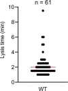
Figure 4—figure supplement 3.. Time to lysis after cluster formation.
Time to lysis was determined by first monitoring cluster formation and then loss of contrast by the prey cell. The measurements were performed over two biological replicates (n=61). The median is shown as a red bar.
Figure 4—figure supplement 4.
Figure 4—figure supplement 4.. Stable expression of NG-KilD in different mutant backgrounds.
NG-KilD is detected at the expected molecular weight by the anti-neon Green antibody. (-) NG: DZ2 Myxococcus cell extracts that do not express neon Green. Dotted line indicates gel splicing.
Figure 5.
Figure 5.. The kil genes are required for M.xanthus nutrition over prey cells.
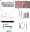
(a) The Kil system is essential for predation. Core deletion mutants in Tad-like genes, ∆kilC (Secretin), ∆kilF (ATPase), ∆kilH (IM platform), and ∆kilG (IM platform) were mixed with E. coli and spotted on CF agar plates (+ 0.07% glucose). After 24 hr of incubation, the mutants carrying the empty vector (EV) pSWU19 were strongly deficient in predation. The same kil mutants ectopically expressing the different kil genes under the control of the pilA promoter presented a restored predation phenotype similar to the WT-EV control. (b) A kil mutant can invade but cannot lyse E. coli prey colonies. mCherry-labeled WT and triple kilACF mutant are shown for comparison. Note that invading WT cells form corridors (yellow arrowheads) in the prey colony and ghost E. coli cells as well as cell debris (blue arrowheads) are left behind the infiltrating Myxococcus cells. In contrast, while the ∆kilACF penetrates the prey colony, corridors and prey ghost cells are not observed. Scale bar = 10 µm. See corresponding Video 9 for the full time lapse. (c) The kil genes are essential for prey killing. E. coli mCherry cells were measured by flow cytometry at time 0, 24, 48, and 72 hr after the onset of predation. The E. coli survival index was calculated by dividing the percentage of ‘E. coli events’ at t=24, 48, or 72 hr by the percentage of ‘E. coli events’ at the beginning of the experiment (t=0). This experiment was independently performed three times (n=6 per strain and time point). For each sample, 500,000 events were analyzed. Each data point indicates the mean ± the standard deviation. For each time point, unpaired t-test (with Welch’s correction) statistical analysis was performed to evaluate if the differences observed, relative to wild-type, were significant (***: p≤0.001) or not (ns: p>0.05). (d) E. coli survival in presence of the different kil mutant strains at 48 hr. E. coli mCherry cells were measured by flow cytometry at time 0 and 48 hr after predation. This experiment was independently performed three times (n=9 per strain and time point). Events were counted as in (c) and each data point indicates the mean ± the standard deviation. One-way ANOVA statistical analysis followed by Dunnett’s posttest was performed to evaluate if the differences observed, relative to wild-type, were significant (****: p≤0.0001). (e, f) The kil genes are essential for Myxococcus growth on prey. (e) Cell growth during invasion. Cell length is a function of cell age during invasion and can be monitored over time in WT cells (in red). In contrast, cell length tends to decrease in a ∆kilACF mutant (in green) showing that they are not growing in presence of prey. See associated Video 10 for the full time lapse. (f) Quantification of cell growth in WT and in ∆kilACF mutant backgrounds. Each individual cell was tracked for 5 hr in two biological replicates for each strain. Violin plot of the growth distributions (shown as the cell size increase slopes) are shown. Statistics: Student t-test, ***: p<0.001.
Figure 5—figure supplement 1.
Figure 5—figure supplement 1.. Growth and motility of WT and ∆kilACF strains on agar supporting both A- and S-motility (1.5%) and S-motility only (0.5%).
Scale bar = 2 mm.
Figure 5—figure supplement 2.
Figure 5—figure supplement 2.. Growth curves of WT and ∆kilACF in CYE-rich medium.
The measurements were performed over three biological replicates. Error bars represent the standard deviation to the mean.
Figure 6.
Figure 6.. The Kil system mediates killing against diverse bacterial species.
(a) The kil genes are predation determinants against various species. To evaluate if M. xanthus kil mutant had lost the ability to lyse by direct contact different preys, prey-cell suspensions were directly mixed with M. xanthus WT or ∆kilACF and spotted on CF agar (+ 0.07% glucose). After 24 and 48 hr of incubation, pictures of the spots corresponding to the different predator/prey couples were taken. Note that Pseudomonas aeruginosa is resistant in this assay. Scale bar = 3.5 mm. (b, c, d, e, f) Prey cell survival upon predation was evaluated by CFU counting. The different preys (KanR) were mixed with M. xanthus WT (blue circles) or ∆kilACF (orange circles) strains and spotted on CF agar (+ 0.07% glucose). Spots were harvested after 0, 8, 24, and 48 hr of predation, serially diluted and platted on agar plates with kanamycin for CFU counting. The prey alone (black circles) was used as a control. Two experimental replicates were used per time point. This experiment was independently performed three times. Error bars represent the standard deviation to the mean. (g) NG-KilD cluster formation and subsequent contact-dependent killing of Caulobacter crescentus. Scale bar = 2 µm. See corresponding Video 11 for the full time lapse. (h) NG-KilD cluster formation and subsequent contact-dependent killing of Salmonella enterica Typhimurium. Scale bar = 2 µm. See corresponding Video 12 for the full time lapse. (i) NG-KilD cluster formation and subsequent contact-dependent killing of B. subtilis. See corresponding Video 13 for the full time lapse. Scale bar = 2 µm.
Figure 7.
Figure 7.. The Kil system is conserved in predatory delta-proteobacteria.
Phylogenetic tree of the Type-IV filamentous system that gave rise to the M. xanthus Kil system. Only the four well-conserved Kil system components were used for constructing the phylogenetic tree. Dots indicate stable bootstrap values (> 75), classes are indicated next to species names. The M. xanthus Kil system is also found in other Myxococcales and closely related systems are also present in Bradymonadales and Bdellovibrionales, suggesting a functional specialization related to predation. The genetic organization of kil-like genes is shown for example members of each orders, Bradymonas sediminis and Bdellovibrio bacteriovorus (see also Supplementary file 2). The nomenclature and color code for Kil homologs are the same as in Figure 3. Gene accession numbers (KEGG) are shown above gene symbols.
Figure 7—figure supplement 1.
Figure 7—figure supplement 1.. Predation phenotype of a Myxococcus D,D-decarboxylase mutant (Zhang et al., 2020a).
Colony plate assays showing invasion of an E. coli prey colony 48 hr after plating by a dacB mutant. Scale bar = 2 mm.

Comment in

References

    1. Almawi AW, Matthews LA, Guarné A. FHA domains: phosphopeptide binding and beyond. Progress in Biophysics and Molecular Biology. 2017;127:105–110. doi: 10.1016/j.pbiomolbio.2016.12.003. - DOI - PubMed
    1. Altschul SF, Madden TL, Schäffer AA, Zhang J, Zhang Z, Miller W, Lipman DJ. Gapped BLAST and PSI-BLAST: a new generation of protein database search programs. Nucleic Acids Research. 1997;25:3389–3402. doi: 10.1093/nar/25.17.3389. - DOI - PMC - PubMed
    1. Avidan O, Petrenko M, Becker R, Beck S, Linscheid M, Pietrokovski S, Jurkevitch E. Identification and characterization of Differentially-Regulated type IVb pilin genes necessary for predation in obligate bacterial predators. Scientific Reports. 2017;7:1013. doi: 10.1038/s41598-017-00951-w. - DOI - PMC - PubMed
    1. Basler M, Ho BT, Mekalanos JJ. Tit-for-tat: type VI secretion system counterattack during bacterial cell-cell interactions. Cell. 2013;152:884–894. doi: 10.1016/j.cell.2013.01.042. - DOI - PMC - PubMed
    1. Berry JL, Pelicic V. Exceptionally widespread nanomachines composed of type IV pilins: the prokaryotic swiss army knives. FEMS Microbiology Reviews. 2015;39:1–21. doi: 10.1093/femsre/fuu001. - DOI - PMC - PubMed

Publication types

MeSH terms

Substances

Associated data