. 2021 Oct 22;6(20):e151527.
doi: 10.1172/jci.insight.151527.

Tie2 activation protects against prothrombotic endothelial dysfunction in COVID-19

Affiliations

Tie2 activation protects against prothrombotic endothelial dysfunction in COVID-19

Alec A Schmaier et al. JCI Insight. .

Abstract

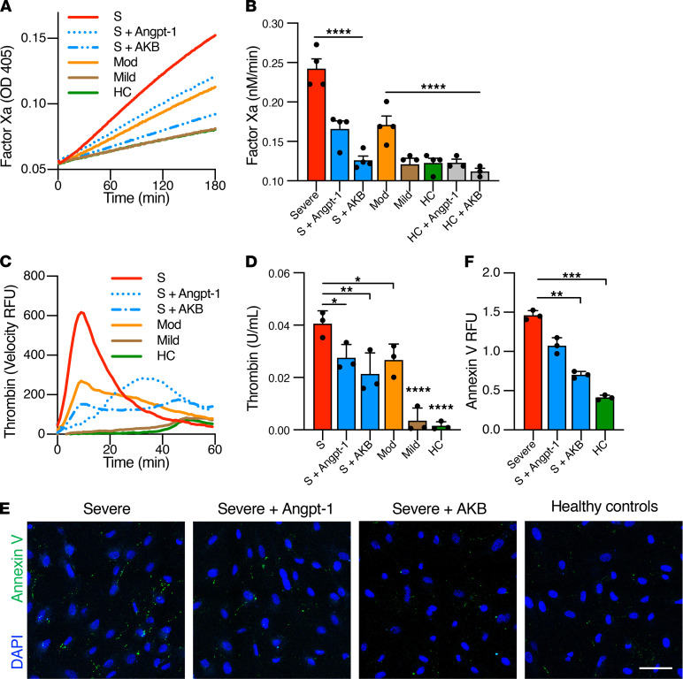
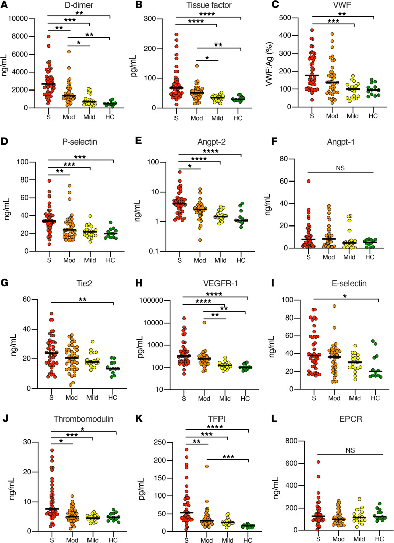
Endothelial dysfunction accompanies the microvascular thrombosis commonly observed in severe COVID-19. Constitutively, the endothelial surface is anticoagulant, a property maintained at least in part via signaling through the Tie2 receptor. During inflammation, the Tie2 antagonist angiopoietin-2 (Angpt-2) is released from endothelial cells and inhibits Tie2, promoting a prothrombotic phenotypic shift. We sought to assess whether severe COVID-19 is associated with procoagulant endothelial dysfunction and alterations in the Tie2/angiopoietin axis. Primary HUVECs treated with plasma from patients with severe COVID-19 upregulated the expression of thromboinflammatory genes, inhibited the expression of antithrombotic genes, and promoted coagulation on the endothelial surface. Pharmacologic activation of Tie2 with the small molecule AKB-9778 reversed the prothrombotic state induced by COVID-19 plasma in primary endothelial cells. Lung autopsies from patients with COVID-19 demonstrated a prothrombotic endothelial signature. Assessment of circulating endothelial markers in a cohort of 98 patients with mild, moderate, or severe COVID-19 revealed endothelial dysfunction indicative of a prothrombotic state. Angpt-2 concentrations rose with increasing disease severity, and the highest levels were associated with worse survival. These data highlight the disruption of Tie2/angiopoietin signaling and procoagulant changes in endothelial cells in severe COVID-19. Our findings provide rationale for current trials of Tie2-activating therapy with AKB-9778 in COVID-19.

Keywords: COVID-19; Coagulation; Vascular Biology.


Conflict of interest statement

Conflict of interest: SMP serves on the advisory board for Aerpio Pharmaceuticals, Inc. KP is an employee of Aerpio Pharmaceuticals, Inc.

Figures

Figure 1
Figure 1. Plasma from patients with COVID-19 induces thromboinflammatory gene expression in endothelial cells.
HUVECs were cultured overnight in the presence of 10% pooled plasma from patients with severe (S, ICU patients), moderate (Mod, non-ICU hospitalized patients), or mild (nonhospitalized outpatients) COVID-19 or healthy controls (HC) and analyzed for relative fold mRNA expression change of tissue factor: (A) F3, (B) E selectin (SELE), (C) ANGPT2, (D) TIE2, (E) VE-PTP (PTPRB), (F) EPCR (PROCR), (G) TFPI, and (H) thrombomodulin (THBD). Where indicated, cells were pretreated with Angpt-1 (300 ng/mL) or AKB-9778 (5 μM) for 30 minutes prior to incubation with plasma (n = 3–4 individual biologic replicates performed in technical duplicate). Gene expression was normalized to that of actin and changes are shown relative to HC. Graphs represent the mean ± SD. Significance in comparison with severe (S) was determined by 1-way ANOVA using Dunnett’s post hoc test, *P < 0.05, **P < 0.01, ***P < 0.001, ****P < 0.0001.
Figure 2
Figure 2. Plasma from patients with COVID-19 promotes activation of coagulation on endothelial cells.
HUVECs were cultured overnight in the presence of 10% pooled plasma from patients with severe (S, ICU), moderate (Mod, non-ICU), or mild (outpatient) COVID-19 or healthy controls (HC). (AD) Cells were analyzed for their ability to generate factor Xa or thrombin. Where indicated, cells were pretreated with Angpt-1 (300 ng/mL) or AKB-9778 (5 μM) for 30 minutes prior to incubation with plasma. (A and C) Representative experiments are depicted as mean absorbance (405 nm) or the first derivative of arbitrary fluorescence units as a function of time. (B and D) The rate of reaction for factor Xa and thrombin were converted to nM/min and U/mL, respectively, by comparison to standard curve. For factor Xa and thrombin generation assays, each data point represents the mean of 3 technical replicates, with 3–5 biologic replicates performed in total. (E and F) Cells were stained with annexin V to assess for phosphatidylserine externalization (n = 3 per group). Total fluorescent area was quantified and normalized for number of nuclei. (F) Graph represents the mean total fluorescence per 3 × 3 tile scan image ± SD. Scale bar: 50 μm. Significance was determined by 1-way ANOVA using Dunnett’s post hoc test, *P < 0.05, **P < 0.01, ***P < 0.001, ****P < 0.0001.
Figure 3
Figure 3. Procoagulant tissue signature of COVID-19 lung endothelium.
Lung specimens were obtained during limited autopsy immediately after death in patients who died from COVID-19 (n = 5). Control samples (n = 4) were obtained from tumor-free margins of lung tumor resections and processed in identical fashion regarding timing and method of fixation. Lung specimens from patients with COVID-19 demonstrated (A) an increase in the prothrombotic endothelial protein vWF and (B and C) a loss of the antithrombotic factors thrombomodulin (THBD) and EPCR. In each tissue section, 2 images were obtained and mean fluorescence intensity was analyzed for each tile-scanned image and normalized to background intensity. Scale bar: 100 μm. For graphs, mean is represented by the bar with each dot as a replicate; error bars indicate SD. Significance was determined by a 2-tailed Mann-Whitney test, **P < 0.01, ****P < 0.0001.
Figure 4
Figure 4. Measurement of thrombotic and endothelial markers in patients with COVID-19 by disease severity.
Markers included (A) D-dimer, (B) tissue factor, (C) vWF, (D) P selectin, (E) Angpt-2, (F) Angpt-1, (G) Tie2, (H) VEGFR-1, (I) E selectin, (J) thrombomodulin, (K) TFPI, and (L) EPCR. Each data point represents individual patient plasma measurements corresponding to disease severity. Bar indicates median value. Significance among groups determined by Kruskal-Wallis with Dunn’s multiple comparison test, *P < 0.05, **P < 0.01, ***P < 0.001, ****P < 0.0001. S, severe (ICU patients); Mod, moderate (hospitalized, non-ICU); Mild (COVID-19–positive outpatients); and HC, healthy controls.
Figure 5
Figure 5. Markers of thrombotic and endothelial activation are associated with worse survival among patients with COVID-19 in the ICU.
Kaplan-Meier curves of survival according to the top tertile (n = 14) versus bottom 2 tertiles (n = 28) of the plasma concentration of indicated analytes: (A) Angpt-2, (B) E selectin, (C) P selectin, (D) vWF:Ag, and (E) thrombomodulin. Significance was determined by log-rank test.

Update of

References

    1. Chen G, et al. Clinical and immunological features of severe and moderate coronavirus disease 2019. J Clin Invest. 2020;130(5):2620–2629. doi: 10.1172/JCI137244. - DOI - PMC - PubMed
    1. Gupta A, et al. Extrapulmonary manifestations of COVID-19. Nat Med. 2020;26(7):1017–1032. doi: 10.1038/s41591-020-0968-3. - DOI - PMC - PubMed
    1. Zhou F, et al. Clinical course and risk factors for mortality of adult inpatients with COVID-19 in Wuhan, China: a retrospective cohort study. Lancet. 2020;6736(20):1054–1062. - PMC - PubMed
    1. Colling ME, Kanthi Y. COVID-19-associated coagulopathy: An exploration of mechanisms. Vasc Med. 2020;25(5):471–478. doi: 10.1177/1358863X20932640. - DOI - PMC - PubMed
    1. Berger JS, et al. Prevalence and outcomes of D-dimer elevation in hospitalized patients with COVID-19. Arterioscler Thromb Vasc Biol. 2020;40(10):2539–2547. doi: 10.1161/ATVBAHA.120.314872. - DOI - PMC - PubMed

Publication types

MeSH terms