. 2021 Nov 8;31(21):4839-4844.e4.
doi: 10.1016/j.cub.2021.08.043. Epub 2021 Sep 9.

Functionally homologous representation of vocalizations in the auditory cortex of humans and macaques

Affiliations

Functionally homologous representation of vocalizations in the auditory cortex of humans and macaques

Clémentine Bodin et al. Curr Biol. .

Abstract

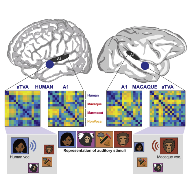
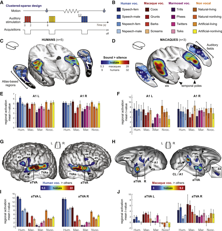
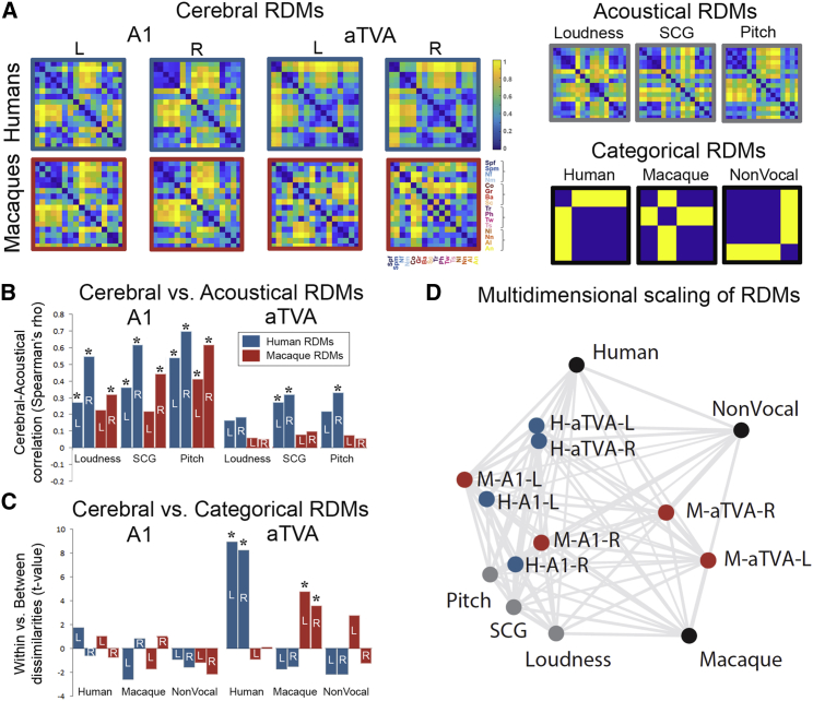
How the evolution of speech has transformed the human auditory cortex compared to other primates remains largely unknown. While primary auditory cortex is organized largely similarly in humans and macaques,1 the picture is much less clear at higher levels of the anterior auditory pathway,2 particularly regarding the processing of conspecific vocalizations (CVs). A "voice region" similar to the human voice-selective areas3,4 has been identified in the macaque right anterior temporal lobe with functional MRI;5 however, its anatomical localization, seemingly inconsistent with that of the human temporal voice areas (TVAs), has suggested a "repositioning of the voice area" in recent human evolution.6 Here we report a functional homology in the cerebral processing of vocalizations by macaques and humans, using comparative fMRI and a condition-rich auditory stimulation paradigm. We find that the anterior temporal lobe of both species possesses cortical voice areas that are bilateral and not only prefer conspecific vocalizations but also implement a representational geometry categorizing them apart from all other sounds in a species-specific but homologous manner. These results reveal a more similar functional organization of higher-level auditory cortex in macaques and humans than currently known.

Keywords: auditory cortex; comparative approach; conspecific vocalizations; functional MRI; humans; macaques; speech evolution; temporal voice areas; voice.


Conflict of interest statement

Declaration of interests The authors declare no competing interest.

Figures

None
Graphical abstract
Figure 1
Figure 1
Auditory cerebral activation in humans and macaques (A) Scanning protocol. Auditory stimuli were repeated three times in rapid succession during silent intervals between scans; macaques were rewarded with juice after 8-s periods of immobility. (B) Auditory stimuli. Stimuli consisted of 96 complex sounds from 4 large categories divided into 16 subcategories. (C and D) Areas with significant (p < 0.05, corrected) activation to sounds versus the silent baseline. t value threshold as indicated under the color bar. (C) Humans. PT, planum temporale; HG, Heschl’s gyrus (from Harvard Oxford atlas); Tp, temporal pole; sts, superior temporal sulcus. (D) Macaques. A1, R, core auditory areas; CL, CM, RT, belt auditory areas from Petkov et al. (E and F) Group-averaged regional mean activation (t values) for the 16 sound subcategories compared to silence in (E) humans and (F) macaques. Error bars indicate SEM. (G and H) CV-selective areas showing greater fMRI signal in response to CVs versus all other sounds. White circles indicate the location of bilateral anterior temporal voice areas in both species. (G) Human voc. > others at p < 0.05 corrected; TVAS, temporal voice areas; FVAs, frontal voice areas; 44-45, corresponding Brodmann areas from Harvard Oxford Atlas. (H) Macaque voc. > others at p < 0.001 uncorrected, p < 0.05 cluster-size corrected; as, arcuate sulcus; MC, motor cortex; PMC, premotor cortex; 44, corresponding Brodmann area from D99 atlas. (I and J) Group-averaged regional mean activation (t values) for the 16 sound subcategories compared to silence in the aTVAs in (I) humans and (J) macaques. See also Figures S1–S3 and Table S1.
Figure 2
Figure 2
Prefrontal CV-selective activations in macaques The statistical map of the contrast of CVs versus all other sounds in the three macaque subjects (p < 0.05, corrected) in shown in color scale overlaid on a T1-weighted image of the macaque brain in sagittal (top) and axial (bottom) slices. Black rectangles (top) zoom in activations in prefrontal cortex and show them relative to the anatomical parcellation of the D99 template (bottom). Numbers indicate anatomical localization of the maxima of CV selectivity in prefrontal cortex.
Figure 3
Figure 3
Representational similarity analysis in A1 and the aTVAs (A) Representational dissimilarity matrices (RDMs) showing percentile dissimilarities in pairwise fMRI response to the 16 sound subcategories for left and right A1 and aTVAs in both species, along with 3 comparison acoustical RMS (right column, top row) and 3 categorical RDMs (bottom row). (B) Comparison between brain RDMs and acoustical RDMs (Spearman correlation). p < 0.05, Bonferroni-corrected. (C) Comparison between brain RDMs and categorical model RDMs. (D) 2D representation of dissimilarities within brain and comparison RDMs via multidimensional scaling. Large distances indicate large dissimilarities (low correlations). Blue disks, human RDMs; red disks, macaque RDMs; black disks, model RDMs; gray disks, acoustical RDMs; L, left hemisphere; R, right hemisphere.

References

    1. Kaas J.H., Hackett T.A. Subdivisions of auditory cortex and processing streams in primates. Proc. Natl. Acad. Sci. USA. 2000;97:11793–11799. - PMC - PubMed
    1. Rauschecker J.P., Tian B., Hauser M. Processing of complex sounds in the macaque nonprimary auditory cortex. Science. 1995;268:111–114. - PubMed
    1. Belin P., Zatorre R.J., Lafaille P., Ahad P., Pike B. Voice-selective areas in human auditory cortex. Nature. 2000;403:309–312. - PubMed
    1. Pernet C.R., McAleer P., Latinus M., Gorgolewski K.J., Charest I., Bestelmeyer P.E., Watson R.H., Fleming D., Crabbe F., Valdes-Sosa M., Belin P. The human voice areas: Spatial organization and inter-individual variability in temporal and extra-temporal cortices. Neuroimage. 2015;119:164–174. - PMC - PubMed
    1. Petkov C.I., Kayser C., Steudel T., Whittingstall K., Augath M., Logothetis N.K. A voice region in the monkey brain. Nat. Neurosci. 2008;11:367–374. - PubMed

Publication types

LinkOut - more resources