. 2021 Nov 8;31(21):4831-4838.e4.
doi: 10.1016/j.cub.2021.08.042. Epub 2021 Sep 9.

Perturbation-specific responses by two neural circuits generating similar activity patterns

Affiliations

Perturbation-specific responses by two neural circuits generating similar activity patterns

Daniel J Powell et al. Curr Biol. .

Abstract

A fundamental question in neuroscience is whether neuronal circuits with variable circuit parameters that produce similar outputs respond comparably to equivalent perturbations.1-4 Work on the pyloric rhythm of the crustacean stomatogastric ganglion (STG) showed that highly variable sets of intrinsic and synaptic conductances can generate similar circuit activity patterns.5-9 Importantly, in response to physiologically relevant perturbations, these disparate circuit solutions can respond robustly and reliably,10-12 but when exposed to extreme perturbations the underlying circuit parameter differences produce diverse patterns of disrupted activity.7,12,13 In this example, the pyloric circuit is unchanged; only the conductance values vary. In contrast, the gastric mill rhythm in the STG can be generated by distinct circuits when activated by different modulatory neurons and/or neuropeptides.14-21 Generally, these distinct circuits produce different gastric mill rhythms. However, the rhythms driven by stimulating modulatory commissural neuron 1 (MCN1) and bath-applying CabPK (Cancer borealis pyrokinin) peptide generate comparable output patterns, despite having distinct circuits that use separate cellular and synaptic mechanisms.22-25 Here, we use these two gastric mill circuits to determine whether such circuits respond comparably when challenged with persisting (hormonal: CCAP) or acute (sensory: GPR neuron) metabotropic influences. Surprisingly, the hormone-mediated action separates these two rhythms despite activating the same ionic current in the same circuit neuron during both rhythms, whereas the sensory neuron evokes comparable responses despite acting via different synapses during each rhythm. These results highlight the need for caution when inferring the circuit response to a perturbation when that circuit is not well defined physiologically.

Keywords: Cancer borealis pyrokinin; central pattern generator; circuit flexibility; crustacean cardioactive peptide; degenerate circuits; dynamic clamp; gastric mill rhythm; gastro-pyloric receptor neurons; neuromodulation; stomatogastric ganglion.


Conflict of interest statement

Declaration of interests The authors declare no competing interests.

Figures
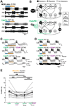
Figure 1.
Figure 1.. The peptide hormone CCAP reduces the retraction duration of the CabPK-gastric mill rhythm.
(A) Similar gastric mill rhythms driven by the projection neuron MCN1 and bath-applied CabPK peptide. Blue, protraction phase; orange, retraction phase. (B) MCN1- and CabPK-gastric mill circuit diagrams. Top row, protraction neurons; bottom row, retraction neurons. The excitatory action of bath-applied CabPK is indicated by the excitatory synapse symbols. Circle shading legend indicates the role of each neuron in gastric mill rhythm generation. All synapses shown are in the STG. Modified from , . (C) Intracellular recording of the same LG neuron during the CabPK-gastric mill rhythm with: saline (top), CCAP (middle), and dynamic clamp injection of CCAP-activated IMI (IMI-CCAP) into LG during saline superfusion (bottom). The latter recording includes the IMI-CCAP current injected into LG. Calibration bars are consistent for all conditions. (D) Intracellular recording of the same LG neuron during the CabPK-rhythm with: saline (top), added CCAP (middle), and CCAP plus injection of -IMI-CCAP (bottom). Calibration bars are the same for all panels. (E) CabPK-rhythm retraction duration across experiments in saline, CCAP, IMI-CCAP, and CCAP + -IMI-CCAP. Small unfilled circles, mean values per preparation; large filled circles, mean values across preparations (RM-ANOVA: p < 0.001, N = 13; Holm-Sidak post-hoc: **p < 0.01; all other comparisons were not significantly different). Lines connect data points from same preparation.
Figure 2.
Figure 2.. CCAP application decreases the retraction duration of the CabPK-gastric mill rhythm and prolongs the MCN1-gastric mill protraction phase.
(A,B) Example gastric mill rhythms, represented by an intracellular LG neuron recording, driven by (A) CabPK application and (B) MCN1 stimulation (6 Hz) in the absence (top) and presence (bottom) of CCAP. Blue and orange bars underneath each LG recording denote duration of protraction and retraction phases, respectively. Orange or blue bars for each control condition are also displayed under the bars for the CCAP-modulated rhythms. Values in bottom-right calibration bars pertain to all traces in (A) and (B). Both panels are from the same preparation. (C) Mean retraction duration value for each CabPK- and MCN1-gastric mill rhythm without and with CCAP (repeated paired t-tests: **p = 0.0088; Bonferroni correction: α = 0.0167, n.s. not significantly different, N = 6). Same colors for CabPK- and MCN1 data points are from the same preparation; filled circles (black) are mean values across preparations. (D) Mean protraction duration values for both gastric mill rhythms without and with CCAP application (repeated paired t-tests: ***p = 0.00095; Bonferroni correction: α = 0.0167, N = 6). Labeling as in (C). (E) LG neuron burst offset phase during CabPK- and MCN1-gastric mill rhythms, without and with CCAP application (repeated paired t-tests: ***p = 0.00054; Bonferroni correction: α = 0.0167, N = 6). Labeling as in (C). The same color in panels C-E represent data from the same preparation. See also Table S1.
Figure 3.
Figure 3.. GPR stimulation during the MCN13-6 Hz- and CabPK-gastric mill rhythm protraction or retraction phase shortens protraction and prolongs retraction, respectively.
(A) Intracellular LG recording during the MCN1-rhythm without GPR stimulation (top), and with GPR stimulation during one retraction- (middle) or protraction phase (bottom). All panels: GPR stimulations @ 6-8 Hz. Red bars: GPR stimulus duration. All traces are from the same preparation. Calibration bar values are consistent across recordings. (B) Mean MCN1-rhythm retraction duration before, during, and after GPR stimulation during the retraction phase (RM-ANOVA: p < 0.0001, N = 10; Bonferroni post-hoc: ****p < 0.0001). Lines between conditions link mean values from the same experiment. Black horizontal lines indicate the means for each condition in (B, C, E, F, I, J). (C) Mean protraction duration across experiments for the MCN1-rhythm before, during, and after GPR stimulation during protraction (RM-ANOVA: p < 0.0001, N = 10; Bonferroni post-hoc: ****p < 0.0001). (D) Intracellular LG recordings during the CabPK (10−6 M)-rhythm without (top) and with GPR stimulation (6 Hz, red bars) during retraction (middle) or protraction (bottom). All traces in (D) are from the same preparation. (E) Mean retraction duration of the CabPK-rhythm across experiments before, during, and after GPR stimulation during retraction (RM-ANOVA: p < 0.0001, N = 16; Bonferroni post-hoc: **p < 0.01; ****p < 0.0001). (F) Mean protraction duration of the CabPK-rhythm across experiments before, during, and after GPR stimulation during protraction (RM-ANOVA: p < 0.0001, N = 16; Bonferroni post-hoc: **p < 0.01; ***p ≤ 0.001). (G) Simplified gastric mill circuit schematic, including the pertinent GPR synapses, with ionotropic glutamate synapses functioning in saline (left; filled black circles) and suppressed with picrotoxin (PTX: 10−5 M; filled gray circles) (i, ionotropic; m, metabotropic)., (H) LG response to GPR stimulation (red bar) when LG is functionally isolated from STG inputs with PTX saline, and two electrode current clamp is used to inject constant amplitude and duration depolarizing current steps into LG. Stimulation artifacts are evident in the LG recording. Dashed lines indicate baseline Vm in LG before (black) and during (blue) GPR stimulation. (I) Mean baseline LG membrane potential (Vm) before and during GPR stimulation (paired t-test: ***p = 0.00015, N = 7). J, Mean number of LG spikes per depolarization per experiment before, during, and after GPR stimulation (repeated paired t-tests: ***p = 0.0004, **p = 0.0011, n.s. p = 0.48; Bonferroni correction: α = 0.0167; N = 7).
Figure 4.
Figure 4.. Schematic illustrating that different circuits generating similar outputs can lose or retain this relationship when influenced by the same input.
(Left) CabPK peptide application (top) and MCN1 projection neuron stimulation (bottom) configure different gastric mill circuits but elicit comparable gastric mill rhythms, represented by schematic activity patterns of the rhythm generator neurons LG and Int1. (Middle) Continuous application of a peptide hormone (purple) alters the projection neuron rhythm by prolonging the burst duration, while it increases the speed of the CabPK-rhythm by reducing the time between bursts. (Right) Input from a sensory neuron during the interburst phase (red) slows both rhythms by prolonging the interburst, albeit via different synapses.

Comment in

References

    1. Cropper EC, Dacks AM, and Weiss KR (2016). Consequences of degeneracy in network function. Curr. Opin. Neurobiol 41, 62–67. - PMC - PubMed
    1. Wang Y, Weiss KR, and Cropper EC (2019). Network degeneracy and the dynamics of task switching in the feeding circuit in Aplysia. J. Neurosci 39, 8705–8716. - PMC - PubMed
    1. Yeon J, Takeishi A, and Sengupta P (2021). Chronic vs acute manipulations reveal degeneracy in a thermosensory neuron network. MicroPubl. Biol 2021. - PMC - PubMed
    1. Goaillard JM, and Marder E (2021). Ion channel degeneracy, variability, and covariation in neuron and circuit resilience. Annu. Rev. Neurosci 44, 335–357. - PubMed
    1. Prinz AA, Bucher D, and Marder E (2004). Similar network activity from disparate circuit parameters. Nat. Neurosci 7, 1345–1352. - PubMed

Publication types

LinkOut - more resources