. 2021 Nov 9;54(11):2595-2610.e7.
doi: 10.1016/j.immuni.2021.08.009. Epub 2021 Sep 9.

Candida albicans elicits protective allergic responses via platelet mediated T helper 2 and T helper 17 cell polarization

Affiliations

Candida albicans elicits protective allergic responses via platelet mediated T helper 2 and T helper 17 cell polarization

Yifan Wu et al. Immunity. .

Abstract

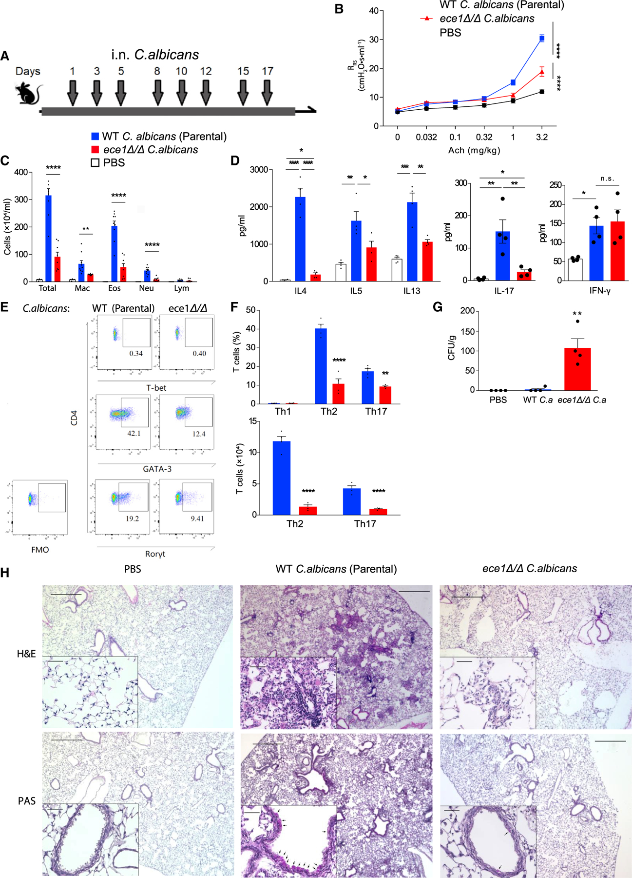
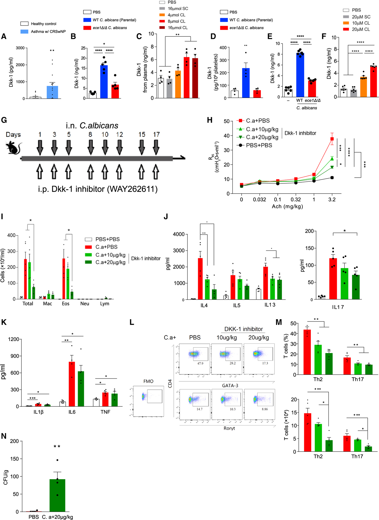
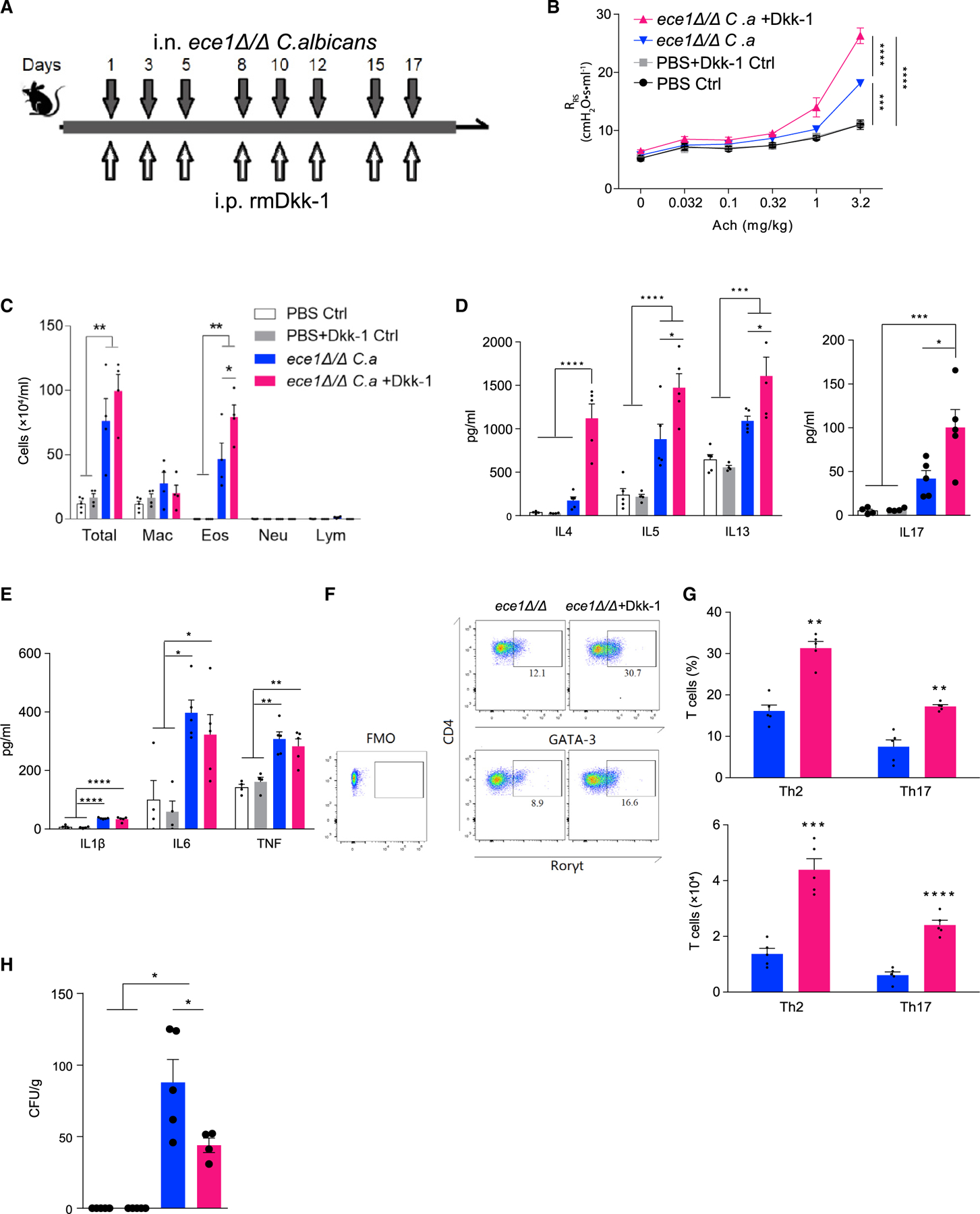
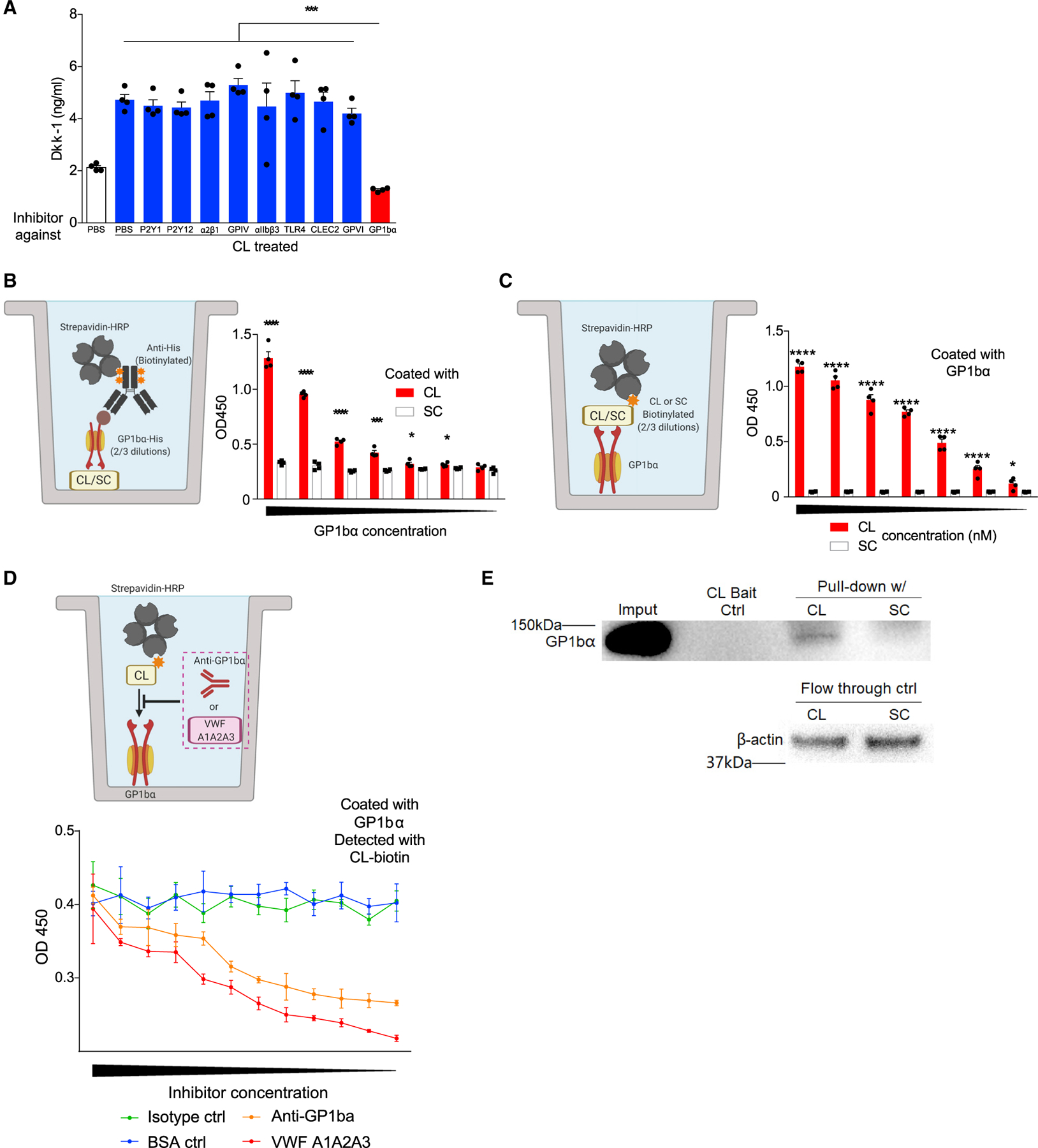
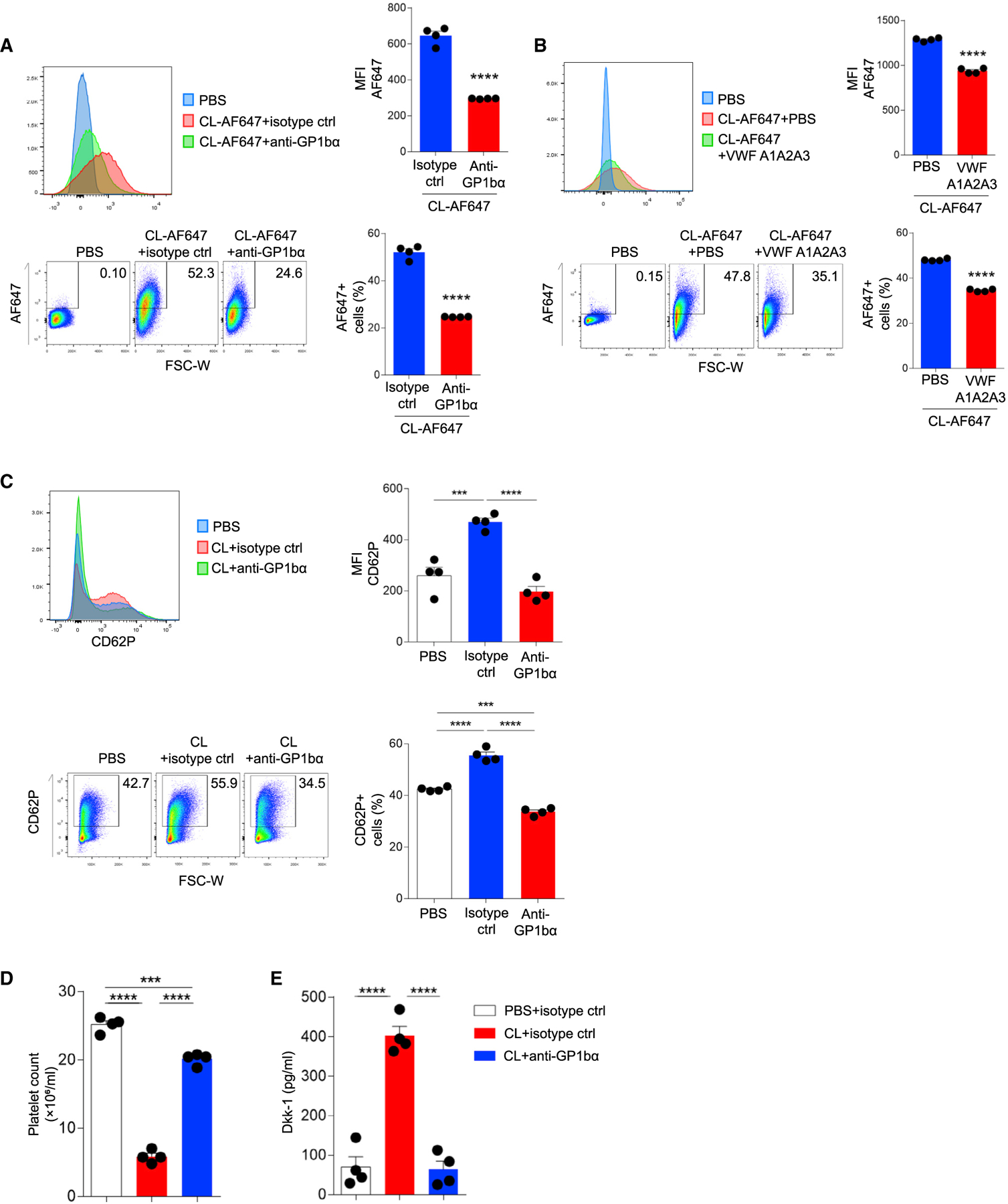
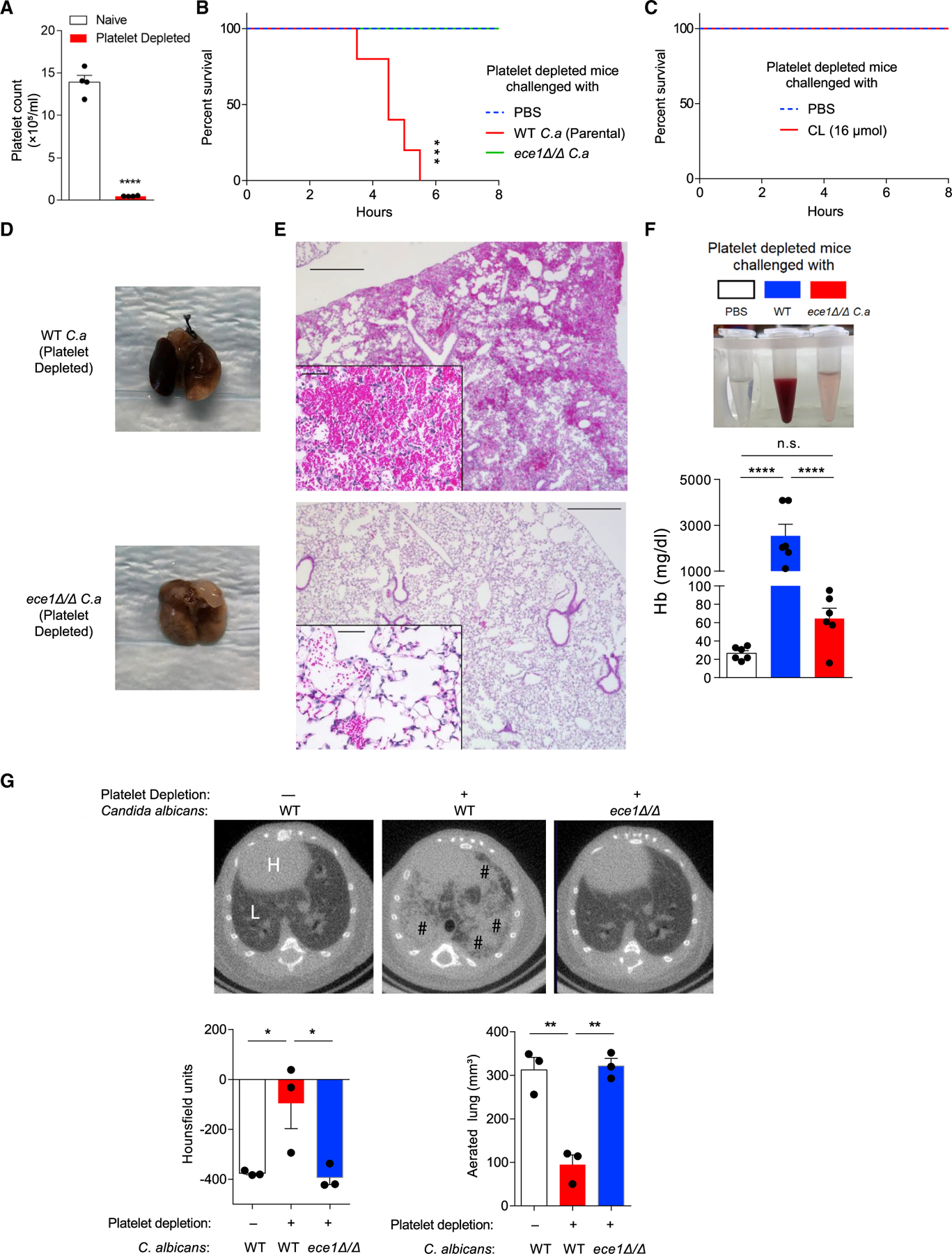
Fungal airway infection (airway mycosis) is an important cause of allergic airway diseases such as asthma, but the mechanisms by which fungi trigger asthmatic reactions are poorly understood. Here, we leverage wild-type and mutant Candida albicans to determine how this common fungus elicits characteristic Th2 and Th17 cell-dependent allergic airway disease in mice. We demonstrate that rather than proteinases that are essential virulence factors for molds, C. albicans instead promoted allergic airway disease through the peptide toxin candidalysin. Candidalysin activated platelets through the Von Willebrand factor (VWF) receptor GP1bα to release the Wnt antagonist Dickkopf-1 (Dkk-1) to drive Th2 and Th17 cell responses that correlated with reduced lung fungal burdens. Platelets simultaneously precluded lethal pulmonary hemorrhage resulting from fungal lung invasion. Thus, in addition to hemostasis, platelets promoted protection against C. albicans airway mycosis through an antifungal pathway involving candidalysin, GP1bα, and Dkk-1 that promotes Th2 and Th17 responses.

Keywords: Candida; Dkk-1; GP1bα; allergy; asthma; candidalysin; platelets.


Conflict of interest statement

Declaration of interests D.B.C is a scientific consultant to Atrapos Therapeutics, LLC and Pulmocide, LLC.

Figures

Figure 1.
Figure 1.. Candidalysin is necessary for the induction of allergic airway disease in mice
(A) C57BL/6 mice were challenged intranasally with 105 viable cells of wildtype parental strain or ece1Δ/Δ C. albicans as indicated in the timeline. (B) Respiratory system resistance (RRS) was assessed after intravenous injection of increasing doses of acetylcholine (Ach). (C) Quantitation of cells from bronchoalveolar lavage fluid samples (mac, macrophages; eos, eosinophils; neu, neutrophils; lym, lymphocytes). (D) Cytokines quantitated by ELISA from deaggregated lung supernatants. (E and F) T cells from lungs analyzed by flow cytometry. (E) Representative flow plot of TH1 (T-bet positive), Th2 (GATA3 positive), and Th17 (RORγt positive) cells from lungs after challenge. (F) Aggregate T cell data expressed as percentages and absolute cell numbers. (G) C. albicans colony-forming units (CFU) cultured from lungs. (H) Hematoxylin and eosin (H&E) and periodic acid-Schiff (PAS) staining of 5 mm lung sections from mice challenged under the indicated conditions. n ≥ 4, mean ± SEM. n.s., not significant; *p < 0.05, **p < 0.01, ***p < 0.001, ****p < 0.0001, using two-tailed Student’s t test (F) or one-way ANOVA followed by Tukey’s test (A–D and G) for multiple comparison. Magnification: 40× and 200×. Scale bars, 500 or 50 μm, respectively. Data are representative of three independent experiments. See also Figures S1 and S2.
Figure 2.
Figure 2.. Dkk-1 is secreted by mouse and human platelets in response to candidalysin and is required for robust allergic airway disease and C. albicans clearance from lung
(A) Plasma Dkk-1 concentrations from patients with asthma and CRS as compared with non-allergic healthy controls. (B and C) Dkk-1 concentrations quantitated from plasma of mice challenged intranasally with (B) wild-type parental strain or ece1Δ/Δ C. albicans or (C) candidalysin (CL) or scrambled control (SC). (D) Dkk-1 was quantitated from platelets of mice challenged intranasally with wild-type or ece1Δ/Δ C. albicans. (E and F) Human platelets in plasma were incubated with either (E) C. albicans or (F) CL/SC after which secreted Dkk-1 was quantitated. (G) C57BL/6 mice were challenged intranasally with C. albicans (C.a) and intraperitoneally with Dkk-1 inhibitor (WAY262611) as shown. (H) Respiratory system resistance (RRS) was quantitated as in Figure 1. (I) Quantitation of cells from the bronchoalveolar lavage fluid (mac, macrophages; eos, eosinophils; neu, neutrophils; lym, lymphocytes). (J and K) Cytokines assessed by ELISA from lung homogenate supernatants including (J) IL-4, IL-5, IL-13, and IL-17 and (K) IL-1b, IL-6, and TNF. (L and M) T cell quantitation from lungs as determined by flow cytometry. (L) Gating strategy for quantitation of Th1, Th2 and Th17 cells from lungs. (M) Quantitation of T cells assessed as percentages and absolute cell counts. (N) Lung fungal burdens. n ≥ 4, mean ± SEM. n.s., not significant; *p < 0.05, **p < 0.01, ***p < 0.001, ****p < 0.0001, using Mann-Whitney test (A), one-way ANOVA followed by Tukey’s test for multiple comparisons (B–M) or two-tailed Student’s t test (N). Data are representative of two independent experiments. See also Figure S3.
Figure 3.
Figure 3.. Recombinant Dkk-1 enhances allergic airway disease in ece1Δ/Δ C. albicans-challenged mice
(A) Wild-type mice were challenged intranasally with ece1Δ/Δ C. albicans (C.a) and intraperitoneally with recombinant mouse Dkk-1 as shown. (B) Respiratory system resistance (RRS) was assessed by increasing intravenous acetylcholine challenge. (C) Quantitation of cells from bronchoalveolar lavage fluid. (D and E) Cytokines assessed by ELISA from lung homogenate supernatants including (D) IL-4, IL-5, IL-13, and IL-17 and (E) IL-1β, IL-6, and TNF. (F and G) T cell quantification from lungs as assessed by flow cytometry. (F) Representative flow cytometry plot of Th1, Th2, and Th17 cells from lungs after challenge. (G) Quantitation of T cells as expressed as percentages and absolute cell numbers. (H) C. albicans CFU retrieved from whole lung. n ≥ 4, mean ± SEM. n.s., not significant; *p < 0.05, **p < 0.01, ***p < 0.001, ****p < 0.0001, using two-tailed Student’s t test (G) or one-way ANOVA followed by Tukey’s test (A–E and H) for multiple comparisons. Data are representative of two independent experiments.
Figure 4.
Figure 4.. Candidalysin primes human platelets to release Dkk-1 via GP1bα
(A) Human platelets were incubated with PBS or candidalysin (CL) at 10 μM and with blocking reagents to the indicated platelet receptors as indicated after which secreted Dkk-1 was quantitated. (B and C) Schematic diagrams and aggregate data depicting in vitro assays in which the dose-dependent binding of either plate-bound candidalysin or scrambled control peptide (SC) (B) or GP1bα (C) to the other reagent was determined colorimetrically (OD, optical density). (D) Schematic diagram and aggregate data depicting in vitro binding assays with GP1ba blocking reagents (anti-GP1bα antibody; VWF A1A2A3 tridomain) in which the dose-dependent inhibition of binding of candidalysin is colorimetrically quantitated against plates coated with GP1bα. (E) Pull-down assay of GP1ba from human platelet lysates using biotinylated candidalysin or SC as bait. n ≥ 4, mean ± SEM. *p < 0.05, **p < 0.01, ***p < 0.001 and ****p < 0.0001 using one-way ANOVA followed by Tukey’s test for multiple comparisons (A) or two-tailed Student’s t test (B–C). . Illustrative figures generated at biorender.com. Data are representative of two independent experiments.
Figure 5.
Figure 5.. Candidalysin directly binds to human platelets via GP1bα
(A and B) Flow cytometric analysis of human platelets incubated with AF647-conjugated CL (10 μM) with or without (A) anti-GP1bα antibody or (B) VWF A1A2A3 tridomain. (C) Flow cytometric analysis of P-selectin (CD62P) on human platelets after incubation with CL (10 μM) without or with anti-GP1bα antibody. Representative histograms, percentage quantification, and median fluorescence intensity (MFI) data are shown. (D and E) Human platelets were washed and then resuspended in PBS with blocking anti-GP1bα antibody or isotype control antibody prior to addition of candidalysin (CL; 10 μM) after which (D) platelet counts and (E) Dkk-1 concentrations from supernatants were determined. n = 4, mean ± SEM. ***p < 0.001 and ****p < 0.0001 using two-tailed Student’s t test (A and B) or one-way ANOVA followed by Tukey’s test for multiple comparisons (C and D). Data are representative of two independent experiments. See also Figure S4.
Figure 6.
Figure 6.. Thrombocytopenic mice rapidly succumb to C. albicans airway challenge
(A) Total platelet count in whole blood from mice 2 h after platelet depletion with anti-GP1bα antibody. (B) Survival curves (h) of platelet-depleted mice challenged once intranasally with PBS or 105 wild-type parental strain or ece1Δ/Δ C. albicans. (C) Survival curve of platelet-depleted mice challenged intranasally with 16 μmol candidalysin or PBS. (D–F) Bronchoalveolar lavage fluid and whole lungs were collected 4 h after platelet-depleted mice were challenged intranasally with wild-type or ece1Δ/Δ C. albicans. (D) Gross appearance of lungs. (E) Microscopic appearance of lungs (H&E staining) and (F) quantitation of hemoglobin from BALF. (G) Representative microCT-based imaging of platelet-sufficient and platelet-depleted mice challenged with either wild-type or ece1 Δ/Δ C. albicans as indicated. Bar graphs depict lung density as measured in Hounsfield units and aerated lung volume. H, heart; L, lung; #, areas of abnormal alveolar filling. n ≥ 4, mean ± SEM. *p < 0.05, **p < 0.01, ***p < 0.001 and ****p < 0.0001 using two-tailed Student’s t test (A), one-way ANOVA followed by Tukey’s test for multiple comparison (F–G), or Log-rank test for survival curves (B and C). Data are representative of two independent experiments. See also Figure S5.

Comment in

References

    1. Auton M, Cruz MA, and Moake J (2007). Conformational stability and domain unfolding of the Von Willebrand factor A domains. J. Mol. Biol 366, 986–1000. - PubMed
    1. Azuma H, Dent JA, Sugimoto M, Ruggeri ZM, and Ware J (1991). Independent assembly and secretion of a dimeric adhesive domain of von Willebrand factor containing the glycoprotein Ib-binding site. J. Biol. Chem 266, 12342–12347. - PubMed
    1. Baum GL (1960). The significance of Candida albicans in human sputum. N. Engl. J. Med 263, 70–73. - PubMed
    1. Baurand A, Raboisson P, Freund M, Léon C, Cazenave J-P, Bourguignon J-J, and Gachet C (2001). Inhibition of platelet function by administration of MRS2179, a P2Y1 receptor antagonist. Eur. J. Pharmacol 412, 213–221. - PubMed
    1. Beckert H, Meyer-Martin H, Buhl R, Taube C, and Reuter S (2018). The Canonical but Not the Noncanonical Wnt Pathway Inhibits the Development of Allergic Airway Disease. J. Immunol 201, 1855–1864. - PubMed

Publication types

MeSH terms

LinkOut - more resources