. 2021 Sep:135:105044.
doi: 10.1016/j.yhbeh.2021.105044. Epub 2021 Sep 8.

Alterations in corticotropin-releasing factor receptor type 1 in the preoptic area and hypothalamus in mice during the postpartum period

Affiliations

Alterations in corticotropin-releasing factor receptor type 1 in the preoptic area and hypothalamus in mice during the postpartum period

Rose M De Guzman et al. Horm Behav. 2021 Sep.

Abstract

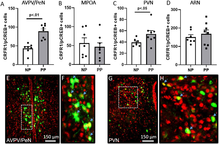
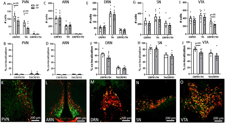
Corticotropin-releasing factor (CRF) signaling through CRF receptor 1 (CRFR1) regulates autonomic, endocrine, and behavioral responses to stress, as well as behavioral changes during the maternal period. Previous work in our lab reported higher levels of CRFR1 in female, compared to male, mice within the rostral anteroventral periventricular nucleus (AVPV/PeN), a brain region involved in maternal behaviors. In this study, we used CRFR1-GFP reporter mice to investigate whether the reproductive status (postpartum vs. nulliparous) of acutely stressed females affects levels of CRFR1 in the AVPV/PeN and other regions involved in maternal functions. Compared to nulliparous, postpartum day 14 females showed increased AVPV/PeN CRFR1-GFP immunoreactivity and an elevated number of restraint stress-activated AVPV/PeN CRFR1 cells as assessed by immunohistochemical co-localization of CRFR1-GFP and phosphorylated CREB (pCREB). The medial preoptic area (MPOA) and paraventricular hypothalamus (PVN) of postpartum mice showed modest decreases in CRFR1-GFP immunoreactivity, while increased CRFR1-GFP/pCREB co-expressing cells were found in the PVN following restraint stress relative to nulliparous mice. Tyrosine hydroxylase (TH) and CRFR1-GFP co-localization was also assessed in the AVPV/PeN and other regions and revealed a decrease in co-localized neurons in the AVPV/PeN and ventral tegmental area of postpartum mice. Corticosterone analysis of restrained mice revealed blunted peak, but elevated recovery, levels in postpartum compared to nulliparous mice. Finally, we investigated projection patterns of AVPV/PeN CRFR1 neurons using female CRFR1-Cre mice and revealed dense efferent projections to several preoptic, hypothalamic, and hindbrain regions known to control stress-associated and maternal functions. Together, these findings contribute to our understanding of the neurobiology that might underlie changes in stress-related functions during the postpartum period.

Keywords: Corticotropin-releasing factor; Corticotropin-releasing factor receptor type 1; Hypothalamus; Paraventricular nucleus; Postpartum; Preoptic area; Rostral anteroventral periventricular nucleus; Sex differences; Tyrosine hydroxylase.


Conflict of interest statement

Conflict of Interest

None declared.

Figures

Figure 1.
Figure 1.. CRFR1-GFP levels in the AVPV/PeN.
(A) There were significant differences in dark (postpartum > nulliparous) and light (nulliparous > postpartum CRFR1-GFP neurons, although no significant differences in total CRFR1-GFP neurons were found. (B) Density analysis revealed increased CRFR1-GFP labeling density within the AVPV/PeN of postpartum mice. Collectively, CRFR1-GFP immunoreactivity is increased in the postpartum day 14 mouse brain. (C-D) Representative images of a nulliparous (NP) and postpartum day 14 (PP14) AVPV/PeN. (E-F) High magnification images of inset boxes in C-D that indicate examples of light (yellow asterisk) and dark (red asterisk) labeled cells.
Figure 2.
Figure 2.. CRFR1-GFP levels in the MPOA, PVN, and ARN.
(A) In the MPOA, there was a significant decrease in darkly labeled CRFR1-GFP neurons and a trend toward a decrease in total CRFR1-GFP neurons in postpartum mice compared to nulliparous mice. (B) In the PVN, there was a trend toward a decrease in darkly labeled neurons in postpartum mice. (C) There were no differences in the ARN. Density analysis revealed a trend toward decreased CRFR1-GFP labeling density in the MPOA (D), and a significant decrease in the PVN (E), of postpartum relative to nulliparous mice. No difference in CRFR1-GFP density was found in the ARN (F). NP, nulliparous; PP postpartum. Representative images of CRFR1-GFP within the MPOA (G,H) and PVN (I,J) are shown. 3v, 3rd ventricle.
Figure 3.
Figure 3.. CRFR1-GFP/pCREB co-localization in postpartum and nulliparous mice after restraint stress.
Postpartum mice had greater levels of CRFR1-GFP neurons (green) expressing pCREB (red) in the (A) AVPV/PeN and (C) PVN compared to nulliparous. PP14 and nulliparous mice did not differ in the number of CRFR1 expressing pCREB in the (B) MPOA or (D) ARN. Representative images of AVPV/PeN (E,F) and PVN (G,H) are shown. High magnification images of inset boxes of E and G are shown in F and H. Arrows indicate examples of co-labeled neurons (yellow nuclear label). Data presented as mean ± SEM.
Figure 4.
Figure 4.. AVPV/PeN CRFR1-GFP/TH co-localization.
Postpartum mice had lower levels of (A) TH and CRFR1-GFP/TH co-localization compared to nulliparous mice. (B) Postpartum mice also had a lower percentage of CRFR1 neurons co-localized with TH, and a trend toward lower percentage of TH neurons co-localized with CRFR1, compared to nulliparous mice. Representative images of AVPV/PeN CRFR1-GFP (green), TH (red), and CRFR1-GFP/TH+ (orange/yellow) co-localization levels in nulliparous (C-F) and PP14 (G-J) mice. High magnification of inset boxes in E and I are shown in F and J. Blue arrows indicate examples of co-labeled neurons. Data presented as mean ± SEM.
Figure 5.
Figure 5.. CRFR1-GFP/TH co-localization in the PVN, ARN, DRN, SN, and VTA.
(A) Postpartum mice had lower levels of CRFR1-GFP and TH neurons in the PVN compared to nulliparous mice. (B) Postpartum and nulliparous mice did not differ in levels and percentage of CRFR1-GFP/TH and TH/CRFR1 co-localization in the PVN. (C-H) Postpartum and nulliparous mice did not differ in CRFR1, TH, and CRFR1-GFP/TH neurons, or percentages of CRFR1 neurons co-localized with TH and TH neurons co-localized with CRFR1, in the ARN, DRN, and SN. The number of CRFR1/TH co-labeled cells (I) and percentage of CRFR1 cells expressing TH (J) were decreased in the VTA of PP14 compared to nulliparous mice. (K-O) Representative images of PVN, ARN, DRN, SN, and VTA co-labeling in a PP14 mouse. Data presented as mean ± SEM.
Figure 6.
Figure 6.. Baseline, restraint stress-induced, and recovery corticosterone levels.
Corticosterone levels at baseline, stress-induced (after a 30-minute restraint), and recovery corticosterone (90-min after restraint initiation and 1-hr left undisturbed) reveal attenuated corticosterone levels at 30 minutes after restraint onset and elevated corticosterone at 90 minutes in PP14 compared to nulliparous mice.
Figure 7.
Figure 7.. Representative images of anterograde projection sites from AVPV/PeN CRFR1 neurons.
(A) The injection site showing labeling of CRFR1-Cre neurons in the AVPV. Afferent labeling is found in numerous regions including the (B) ventral lateral septum (LSv), (C) dorsal bed nucleus of the stria terminalis (BSTd), (D) principal nucleus of the BST (BSTpr), (E) paraventricular nucleus (PVN), (F) dorsomedial hypothalamus (DMH), arcuate nucleus (ARN), and tuberal nucleus (TN), (G) lateral hypothalamus (lat hyp), (H) paraventricular thalamus (PVT), (I) medial amygdala (MeA), (J) periaqueductal gray (PAG), (K) dorsal raphe nucleus (DRN), (L) Barrington’s nucleus (BN), and (M) parabrachial nucleus (PBN). LV; lateral ventricle, 3 V; 3rd ventricle, 4v; 4th ventricle, OT; optic tract. Arrows are shown within regions with moderate to low labeling intensity to indicate specific sites that show fiber labeling.

References

    1. Agrati D, Browne D, Jonas W, Meaney M, Atkinson L, Steiner M, Fleming AS, 2015. Maternal anxiety from pregnancy to 2 years postpartum: transactional patterns of maternal early adversity and child temperament. Arch Womens Ment Health 18, 693–705. - PubMed
    1. Altemus M, Deuster PA, Galliven E, Carter CS, & Gold PW, 1995. Suppression of hypothalmic-pituitary-adrenal axis responses to stress in lactating women. The Journal of Clinical Endocrinology & Metabolism, 80(10), 2954–2959. - PubMed
    1. Barnett B, Schaafsma MF, Guzman AM and Parker GB, 1991. Maternal anxiety: a 5-year review of an intervention study. Journal of Child Psychology and Psychiatry, 32(3), 423–438. - PubMed
    1. Berghorn KA, Le WW, Sherman TG and Hoffman GE, 2001. Suckling stimulus suppresses messenger RNA for tyrosine hydroxylase in arcuate neurons during lactation. Journal of Comparative Neurology, 438(4), 423–432. - PubMed
    1. Brand SR and Brennan PA, 2009. Impact of antenatal and postpartum maternal mental illness: how are the children? Clinical obstetrics and gynecology, 52(3), 441–455. - PubMed

Publication types

Substances