. 2021 Sep 9;134(20):2475-2482.
doi: 10.1097/CM9.0000000000001689.

Pro-pigmentary action of 5-fluorouracil through the stimulated secretion of CXCL12 by dermal fibroblasts

Affiliations

Pro-pigmentary action of 5-fluorouracil through the stimulated secretion of CXCL12 by dermal fibroblasts

Zhi-Kai Liao et al. Chin Med J (Engl). .

Abstract

Background: There is growing evidence that 5-fluorouracil (5-FU) combined with therapeutic trauma can effectively induce skin repigmentation in vitiligo patients who are unresponsive to conventional treatments. Previous studies have mainly focused on identifying the antimitotic activity of 5-FU for the treatment of skin cancer, but few studies have investigated its extra-genotoxic actions favoring melanocyte recruitment.

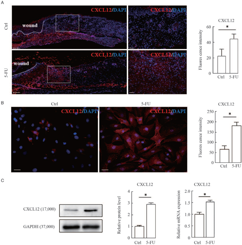
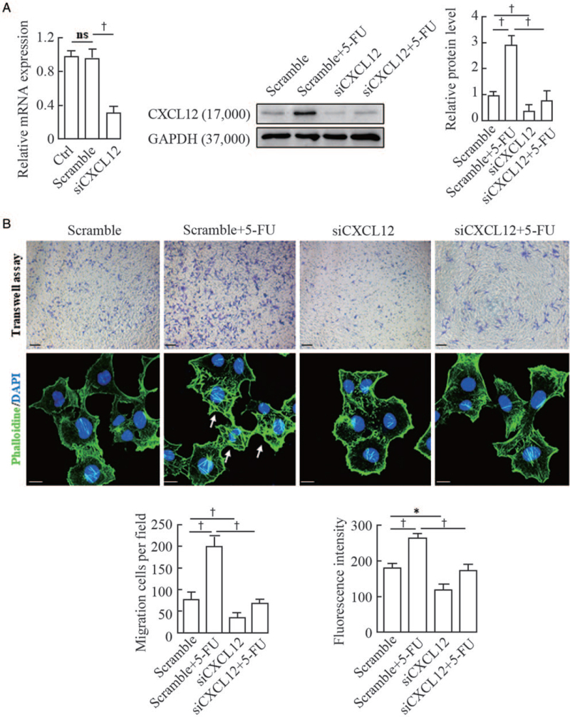
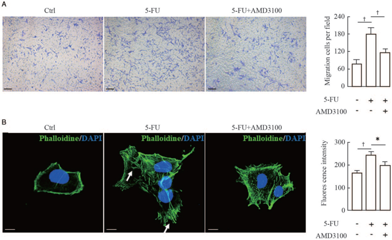
Methods: We utilized the full thickness excisional skin wound model in Dct-LacZ transgenic mice to dynamically assess the migration of melanocytes in the margins of wounds treated with or without 5-FU. The in-situ expression of CXCL12 was examined in the wound beds using immunofluorescence staining. Quantitative real-time polymerase chain reaction and Western blotting analyses were performed to detect the expression levels of CXCL12 mRNA and protein in primary mouse dermal fibroblasts treated with or without 5-FU. Transwell assays and fluorescein isothiocyanate (FITC)-phalloidin staining were used to observe cell migration and filamentous actin (F-actin) changes of melan-a murine melanocytes.

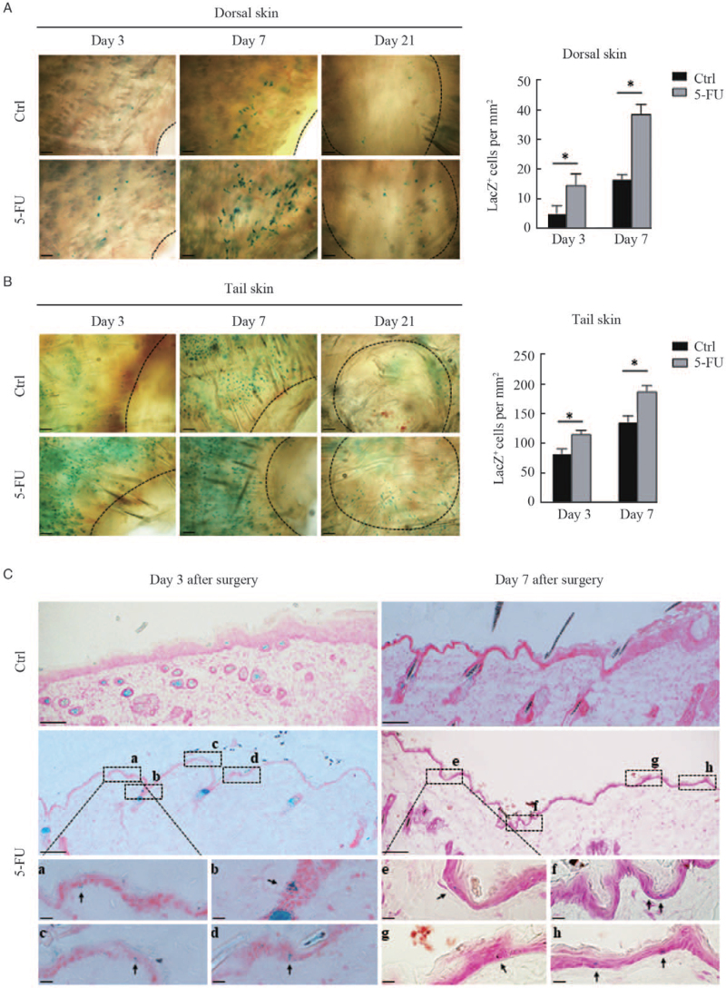
Results: Whole mount and cryosection X-gal staining showed that the cell numbers of LacZ-positive melanocytes were much higher in the margins of dorsal and tail skin wounds treated with 5-FU compared with the controls. Meanwhile, CXCL12 immunostaining was significantly increased in the dermal compartment of wounds treated with 5-FU (control vs. 5-FU, 22.47 ± 8.85 vs. 44.69 ± 5.97, P < 0.05). Moreover, 5-FU significantly upregulated the expression levels of CXCL12 mRNA (control vs. 5-FU, 1.00 ± 0.08 vs. 1.54 ± 0.06, P < 0.05) and protein (control vs. 5-FU, 1.00 ± 0.06 vs. 2.93 ± 0.10, P < 0.05) in cultured fibroblasts. Inhibition of the CXCL12/CXCR4 axis suppressed melanocyte migration in vitro using a CXCL12 small interfering RNA (siRNA) or a CXCR4 antagonist (AMD3100).

Conclusion: 5-FU possesses a pro-pigmentary activity through activation of the CXCL12/CXCR4 axis to drive the chemotactic migration of melanocytes.


Conflict of interest statement

None.

Figures

Figure 1
Figure 1
Visualization of LacZ+ cells in the margins of wounds treated with or without 5-FU. Full-thickness excisional wounds were created in the dorsal and tail skins of Dct-lacZ transgenic mice. The skin was biopsied from the wound margins on the third, seventh, and 21st day after surgery. Representative images of LacZ+ cells in dorsal skin (A, upper panel) and tail skin (B, lower panel) by whole-mount X-Gal staining are shown. The black dashed lines indicate the leading edge of wounds. Scale bars: 100 μm. Histograms showing the number of LacZ+ melanocytes in the wound margins of dorsal and tail skin on the third and seventh day after surgery, expressed as means ± SD of three independent experiments, P < 0.01, compared to controls. (C) Representative images of LacZ+ cells in cryosections of dorsal skin with X-Gal staining are shown. Scale bars: 100 μm. Black rectangles (a–h) indicate regions enlarged below. Scale bar: 20 μm (enlarged images). Black arrows indicate typical LacZ+ melanocytes. 5-FU: 5-fluorouracil; SD: Standard deviation.
Figure 2
Figure 2
Upregulation of chemokine CXCL12 expression in wound margins and cultured fibroblasts by 5-FU. (A) Representative images of immunofluorescence staining of CXCL12 (red) and counterstaining with DAPI (blue) in cryosections of dorsal skin wounds are shown. White dashed lines indicate the leading edge of wounds. Scale bars: 200 μm. White rectangles indicate regions enlarged on the right. Scale bar: 50 μm (enlarged images). Histograms showing the comparison of fluorescence intensity for wound margins treated with or without 5-FU are shown on the right. Immunofluorescence staining (B), Western blotting and qRT-PCR (C) were used to examine the expression levels of CXCL12 protein and mRNA in cultured fibroblasts treated with 50 μmol/L 5-FU for 24 h. Representative blots are shown. Data are shown as means ± SD of three independent experiments. P < 0.01 vs. control. 5-FU: 5-fluorouracil; DAPI: diamidino-2-phenylindole; qRT-PCR: Quantitative real-time polymerase chain reaction; SD: Standard deviation.
Figure 3
Figure 3
Effect of siRNA-mediated knockdown of CXCL12 in murine fibroblasts on the migration and F-actin distribution of melan-a melanocytes. (A) Murine fibroblasts were transfected with CXCL12 siRNA (siCXCL12) and a scrambled control for 48 h. The efficiency of CXCL12 knockdown by siRNAs was tested using qRT-PCR and Western blotting. Representative blots are shown in the middle. The histogram (on the right) shows the densitometric quantification of data with means ± SD of three independent experiments. (B) Melan-a melanocytes were seeded in the upper chambers, and the lower chambers were seeded with siCXCL12-transfected fibroblasts for 24 h of coculture, as described in the Materials and Methods Section. Cells that migrated to the lower surface of the inserts were counted in five microscopic fields using a 20 × objective in at least three independent experiments. A representative image of migrated cells under microscope (upper panel) is shown. Scale bars: 50 μm. Melan-a melanocytes growing on glass coverslips were treated with the conditioned medium from transfected fibroblasts and were subsequently stained for actin with FITC-phalloidin (green). Nuclei were counterstained with DAPI (blue). Representative images of F-actin staining are shown (lower panel). White arrows indicate the distribution of coarse actin bundles. Scale bar: 10 μm. Histograms showing the migrated cells counted in five random fields under microscope (on the left) and the fluorescent intensities of 20 cells (on the right). Data represent means ± SD of three independent experiments. P < 0.05 and P < 0.01 vs. scramble or scramble + 5-FU. 5-FU: 5-fluorouracil; DAPI: Diamidino-2-phenylindole; F-actin: Filamentous actin; FITC: Fluorescein isothiocyanate; qRT-PCR: Quantitative real-time polymerase chain reaction; siRNA: Small interfering RNA; SD: Standard deviation.
Figure 4
Figure 4
Effect of AMD3100 (a specific CXCR4 antagonist) on the migration and F-actin distribution of melanocytes. (A) Melan-a melanocytes were seeded into the upper chambers and pretreated with 10 μmol/L AMD3100 for 1 h, and the lower chambers were seeded with murine fibroblasts for 24 h of coculture, as described in the Materials and Methods Section. Cells that migrated to the lower surface of the inserts were counted in five microscopic fields using a 20 × objective in at least three independent experiments. Representative micrographs of migrating cells are shown. Scale bars: 50 μm. Histogram (on the right) shows the number of migrated cells in five random microscopical fields. (B) Melan-a melanocytes growing on glass coverslips were treated with or without 10 μmol/L AMD3100 for 1 h, and were then treated with the conditioned medium from 5-FU-treated fibroblasts and subsequently stained for actin with FITC-phalloidin (green). Nuclei were counterstained with DAPI (blue). Representative images of F-actin staining are shown. White arrows indicate the distribution of coarse actin bundles in the cytoplasm. Scale bars: 10 μm. Histogram (on the right) shows the fluorescent intensities of 20 cells. Data represent means ± SD of three independent experiments. P < 0.05 and P < 0.01 vs. 5-FU group. 5-FU: 5-fluorouracil; DAPI: Diamidino-2-phenylindole; F-actin: Filamentous actin; SD: Standard deviation.

References

    1. Ezzedine K, Eleftheriadou V, Whitton M, van Geel N. Vitiligo. Lancet 2015; 386:74–84. doi: 10.1016/s0140-6736(14)60763-7. - PubMed
    1. Speeckaert R, van Geel N. Vitiligo: an update on pathophysiology and treatment options. Am J Clin Dermatol 2017; 18:733–744. doi: 10.1007/s40257-017-0298-5. - PubMed
    1. Bae JM, Jung HM, Hong BY, Lee JH, Choi WJ, Lee JH, et al. . Phototherapy for Vitiligo: a systematic review and meta-analysis. JAMA Dermatol 2017; 153:666–674. doi: 10.1001/jamadermatol.2017.0002. - PMC - PubMed
    1. Zohdy HA, Hussein MS. Intradermal injection of fluorouracil versus triamcinolone in localized vitiligo treatment. J Cosmet Dermatol 2018; Epub ahead of print. doi: 10.1111/jocd.12820. - PubMed
    1. Mackenzie MA, Jordan SA, Budd PS, Jackson IJ. Activation of the receptor tyrosine kinase Kit is required for the proliferation of melanoblasts in the mouse embryo. Dev Biol 1997; 192:99–107. doi: 10.1006/dbio.1997.8738. - PubMed