

A big white dot after CPR
- PMID: 34507522
- PMCID: PMC8432442
- DOI: 10.1186/s12245-021-00376-3
A big white dot after CPR
Abstract
Introduction: Patients may remain comatose after the resumption of spontaneous circulation with cardiopulmonary resuscitation. A primary neurologic event may precede a cardiac standstill.
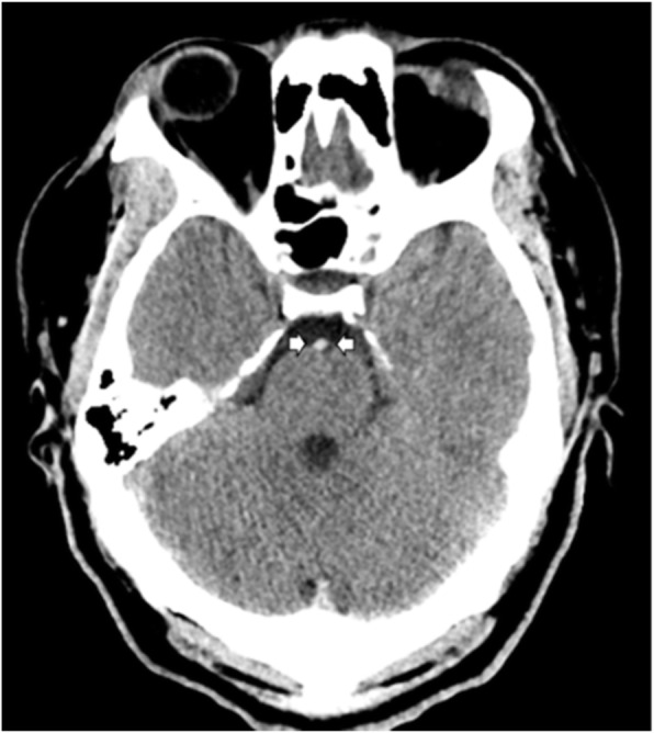
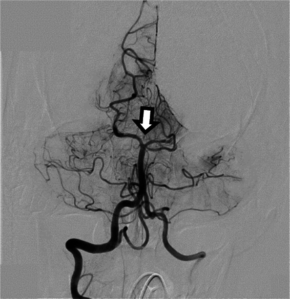
Case report: We present a 33-year-old patient with successful resuscitation for pulseless electrical activity and a "normal computed tomography (CT) scan." Further scrutiny showed a hyperdense basilar artery sign ('big white dot') that led to a CT angiogram confirming an embolus to the proximal basilar artery. His examination showed fixed and dilated midsize (mesencephalic) pupils and extensor posturing. Endovascular retrieval of the clot was successful, but there was a devastating ischemic injury to the brainstem.
Conclusion: This case reminds us to consider neurologic causes of cardiac arrest.
Keywords: Cerebrovascular disease; Interventional; MRI; Neuroradiology; Stroke.
© 2021. The Author(s).
Conflict of interest statement
The authors declare that they have no competing interests.
Figures


References
-
- Arnaout M, Mongardon N, Deye N, Legriel S, Dumas F, Sauneuf B, Malissin I, Charpentier J, Pène F, Baud F, Chiche JD, Mira JP, Cariou A. Out-of-hospital cardiac arrest from brain cause: epidemiology, clinical features, and outcome in a multicenter cohort. Crit Care Med. 2015;43(2):453–460. doi: 10.1097/CCM.0000000000000722. - DOI - PubMed
-
- Joundi RA, Rabinstein AA, Nikneshan D, Tu JV, Fang J, Holloway R, Saposnik G, Stroke Outcomes Research Working Group (SORCan-www.sorcan.ca) Cardiac arrest in acute ischemic stroke: incidence, predisposing factors, and clinical outcomes. J Stroke Cerebrovasc Dis. 2016;25(7):1644–1652. doi: 10.1016/j.jstrokecerebrovasdis.2016.03.010. - DOI - PubMed
LinkOut - more resources
Full Text Sources
