. 2021 Sep 10;9(1):151.
doi: 10.1186/s40478-021-01252-0.

Exogenous Aβ seeds induce Aβ depositions in the blood vessels rather than the brain parenchyma, independently of Aβ strain-specific information

Affiliations

Exogenous Aβ seeds induce Aβ depositions in the blood vessels rather than the brain parenchyma, independently of Aβ strain-specific information

Tsuyoshi Hamaguchi et al. Acta Neuropathol Commun. .

Abstract

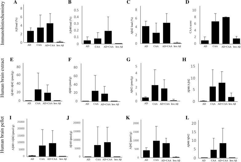
Little is known about the effects of parenchymal or vascular amyloid β peptide (Aβ) deposition in the brain. We hypothesized that Aβ strain-specific information defines whether Aβ deposits on the brain parenchyma or blood vessels. We investigated 12 autopsied patients with different severities of Aβ plaques and cerebral amyloid angiopathy (CAA), and performed a seeding study using an Alzheimer's disease (AD) mouse model in which brain homogenates derived from the autopsied patients were injected intracerebrally. Based on the predominant pathological features, we classified the autopsied patients into four groups: AD, CAA, AD + CAA, and less Aβ. One year after the injection, the pathological and biochemical features of Aβ in the autopsied human brains were not preserved in the human brain extract-injected mice. The CAA counts in the mice injected with all four types of human brain extracts were significantly higher than those in mice injected with PBS. Interestingly, parenchymal and vascular Aβ depositions were observed in the mice that were injected with the human brain homogenate from the less Aβ group. The Aβ and CAA seeding activities, which had significant positive correlations with the Aβ oligomer ratio in the human brain extracts, were significantly higher in the human brain homogenate from the less Aβ group than in the other three groups. These results indicate that exogenous Aβ seeds from different Aβ pathologies induced Aβ deposition in the blood vessels rather than the brain parenchyma without being influenced by Aβ strain-specific information, which might be why CAA is a predominant feature of Aβ pathology in iatrogenic transmission cases. Furthermore, our results suggest that iatrogenic transmission of Aβ pathology might occur due to contamination of brain tissues from patients with little Aβ pathology, and the development of inactivation methods for Aβ seeding activity to prevent iatrogenic transmission is urgently required.

Keywords: Alzheimer’s disease; Amyloid β peptide; Cerebral amyloid angiopathy; Iatrogenic; Strain; Transmission.


Conflict of interest statement

The authors declare no competing interests.

Figures

Fig. 1
Fig. 1
Representative images of the amyloid pathology of the autopsied human brains. The images of the left occipital lobe belong to groups with Alzheimer’s disease (AD) (AD), cerebral amyloid angiopathy (CAA) (EH), AD + CAA (IL), and less amyloid β peptide (Aβ) pathology (MP). Antibodies against Aβ17–24 (4G8, 1:5,000) (A, E, I, and M), Aβ35–40 (1A10, 1:1,000) (B, F, J, and N), and Aβ1–42 (1:100) (C, G, K, and O) were used as primary antibodies for immunohistochemistry, and Congo red staining was also performed (D, H, L, and P). In the AD group, many cored and diffuse plaques with little CAA were observed, and the majority of the Aβ pathologies consisted of Aβ42 (AC). The cored plaques were stained with Congo red and displayed apple-green birefringence in polarized light, but diffuse plaques were not stained with Congo red (D). The small insert shows a Congo red-positive plaque with high magnification (D). In the CAA group, many brain vessels with CAA anad many diffuse plaques were observed despite the absence of cored plaques (EG). CAA consisted of both Aβ40 and Aβ42, and diffuse plaques were stained mainly with Aβ42 (F and G). The vessels with CAA were positive for Congo red, but there were almost no lesions in the brain parenchyma (H). The small insert shows a Congo red-positive vessel at high magnification (H). In the AD + CAA group, both cored and diffuse plaques in the grey matter and many vessels with CAA were observed, and the majority of cored and diffuse plaques were stained with Aβ42 rather than Aβ40 (I-K). CAA consisted of both Aβ40 and Aβ42 (J and K). The cored plaques and vessels with CAA were stained with Congo red (L). The small insert shows Congo red-positive plaque and vessel at high magnification (L). In the less Aβ group, few diffuse plaques and vessels with CAA were observed (M–P). The scale bar represents 100 µm, and 25 µm for small inserts. AGD: argyrophilic grain disease
Fig. 2
Fig. 2
Representative images of the tau pathology of the autopsied human brains. The images of the left medial temporal lobe belong to groups with Alzheimer’s disease (AD) (A and B), cerebral amyloid angiopathy (CAA) (C and D), AD + CAA (E and F), and less amyloid β peptide (Aβ) pathology (G and H). Gallyas-Braak staining (A, C, E, and G) and immunohistochemistry using antibodies against phosphorylated tau (AT8, 1:1,000) as a primary antibody (B, D, F, and H) were performed. In the AD group, many neurofibrillary tangles (NFTs) and neuropil threads with dystrophic neurites were observed (A and B). In the CAA group, only a few pretangles were observed (C and D). In the AD + CAA group, many NFTs, neuropil threads and dystrophic neurites were observed (E and F). In the less Aβ group, many argyrophilic grains with some pretangles were observed (G and H). Scale bar represents 100 µm. AGD: argyrophilic grain disease
Fig. 3
Fig. 3
Quantitative analysis of amyloid β peptide (Aβ) pathology and biochemical studies of the autopsied patients. The images represent the quantitative analysis of Aβ pathology in the autopsied patients (AD), and concentrations of Aβ40, Aβ42, and Aβ40 + Aβ42, as well as the ratio of Aβ40/Aβ42 in the human brain extracts (EH) and human brain pellets (IL). Compared to Aβ, the Aβ40 and Aβ42 loads in the Alzheimer’s disease (AD), cerebral amyloid angiopathy (CAA) and AD + CAA groups, those with less Aβ pathology were much lower (AC). The CAA scores of the CAA and the AD + CAA groups were higher than those of the AD and the less Aβ groups (D). In the human brain extracts, concentrations of Aβ40 in the CAA and AD + CAA groups were much higher than those in the AD and less Aβ groups, and consequently the concentrations of Aβ40 + Aβ42 in the CAA and AD + CAA groups were also much higher (E and F). The concentrations of Aβ42 in the CAA and AD + CAA groups were also higher than those in the AD and less Aβ groups, but the differences were smaller than for the Aβ40 concentrations (G). The ratio of Aβ40/Aβ42 was Aβ42-dominant in the patients with AD group, while that of the other three groups was Aβ40 dominant (H). In the human brain pellets, the proportion of the concentrations of Aβ40 + Aβ42, Aβ40, and Aβ42, as well as the ratio of Aβ40/Aβ42 among the AD, CAA, AD + CAA, and less Aβ groups were similar to those in the human brain extracts (I–L)
Fig. 4
Fig. 4
A11-positive oligomers and high molecular weight (HMW) Aβ oligomers in the human brain extracts. A11-positive oligomers for the Patient 1–12 in the Table 1 were anlayzed by dot blot analyses (A). Quantification of densitometry showed that the value of A11-positive oligomers in Alzheimer’s disease (AD), cerebral amyloid angiopathy (CAA), and less Aβ groups were slightly lower than those of the AD + CAA group (B). Similarly, the concentrations of HMW Aβ oligomers in the AD + CAA group were slightly higher than those in the other three groups (C). The A11-positive oligomer ratio and HMW Aβ oligomer ratio in the less Aβ group were higher than those in the other three groups (D and E)
Fig. 5
Fig. 5
Representative immunohistochemical images of R1.40 mice injected with autopsied human brain extracts. Representative images of the R1.40 mice injected with human brain extracts from the patients with Alzheimer’s disease (AD) (AD), cerebral amyloid angiopathy (CAA) (EH), AD + CAA (IL), and less amyloid β peptide (Aβ) pathology (MP), and PBS (QT). Antibodies against Aβ17–24 (4G8, 1:5,000) (A, E, I, M,, and Q), Aβ35–40 (1A10, 1:1,000) (B, F, J, N, and R), and Aβ1–42 (1:100) (C, G, K, O, and S) were used as primary antibodies for immunohistochemistry, and Congo red staining was also performed (D, H, L, P, and I). Diffuse Aβ plaques, not cored plaques, and CAA were observed in all groups of mice injected with human brain extracts (A, B, C, E, F, G, I, J, K, M, N, and O). The small inserts show diffuse Aβ plaques at high magnification (A, E, I, and M). Vessels with CAA were stained with Congo red, but few Congo red-positive lesions were observed in the brain parenchyma (D, H, L, and P). The small inserts show Congo red-positive vessels with high magnification (D, H, L, and P). In the group of the PBS-injected mice, neither Aβ plaques nor CAA were observed (QT). The scale bar represents 100 µm, and 25 µm for small inserts
Fig. 6
Fig. 6
Quantitative analysis of amyloid β protein (Aβ) pathology and biochemical studies in R1.40 mice. Quantitative analysis of the Aβ pathology of the R1.40 mice injected with human brain extracts from the Alzheimer’s disease (AD) (R1.40 mice-AD), cerebral amyloid angiopathy (CAA) (R1.40 mice-CAA), AD + CAA (R1.40 mice-AD + CAA), and less Aβ pathology groups (R1.40 mice-less Aβ), and PBS (R1.40 mice-PBS) (A–D). Concentrations of Aβ40, Aβ42, and Aβ40 + Aβ42, and ratio of Aβ40/Aβ42 in the mouse brain extracts (E–H) and in the mouse brain pellets (I–L). The Aβ load in the R1.40 mice-AD was significantly higher than that in the R1.40 mice-CAA, R1.40 mice-AD + CAA, and R1.40 mice-PBS despite no significant difference in Aβ loads between R1.40-AD and R1.40 mice-less Aβ (A). The Aβ42 load was significantly higher in the R1.40 mice-AD than in all the other groups of R1.40 mice (C), while the Aββ40 load was not significantly different among them (B). CAA counts were not significantly different among the R1.40 mice-AD, R1.40 mice-CAA, R1.40 mice-AD + CAA, and R1.40 mice-less Aβ, although the CAA count in R1.40 mice-PBS was significantly lower than in all other mice groups (D). In the mouse brain extract, no significant difference in the concentrations of Aβ0, Aβ42, and Aββ40 + Aβ42 in the mouse brain extracts was observed among R1.40 mice-AD, R1.40 mice-CAA, R1.40 mice-AD + CAA, and R1.40 mice-less Aβ (E–G). Compared to R1.40 mice-PBS, concentrations of Aβ40 in R1.40 mice-CAA, R1.40 mice-AD + CAA, and R1.40 mice-less Aβ; that of Aβ42 in R1.40 mice-AD; and that of Aβ40 + Aβ42 in R1.40 mice-AD, R1.40 mice-CAA, R1.40 mice-AD + CAA, and R1.40-less Aβ were significantly higher (E–G). The Aβ40/Aβ42 ratio of R1.40 mice-PBS was significantly higher than that of R1.40 mice-AD, R1.40 mice-CAA, R1.40 mice-AD + CAA and R1.40 mice-less Aβ, although the Aβ40/Aβ42 ratio was not significantly different among the 4 groups of R1.40 mice injected with human brain extracts (H). The Aβ40/Aβ42 ratios of all groups of R1.40 mice were Aβ40 dominant (H). In the mouse brain pellets, the concentrations of Aβ40, Aβ42, or Aβ40 + Aβ42 were not significantly different among R1.40 mice-AD, R1.40 mice-CAA, R1.40 mice-AD + CAA, and R1.40 mice-less Aβ (I–K), although the concentrations of Aβ40 + Aβ42 and Aβ42 in R1.40 mice-PBS were significantly lower than those in all other groups (I and K). The concentration of Aβ40 in R1.40 mice-PBS was significantly lower than that in R1.40 mice-CAA, R1.40 mice-AD + CAA, and R1.40 mice-less Aβ (J). The Aβ40/Aβ42 ratio of R1.40 mice-PBS was significantly higher than that of R1.40 mice-AD, R1.40 mice-CAA and R1.40 mice-AD + CAA, and the Aβ40/Aβ 42 ratio of R1.40 mice-AD was significantly lower than that of R1.40 mice-CAA and R1.40 mice-less Aβ (L). The Aβ40/Aβ42 ratio of all groups of R1.40 mice indicated Aβ40 dominance (L). *p < 0.01, **p < 0.05, ***p < 0.001
Fig. 7
Fig. 7
Amyloid β peptide (Aβ and cerebral amyloid angiopathy (CAA) seeding activities of the autopsied patients. Aβ and CAA seeding activities among the Alzheimer’s disease (AD), CAA, AD + CAA, and less Aβ groups (A and B), and correlations between Aβ or CAA seeding activities and A11-positive oligomer ratios or high molecular weight Aβ oligomers ratios (CF). Aβ and CAA seeding activities in the less Aβ group were significantly higher than those in the other 3 human autopsied patient groups (A and B). In terms of CAA seeding activity, the value in the AD + CAA group was significantly lower than that in the CAA group (B). Aβ and CAA seeding activity was significantly correlated with the A11-positive oligomer ratio (C and D) and the HMW Aβ oligomer ratio (E and F)
Fig. 8
Fig. 8
Proteinase K resistance of amyloid β peptide (Aβ) in human and mouse brain extracts. Human brain extracts of Alzheimer’s disease (AD), cerebral amyloid angiopathy (CAA), AD + CAA and less Aβ groups (A) as well as mouse brain extracts of R1.40 mice-AD, R1.40 mice-CAA, R1.40 mice-AD + CAA, R1.40 mice-less Aβ and R1.40 mice-PBS (B) were digested with 0, 25, 50, and 100 µg/mL of PK, and analyzed by Western blotting using antibodies against Aβ1–16 (6E10, 1:5,000) as the primary antibodies. In the human brain extracts, Aβ oligomers were digested by PK in all four groups (A). However, the signals of the Aβ monomers and dimers were different among the 4 groups; the signals of the Aβ monomers increased in the AD and AD + CAA groups, those of the Aβ dimers increased in the CAA group, and almost no signal of Aβ monomer and dimer signals were detected in the less Aβ group (A). On the other hand, in the mouse brain extracts, Aβ oligomers were digested and the signals of the Aβ dimers were lightly present (B)

References

    1. Albargothy NJ, Johnston DA, MacGregor-Sharp M, Weller RO, Verma A, Hawkes CA, Carare RO. Convective influx/glymphatic system: tracers injected into the CSF enter and leave the brain along separate periarterial basement membrane pathways. Acta Neuropathol. 2018;136:139–152. doi: 10.1007/s00401-018-1862-7. - DOI - PMC - PubMed
    1. Arriagada PV, Growdon JH, Hedley-Whyte ET, Hyman BT. Neurofibrillary tangles but not senile plaques parallel duration and severity of Alzheimer's disease. Neurology. 1992;42:631–639. doi: 10.1212/wnl.42.3.631. - DOI - PubMed
    1. Bakker EN, Bacskai BJ, Arbel-Ornath M, Aldea R, Bedussi B, Morris AW, Weller RO, Carare RO. Lymphatic clearance of the brain: perivascular, paravascular and significance for neurodegenerative diseases. Cell Mol Neurobiol. 2016;36:181–194. doi: 10.1007/s10571-015-0273-8. - DOI - PMC - PubMed
    1. Banerjee G, Adams ME, Jaunmuktane Z, Alistair Lammie G, Turner B, Wani M, Sawhney IMS, Houlden H, Mead S, Brandner S, et al. Early onset cerebral amyloid angiopathy following childhood exposure to cadaveric dura. Ann Neurol. 2019;85:284–290. doi: 10.1002/ana.25407. - DOI - PMC - PubMed
    1. Braak H, Alafuzoff I, Arzberger T, Kretzschmar H, Del Tredici K. Staging of Alzheimer disease-associated neurofibrillary pathology using paraffin sections and immunocytochemistry. Acta Neuropathol. 2006;112:389–404. doi: 10.1007/s00401-006-0127-z. - DOI - PMC - PubMed

Publication types

Substances

LinkOut - more resources