. 2021 Sep 10;11(1):18042.
doi: 10.1038/s41598-021-97197-4.

Phosphopantetheinyl transferase binding and inhibition by amidino-urea and hydroxypyrimidinethione compounds

Affiliations

Phosphopantetheinyl transferase binding and inhibition by amidino-urea and hydroxypyrimidinethione compounds

Coralie Carivenc et al. Sci Rep. .

Abstract

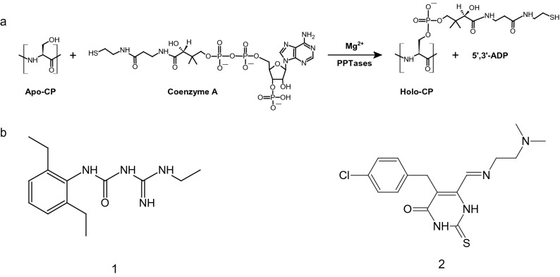
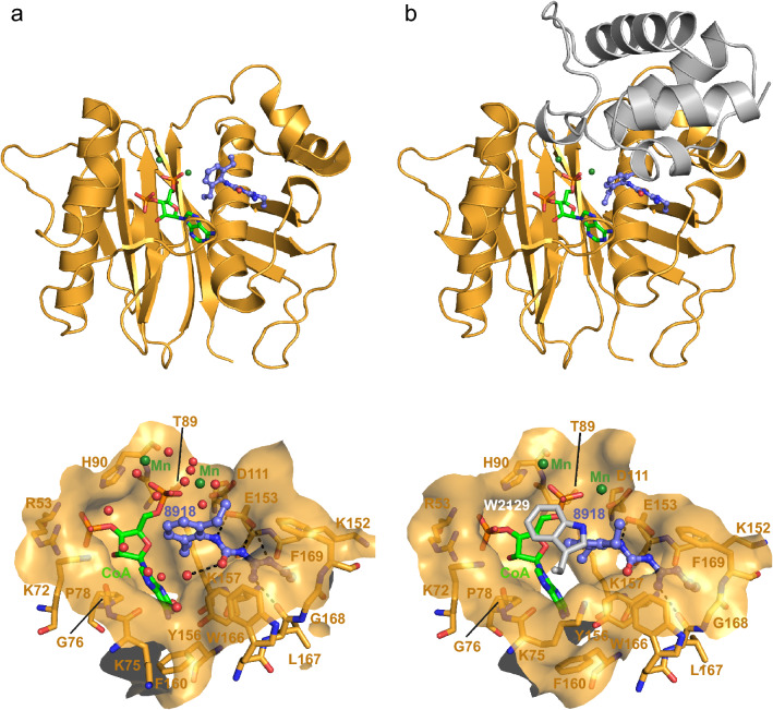
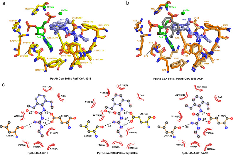
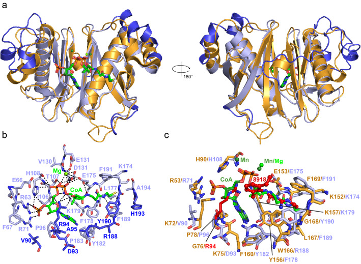
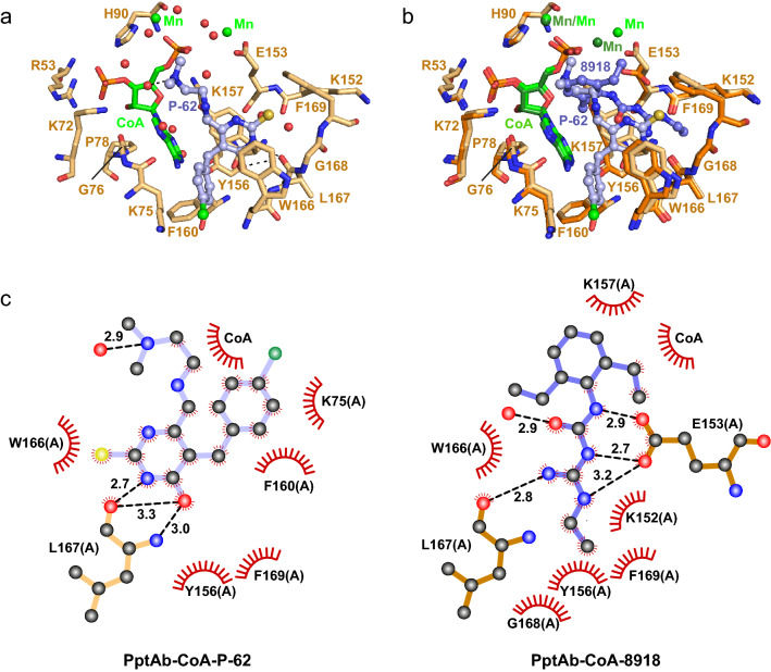
Owing to their role in activating enzymes essential for bacterial viability and pathogenicity, phosphopantetheinyl transferases represent novel and attractive drug targets. In this work, we examined the inhibitory effect of the aminido-urea 8918 compound against the phosphopantetheinyl transferases PptAb from Mycobacterium abscessus and PcpS from Pseudomonas aeruginosa, two pathogenic bacteria associated with cystic fibrosis and bronchiectasis, respectively. Compound 8918 exhibits inhibitory activity against PptAb but displays no activity against PcpS in vitro, while no antimicrobial activity against Mycobacterium abscessus or Pseudomonas aeruginosa could be detected. X-ray crystallographic analysis of 8918 bound to PptAb-CoA alone and in complex with an acyl carrier protein domain in addition to the crystal structure of PcpS in complex with CoA revealed the structural basis for the inhibition mechanism of PptAb by 8918 and its ineffectiveness against PcpS. Finally, in crystallo screening of potent inhibitors from the National Cancer Institute library identified a hydroxypyrimidinethione derivative that binds PptAb. Both compounds could serve as scaffolds for the future development of phosphopantetheinyl transferases inhibitors.


Conflict of interest statement

The authors declare no competing interests.

Figures

Figure 1
Figure 1
Phosphopantetheinylation plays a central role in metabolite biosynthetic pathways. (a) Reaction catalyzed by PPTases. (b) Inhibitors discussed in the present study. The aminido-urea 8918 inhibitor: (1-[(2,6-diethylphenyl)-3-N-ethylcarbamimodoyl]urea; (1)). The NCI compound P-62: (5-[(4-chlorophenyl)methyl]-6-[2-(dimethylamino)ethylimino-methyl]-2-sulfanylidene-1H-pyrimidin-4-one; (2)). This figure was generated using ACD/ChemSketch (Freeware) 2020.2.0 and Adobe Illustrator 25.4.1.
Figure 2
Figure 2
Inhibition by compound 8918. (a) In vitro conversion of apo- to holo-ACP by PptT, PptAb and PcpS after 60 min incubation with 5 µM 8918. The unlabeled lane corresponds to apo-ACP alone (used as a reference). (b) Bacterial viability assessed by MTT assay (serial dilution from 128 to 1 µM 8918). Results are mean and SD from duplicate samples. An uncropped version of the gel can be found in the Supplementary Information file (Supplementary Fig. 1). This figure was generated using Image Lab Version 6.1.0 (Bio-Rad Laboratories, Inc.) and Adobe Illustrator 25.4.1.
Figure 3
Figure 3
Crystallographic scrutinizing of CoA and 8918 binding to PptAb. X-rays structures of: (a) PptAb-CoA-8918 and (b) PptAb-CoA-8918-ACP. Top raw, overall view. Bottom raw, zoom-in the active site. Molecular surfaces include residues of PptAb within 5.0 Å of the ligands. Color scheme: PptAb, bright orange; ACP, dark grey; CoA and divalent cations, green; 8918, purple blue. Hydrogen bonds to 8918 are displayed as black dotted lines. This figure was generated using PyMol Version 2.4.1 (Schrödinger, LCC) and Adobe Illustrator 25.4.1. The 2D interaction maps were designed based on Ligplot+ v.2.2 (https://www.ebi.ac.uk/thornton-srv/software/LigPlus/).
Figure 4
Figure 4
8918 binding to PptAb and PptT. (a) Superposition of PptAb-CoA-8918 (orange) and PptT-CoA-8918 (PDB entry 6CT5, yellow). (b) Superposition of PptAb-CoA-8918 (orange) and PptAb-CoA-8918-ACP (light orange and grey). CoA and divalent cations are in green; 8918 is in purple blue. (c) 2D interaction maps: hydrogen bonds and their length are in black; protein residues/atoms and CoA involved in van der Waals contacts are represented by notched semicircles; letters in parentheses after residue names correspond to protein chain identifiers [one molecule, chain A, in the asymmetric unit of PptAb-CoA-8918; two molecules, chains A and B, for PptT-CoA-8918; for PptAb-CoA-8918-ACP, chains A and B are for PptAb and ACP, respectively]. This figure was generated using PyMol Version 2.4.1 (Schrödinger, LCC) and Adobe Illustrator 25.4.1. The 2D interaction maps were designed based on Ligplot+ v.2.2 (https://www.ebi.ac.uk/thornton-srv/software/LigPlus/).
Figure 5
Figure 5
Structure of PcpS-CoA and its comparison with PptAb-CoA and PptAb-CoA-8918. (a) Overall superimposition of PcpS-CoA and PtpAb-CoA (PDB entry 6QW5). Two perpendicular views along the vertical axis are shown. (b) Zoom-in the active site of PcpS-CoA. Hydrogen bonds of CoA and Mg2+ with protein atoms and water molecules are in black (only direct interactions are displayed). (c) Superimposition of PptAb-CoA-8918 and PcpS-CoA. Color scheme: PptAb, bright orange; PcpS, lightblue; major differences between PcpS and PptAb (panels a,c), blue; CoA and divalent cations, green. In panel (c), R94 of PcpS-CoA and 8918 of PptAb-CoA-8918 are in red and numbering is for PptAb/PcpS. This figure was generated using PyMol Version 2.4.1 (Schrödinger, LCC) and Adobe Illustrator 25.4.1.
Figure 6
Figure 6
Crystallographic analysis of P-62 binding to PptAb-CoA. (a) Zoom-in the active site. (b) Superimposition of PptAb-CoA-P-62 (light orange) and PptAb-CoA-8918 (bright orange). CoA and divalent cations, green; P-62, light blue; 8918, purple blue. (c) 2D interaction map. Hydrogen bonds and their length are in black. Protein residues/atoms and CoA involved in van der Waals contacts are represented by notched semicircles. This figure was generated using PyMol Version 2.4.1 (Schrödinger, LCC) and Adobe Illustrator 25.4.1. The 2D interaction maps were designed based on Ligplot+ v.2.2 (https://www.ebi.ac.uk/thornton-srv/software/LigPlus/).

References

    1. Beld J, Sonnenschein EC, Vickery CR, Noel JP, Burkart MD. The phosphopantetheinyl transferases: Catalysis of a post-translational modification crucial for life. Nat. Prod. Rep. 2014;31(1):61–108. doi: 10.1039/C3NP70054B. - DOI - PMC - PubMed
    1. Kapopoulou A, Lew JM, Cole ST. The MycoBrowser portal: A comprehensive and manually annotated resource for mycobacterial genomes. Tuberculosis (Edinb) 2011;91(1):8–13. doi: 10.1016/j.tube.2010.09.006. - DOI - PubMed
    1. Quadri LE. Biosynthesis of mycobacterial lipids by polyketide synthases and beyond. Crit. Rev. Biochem. Mol. Biol. 2014;49(3):179–211. doi: 10.3109/10409238.2014.896859. - DOI - PubMed
    1. Chalut C, Botella L, de Sousa-D'Auria C, Houssin C, Guilhot C. The nonredundant roles of two 4'-phosphopantetheinyl transferases in vital processes of Mycobacteria. Proc. Natl. Acad. Sci. U.S.A. 2006;103(22):8511–8516. doi: 10.1073/pnas.0511129103. - DOI - PMC - PubMed
    1. Quadri LE, Sello J, Keating TA, Weinreb PH, Walsh CT. Identification of a Mycobacterium tuberculosis gene cluster encoding the biosynthetic enzymes for assembly of the virulence-conferring siderophore mycobactin. Chem. Biol. 1998;5(11):631–645. doi: 10.1016/S1074-5521(98)90291-5. - DOI - PubMed

Publication types

MeSH terms