. 2022 May 4;24(5):796-808.
doi: 10.1093/neuonc/noab213.

A molecularly integrated grade for meningioma

Affiliations

A molecularly integrated grade for meningioma

Joseph Driver et al. Neuro Oncol. .

Abstract

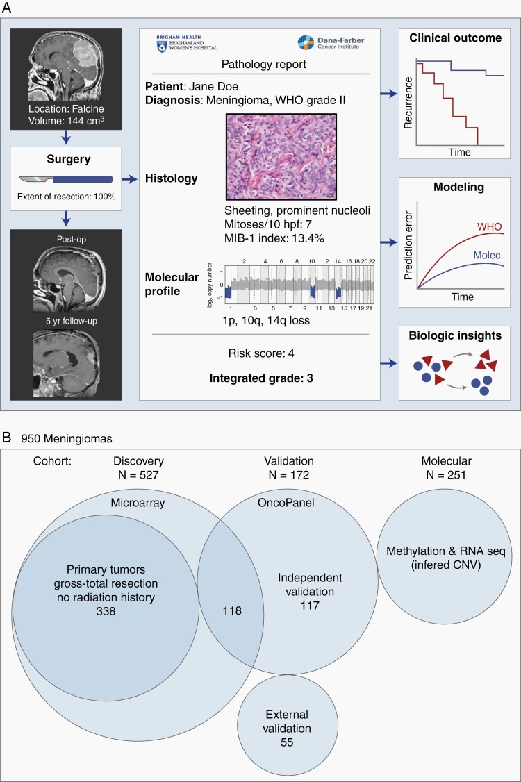
Background: Meningiomas are the most common primary intracranial tumor in adults. Clinical care is currently guided by the World Health Organization (WHO) grade assigned to meningiomas, a 3-tiered grading system based on histopathology features, as well as extent of surgical resection. Clinical behavior, however, often fails to conform to the WHO grade. Additional prognostic information is needed to optimize patient management.

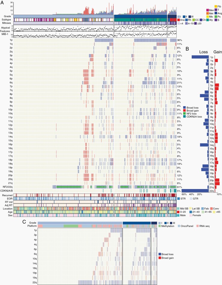
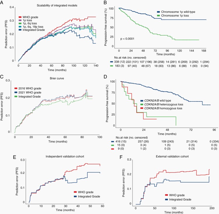
Methods: We evaluated whether chromosomal copy-number data improved prediction of time-to-recurrence for patients with meningioma who were treated with surgery, relative to the WHO schema. The models were developed using Cox proportional hazards, random survival forest, and gradient boosting in a discovery cohort of 527 meningioma patients and validated in 2 independent cohorts of 172 meningioma patients characterized by orthogonal genomic platforms.

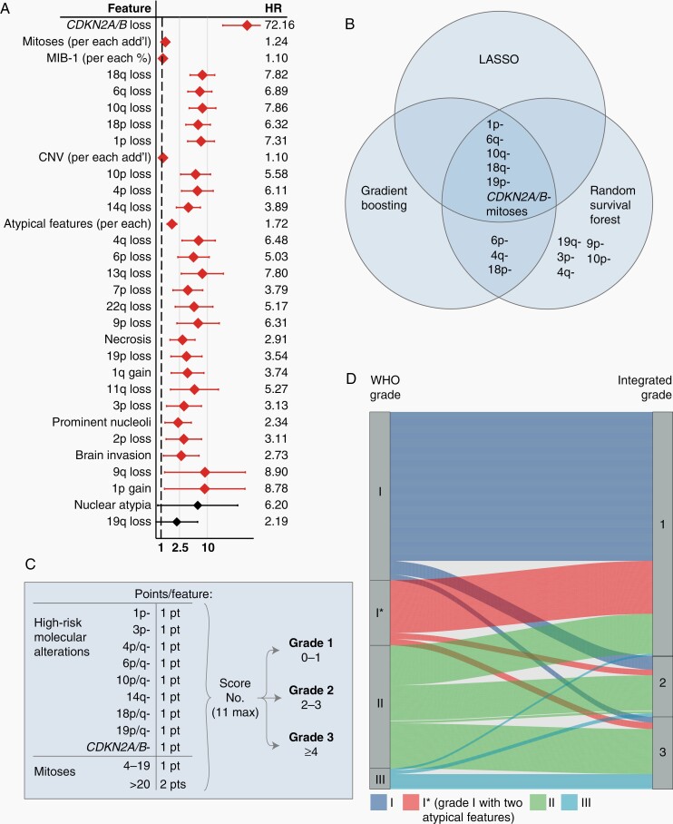
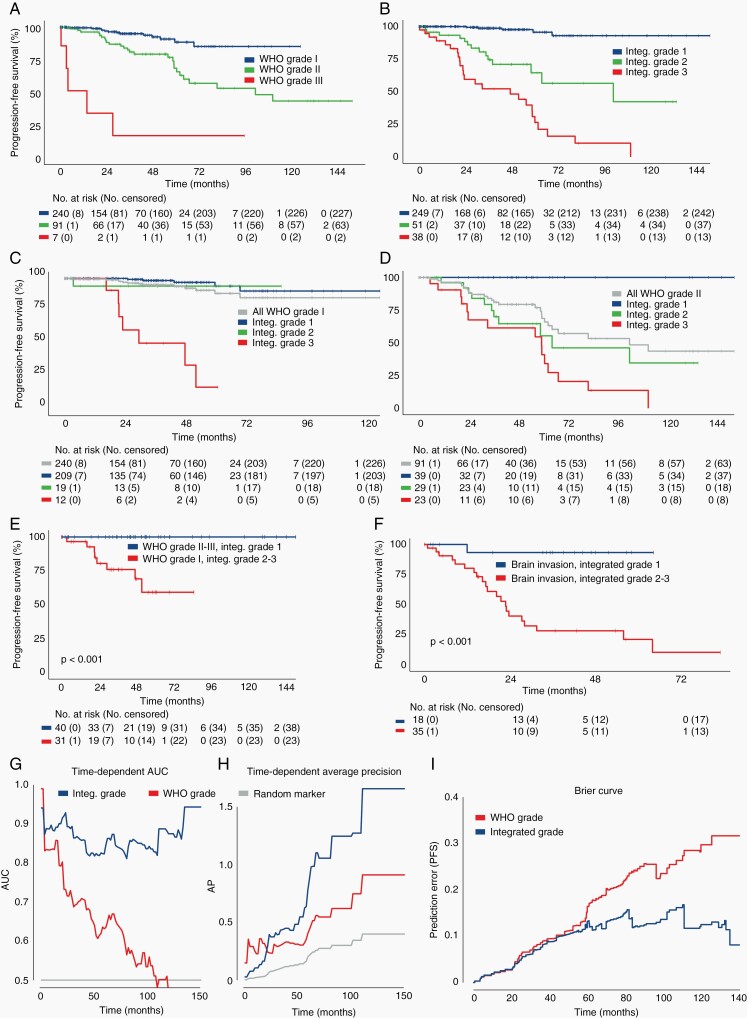
Results: We developed a 3-tiered grading scheme (Integrated Grades 1-3), which incorporated mitotic count and loss of chromosome 1p, 3p, 4, 6, 10, 14q, 18, 19, or CDKN2A. 32% of meningiomas reclassified to either a lower-risk or higher-risk Integrated Grade compared to their assigned WHO grade. The Integrated Grade more accurately identified meningioma patients at risk for recurrence, relative to the WHO grade, as determined by time-dependent area under the curve, average precision, and the Brier score.

Conclusion: We propose a molecularly integrated grading scheme for meningiomas that significantly improves upon the current WHO grading system in prediction of progression-free survival. This framework can be broadly adopted by clinicians with relative ease using widely available genomic technologies and presents an advance in the care of meningioma patients.

Keywords: copy-number alterations; meningioma.


Figures

Fig. 1
Fig. 1
Study overview and description of patient cohorts. (A) A clinical cohort of meningiomas were evaluated for patient demographics, histologic and molecular features, preoperative and postoperative tumor volume, and clinical follow-up including recurrence and death. (B) Description of 950 meningiomas analyzed. The discovery cohort consisted of 527 tumor samples from unique patients, with 338 primary tumors without history of treatment or prior radiotherapy. Validation was performed on 2 independent cohorts of 117 patients and 55 patients. Copy number was additionally derived from a molecular cohort of publicly available methylation and RNAseq datasets.
Fig. 2
Fig. 2
Copy-number profile of meningiomas. (A) Distribution of copy-number alterations, histologic features, treatment factors, and demographic features across 527 meningiomas in the discovery cohort. Tumor subtypes were annotated as: Anaplastic (Ana), Rhabdoid (Rh), Clear Cell (CC), Chordoid (Ch), Atypical (Aty), Microcystic (Mic), Metaplastic (Met), Meningothelial (Men), Transitional (T), Angiomatous (Ang), Psammomatous (Ps), Secretory (S), and Fibroblastic (F). Mitoses were classified as <4, 4-19, or >20. (B) Frequency of copy-number gains and losses across each chromosomal arm among the discovery cohort. (C) Inferred copy-number alterations in high-risk chromosomes of interest among 510 meningiomas with methylation, mutational (OncoPanel), or RNAseq data.
Fig. 3
Fig. 3
Development of a molecularly Integrated Grade. (A) Univariate Cox proportional hazards analysis evaluating tumor-intrinsic, histologic, and molecular features and their associated hazard risk for tumor recurrence across 338 patients with primary meningiomas who underwent GTR without prior radiation. Specific hazard ratios are listed with error bars representing 95% CI. Features are ranked in order from top-down as most significant to least significant, with those with P < .05 denoted by red confidence interval bars. (B) Features most significantly associated with meningioma recurrence, as identified by LASSO, random survival forest, and gradient boosting methods. (C) Integrated Grading scheme: points are assigned for the presence of listed molecular features or elevated mitoses, with the resultant sum categorizing tumors into 1 of the 3 Grades with increasing recurrence risk. (D) Association of WHO grade and Integrated Grade for 527 meningiomas, with 87% concordance between WHO grade I and Integrated Grade 1, 31% concordance between WHO grade II and Integrated Grade 2, and 72% concordance between WHO grade III and Integrated Grade 3. The majority of WHO grade III tumors that reclassified into a different Integrated Grade were rhabdoid meningiomas, while 19/20 anaplastic meningiomas remained Integrated Grade 3. Abbreviation: GTR, gross total resection.
Fig. 4
Fig. 4
Evaluation of Integrated Grade as a predictor of meningioma behavior. (A, B) Progression-free survival of 338 patients with primary meningiomas after GTR, stratified by (A) WHO grade or (B) Integrated Grade. (C, D) Progression-free survival stratified by Integrated Grade among (C) WHO grade I and (D) WHO grade II tumors. (E) Progression-free survival of discrepantly graded meningiomas (P < .01, log-rank test). (F) Progression-free survival among patients with brain invasion on histopathologic analysis, stratified by Integrated Grade 1 vs Integrated Grades 2-3. (G) Time-dependent area under the curve (AUC) of a ROC for Integrated Grade and WHO grade (Integrated Grade: 5-year AUC 0.823, 95% CI 0.724-0.91 vs WHO grade: 5-year AUC 0.632, 95% CI 0.521-0.737). (H) Time-dependent average precision for Integrated Grade and WHO grade (Integrated Grade: 5-year AP 0.781, 95% CI 0.485-1.0; vs WHO grade: 5-year AP 0.405, 95% CI 0.258-0.586). (I) Brier prediction curve for progression-free survival comparing Integrated Grade to WHO grade in the 338 patients (Brier score: 0.098 vs 0.180). Abbreviation: GTR, gross total resection.
Fig. 5
Fig. 5
Validation of integrated grade among multiple patient cohorts. (A) Brier prediction curve for progression-free survival among the 527 patients with varied treatment histories in the discovery cohort (Brier score: 0.157 vs 0.233). (B) Progression-free survival of 527 patients stratified by chromosome 1p status. (C) Progression-free survival of 527 patients stratified by CDKN2A/B status, showing no difference in outcome between patients with homozygous or heterozygous loss. (D) Brier prediction curve for progression-free survival among 244 patients for which mutational data was available in addition to copy-number data. Brier scores for this cohort were determined by the assigned 2016 WHO grading scheme, the new 2021 WHO grading scheme, and the Integrated Grading scheme (Brier score; 2016 WHO 0.163 vs 2021 WHO 0.166 vs Integrated Grade 0.151). (E) Brier prediction curve for progression-free survival calculated for Integrated Grade and WHO grade among the 117-patient internal validation cohort (Brier score 0.178 vs 0.214). (F) Brier prediction curve for progression-free survival calculated for WHO grade and Integrated Grade for the 55-patient external validation cohort (Brier score 0.133 vs 0.181).
Fig. 6
Fig. 6
Impact of treatment and development of nomogram for tumor recurrence. Progression-free survival of 527 meningioma patients in the discovery cohort, stratified by (A–C) extent of resection status (GTR vs STR) for each Integrated Grade, and (D) primary vs recurrent tumor status. (E) Multivariate Cox proportional hazards model incorporating Integrated Grade with extent of resection, tumor volume, and primary vs recurrent tumor status. (F) Nomogram for predicting 1-, 3-, and 5-year recurrence risk based on the four listed parameters. Abbreviation: GTR, gross total resection; STR, subtotal resection.

References

    1. Ostrom QT, Cioffi G, Gittleman H, et al. CBTRUS statistical report: primary brain and other central nervous system tumors diagnosed in the United States in 2012-2016. Neuro Oncol. 2019;21(Suppl 5):v1–v100. - PMC - PubMed
    1. Vernooij MW, Ikram MA, Tanghe HL, et al. Incidental findings on brain MRI in the general population. N Engl J Med. 2007;357(18):1821–1828. - PubMed
    1. Louis DN, Ohgaki H, Wiestler OD, et al. WHO Classification of Tumours of the Central Nervous System. Revised 4th ed. IARC; 2016.
    1. Rogers CL, Perry A, Pugh S, et al. Pathology concordance levels for meningioma classification and grading in NRG Oncology RTOG Trial 0539. Neuro Oncol. 2016;18(4):565–574. - PMC - PubMed
    1. Sayagués JM, Tabernero MD, Maíllo A, et al. Intratumoral patterns of clonal evolution in meningiomas as defined by multicolor interphase fluorescence in situ hybridization (FISH): is there a relationship between histopathologically benign and atypical/anaplastic lesions? J Mol Diagn. 2004;6(4):316–325. - PMC - PubMed

Publication types