

Effect of the soft tissue artifact on marker measurements and on the calculation of the helical axis of the knee during a gait cycle: A study on the CAMS-Knee data set
- PMID: 34509901
- PMCID: PMC8631460
- DOI: 10.1016/j.humov.2021.102866
Effect of the soft tissue artifact on marker measurements and on the calculation of the helical axis of the knee during a gait cycle: A study on the CAMS-Knee data set
Abstract
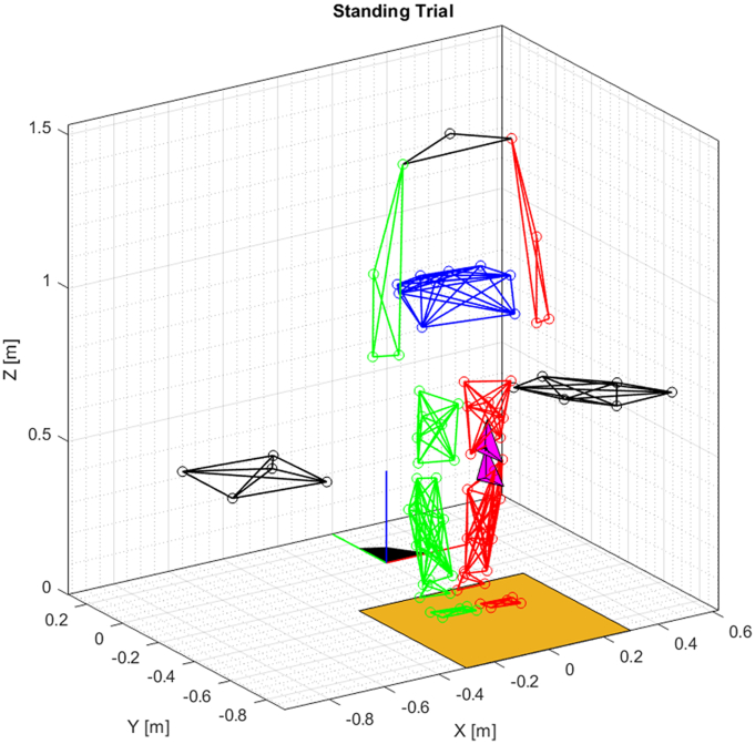
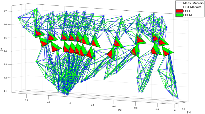
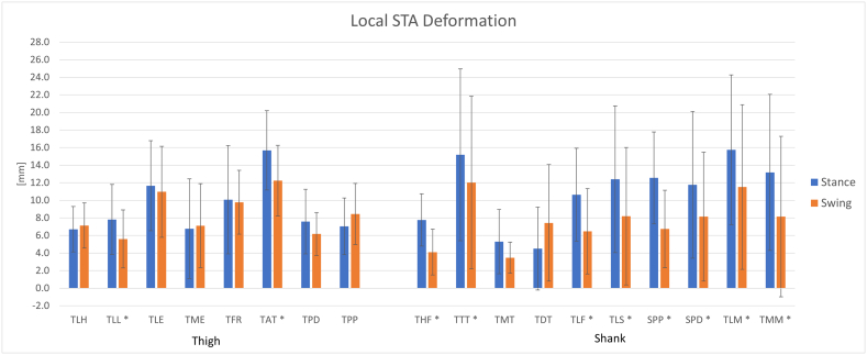
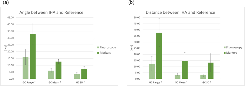
The soft tissue artifact (STA) is a phenomenon occurring when the motion of bones or anatomical segments is measured by means of skin markers: the biological tissues between the markers and the bone produce a relative motion bone-markers that leads to inaccuracies in the estimation of rigid body poses or kinematics. The aim of this study was to quantify the STA by exploiting a recently published gait analysis dataset. The dataset was composed of six adult subjects with a total knee arthroplasty who underwent gait analysis trials. The motion of the knee was concurrently recorded by means of (i) fluoroscopy imaging and (ii) an optoelectronic system and redundant markers attached to the thigh and shank. The STA was studied by comparing the results calculated on the marker sets with the results obtained from the fluoroscopy data. The stance and swing phases were considered separately. Rigid STA motion and local STA deformation were studied separately. In addition to previous studies, the instantaneous helical axis (IHA) of the knee was calculated and the effect of the STA on its calculation was assessed. The largest rigid-motion STA effect was observed on the thigh cluster (~10 deg. and ~ 18 mm). The shank cluster was mainly affected during the swing phase (~7 deg. and ~ 17 mm). The local STA deformation affected differently the markers. The largest effect was ~16 mm and the lowest was ~4 mm. The estimation of the IHA was not reliable when based only on markers, having an estimation error of ~17 deg. and ~ 25 mm. A high variability of results across subjects was observed.
Keywords: Fluoroscopy; Gait analysis; Helical axis; Knee prosthesis; Motion capture; Skin artifact; Soft tissue artifact.
Copyright © 2021 The Authors. Published by Elsevier B.V. All rights reserved.
Conflict of interest statement
The authors declare no conflict of interests.
Figures

References
-
- Ancillao A. 2018. Stereophotogrammetry in Functional Evaluation: History and Modern Protocols; pp. 1–29. (in SpringerBriefs in Applied Sciences and Technology)
-
- Ancillao A., Rossi S., Cappa P. Analysis of knee strength measurements performed by a hand-held multicomponent dynamometer and optoelectronic system. IEEE Transactions on Instrumentation and Measurement. 2017;66(1):85–92. doi: 10.1109/TIM.2016.2620799. - DOI
MeSH terms
LinkOut - more resources
Full Text Sources
