. 2021 Dec;42(12):1624-1636.
doi: 10.1002/humu.24281. Epub 2021 Sep 24.

Noncoding sequence variants define a novel regulatory element in the first intron of the N-acetylglutamate synthase gene

Affiliations

Noncoding sequence variants define a novel regulatory element in the first intron of the N-acetylglutamate synthase gene

Johannes Häberle et al. Hum Mutat. 2021 Dec.

Abstract

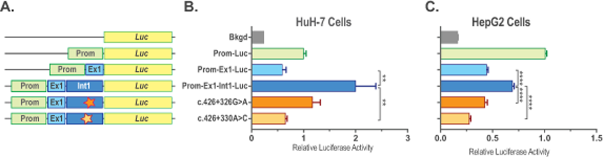
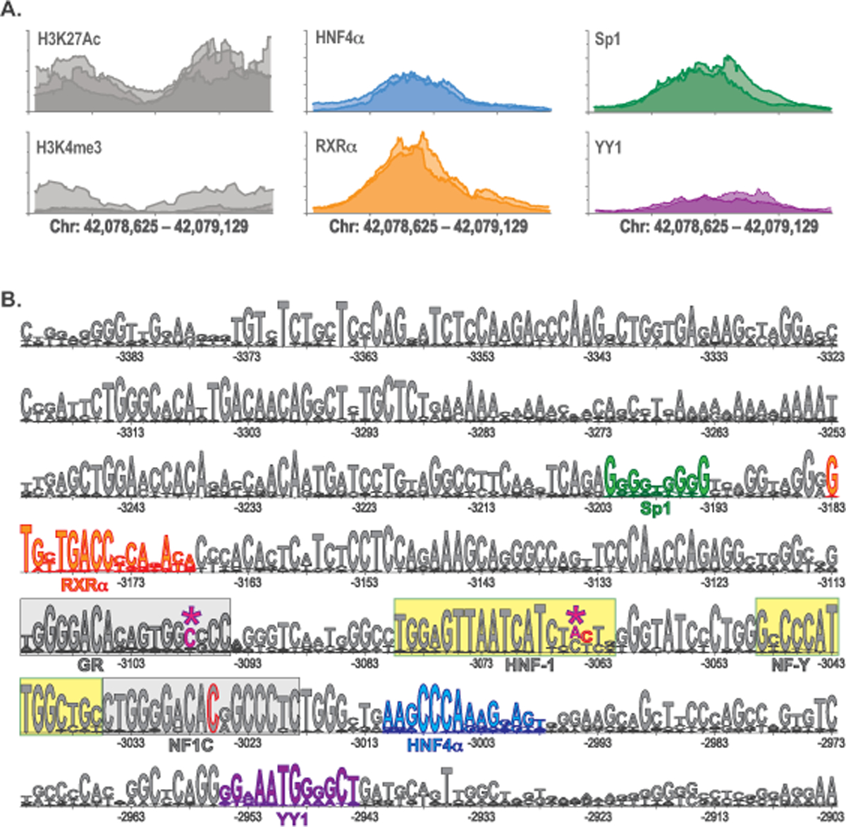
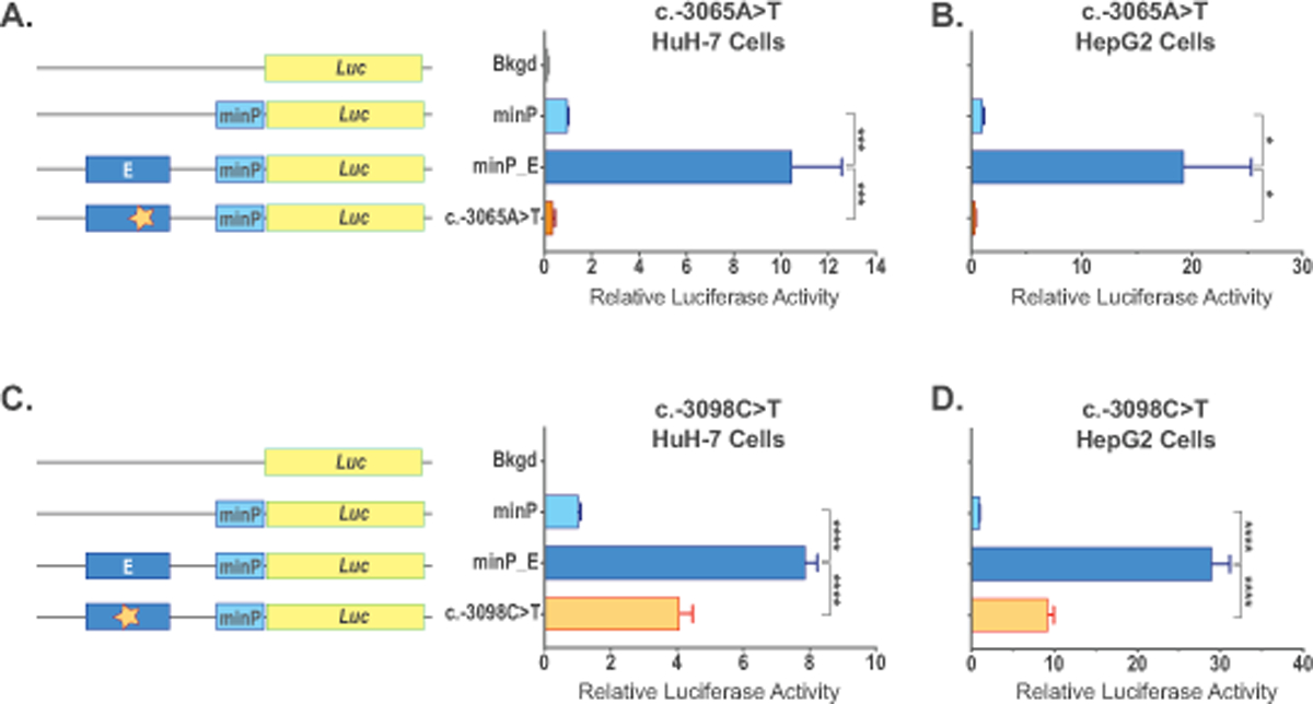
N-acetylglutamate synthase deficiency is an autosomal recessive urea cycle disorder caused either by decreased expression of the NAGS gene or defective NAGS enzyme resulting in decreased production of N-acetylglutamate (NAG), an allosteric activator of carbamylphosphate synthetase 1 (CPS1). NAGSD is the only urea cycle disorder that can be effectively treated with a single drug, N-carbamylglutamate (NCG), a stable NAG analog, which activates CPS1 to restore ureagenesis. We describe three patients with NAGSD due to four novel noncoding sequence variants in the NAGS regulatory regions. All three patients had hyperammonemia that resolved upon treatment with NCG. Sequence variants NM_153006.2:c.427-222G>A and NM_153006.2:c.427-218A>C reside in the 547 bp-long first intron of NAGS and define a novel NAGS regulatory element that binds retinoic X receptor α. Sequence variants NC_000017.10:g.42078967A>T (NM_153006.2:c.-3065A>T) and NC_000017.10:g.42078934C>T (NM_153006.2:c.-3098C>T) reside in the NAGS enhancer, within known HNF1 and predicted glucocorticoid receptor binding sites, respectively. Reporter gene assays in HepG2 and HuH-7 cells demonstrated that all four substitutions could result in reduced expression of NAGS. These findings show that analyzing noncoding regions of NAGS and other urea cycle genes can reveal molecular causes of disease and identify novel regulators of ureagenesis.

Keywords: N-acetylglutamate; N-acetylglutamate synthase; N-acetylglutamate synthase deficiency; intron; mutation analysis; noncoding sequence variants; regulatory element; urea cycle; urea cycle disorders.


Conflict of interest statement

Conflicts of Interest

Mutation analysis (Dr. Häberle and Ms. Rüfenacht) for cases 2 and 3 and functional testing of the four non-coding variants (Dr. Caldovic and Ms. Haskins) were supported by the Recordati Rare Diseases, Inc. that manufactures and sells NCG as Carbaglu® (carglumic acid), which is used for treatment of NAGS deficiency.

Figures

Figure 1.
Figure 1.. Sequence variants in the novel regulatory element in the NAGS intron 1.
A. The phastCons track of the UCSC Genome Browser showing conserved regions in the NAGS gene (top) and map of the NAGS gene (bottom) showing genomic region chr17:42,081,993 – 42,086,412 of the GRCh37/hg19 human genome assembly with exons as gray boxes, introns as gray lines and predicted cis-acting conserved regulatory element in the NAGS intron 1 as purple box. Numbers below gray boxes indicate exon numbers. B. Genomic region chr17:42,082,368–42,083,068 of the GRCh37/hg19 human genome assembly. Results of the ENCODE ChIP-Seq experiments showing binding of transcription factors HNF4α (blue), RXRα (orange) and Sp1 (green) to NAGS intron 1 in the human liver tissue (top) and phastCons track of the UCSC Genome Browser showing conserved region within NAGS intron 1 (bottom). Two shades of blue, orange and green indicate results of the two ChIP-Seq biological replicates in the ENCODE database. C. LOGO alignment of the conserved intronic sequences from 20 mammals. Numbers indicate bp positions in the NAGS intron 1 where G of the donor splice site is +1. Magenta – pathogenic sequence variants. Blue – HNF4α binding site. Orange – RXRα binding site. Green – Sp1 binding site. Asterisks indicate positions of pathogenic sequence variants NM_153006.2:c.427–222G>A and NM_153006.2:c.427–218A>C.
Figure 2.
Figure 2.. Functional testing of the sequence variants found in NAGS intron 1 from patients with NAGSD.
Expression constructs (A) and relative luciferase activity in HuH-7 (B) and HepG2 (C) cells transfected with the promoter-less plasmid pGL4.10[luc2] (Bkgd), plasmid with NAGS promoter controlling luciferase expression (Prom-Luc), plasmid with NAGS promoter controlling luciferase fused to the coding sequence of NAGS exon 1 (Prom-Ex1-Luc), plasmid with NAGS promoter controlling luciferase fused to the NAGS exon and intron 1 (Prom-Ex1-Int1-Luc) and plasmids with intronic mutations found in patients with NAGS deficiency (NM_153006.2:c.427–222G>A and NM_153006.2:c.427–218A>C). Data represent mean ± SEM of n=9 measurements. ** indicate p<0.01, **** indicates p<0.0001.
Figure 3.
Figure 3.. Sequence variants in the −3kb enhancer of the NAGS gene.
A. Genomic region chr17:42,078,635–42,079,129 of the GRCh37/hg19 human genome assembly. Results of the ENCODE ChIP-Seq experiments showing H3K27Ac and H3K4me3 histone modifications (gray) and binding of transcription factors HNF4α (blue), RXRα (orange), Sp1 (green) and YY1 (purple) to −3kb enhancer in the human liver tissue. Shades of gray, blue, orange, green, and purple indicate results of ChIP-Seq biological replicates in the ENCODE database. B. LOGO alignment of the −3kb enhancer sequences from 25 mammals. Confirmed HNF1 and NF-Y binding sites are highlighted in yellow. Conserved GR and NF1C binding sites are highlighted in gray. Magenta and asterisks – pathogenic sequence variants reported here: NC_000017.10:g.42078934C>T (c-3098C>T) and NC_000017.10:g.42078967A>T (c.−3065A>T). Gray with red outline – location of the previously reported sequence variants: NC_000017.10:g.42079006C>T (c.−3026C>T) and NC_000017.10:g.42078968C>A (c.−3064C>A). Blue – HNF4α binding site. Orange – RXRα binding site. Green – Sp1 binding site. Purple – YY1 binding site. Numbers indicate the distance of base pairs in the NAGS −3 kb enhancer from translation initiation codon; its A is +1.
Figure 4.
Figure 4.. Functional testing of the sequence variants found in the NAGS −3kb enhancer from patients with NAGSD.
Expression constructs and effect of the NC_000017.10:g.42078967A>T (c.−3065A>T) sequence variant on luciferase gene expression was tested in HuH-7 (A) and HepG2 (B) cells. Expression constructs and effect of the NC_000017.10:g.42078934C>T (c-3098C>T) sequence variant on luciferase gene expression was tested in HuH-7 (C) and HepG2 (D) cells. Cells were transfected with the promoter-less plasmid pGL4.10[luc2] (Bkgd), plasmid with minimal eukaryotic promoter controlling luciferase expression (minP), plasmid harboring −3kb NAGS enhancer and minimal promoter controlling luciferase expression (minP_E), and plasmids with enhancer mutations found in patients with NAGS deficiency (c.−3065A>T and c.−3098C>T). Data represent mean ± SEM of n=9 measurements. * indicate p<0.05, *** indicate p<0.001, **** indicate p<0.0001.

References

    1. Ah Mew N, & Caldovic L (2011). N-acetylglutamate synthase deficiency: an insight into the genetics, epidemiology, pathophysiology, and treatment. Appl Clin Genet, 4, 127–135. - PMC - PubMed
    1. Al Kaabi EH, & El-Hattab AW (2016). N-acetylglutamate synthase deficiency: Novel mutation associated with neonatal presentation and literature review of molecular and phenotypic spectra. Mol Genet Metab Rep, 8, 94–98. - PMC - PubMed
    1. Beaulieu E, Green L, Elsby L, Alourfi Z, Morand EF, Ray DW, & Donn R (2011). Identification of a novel cell type-specific intronic enhancer of macrophage migration inhibitory factor (MIF) and its regulation by mithramycin. Clin Exp Immunol, 163(2), 178–188. - PMC - PubMed
    1. Bijarnia-Mahay S, Haberle J, Jalan AB, Puri RD, Kohli S, Kudalkar K, . . . Verma IC (2018). Urea cycle disorders in India: clinical course, biochemical and genetic investigations, and prenatal testing. Orphanet J Rare Dis, 13(1), 174. - PMC - PubMed
    1. Bornstein P, McKay J, Morishima JK, Devarayalu S, & Gelinas RE (1987). Regulatory elements in the first intron contribute to transcriptional control of the human alpha 1(I) collagen gene. Proc Natl Acad Sci U S A, 84(24), 8869–8873. - PMC - PubMed

Publication types

MeSH terms

Substances