. 2021 Oct;25(20):9586-9596.
doi: 10.1111/jcmm.16899. Epub 2021 Sep 12.

Simple derivation of skeletal muscle from human pluripotent stem cells using temperature-sensitive Sendai virus vector

Affiliations

Simple derivation of skeletal muscle from human pluripotent stem cells using temperature-sensitive Sendai virus vector

Ghee Wan Tan et al. J Cell Mol Med. 2021 Oct.

Abstract

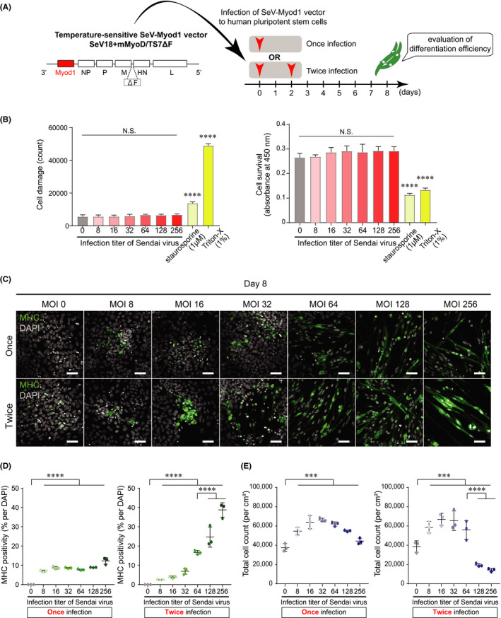
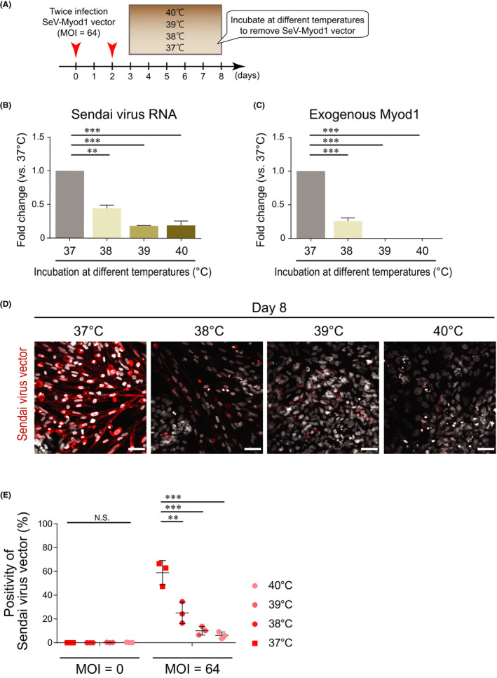
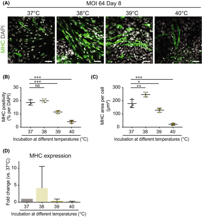
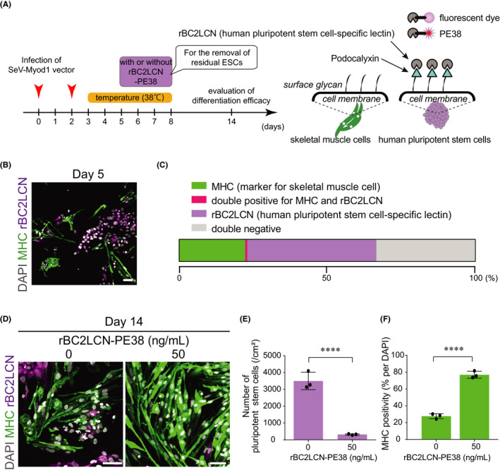
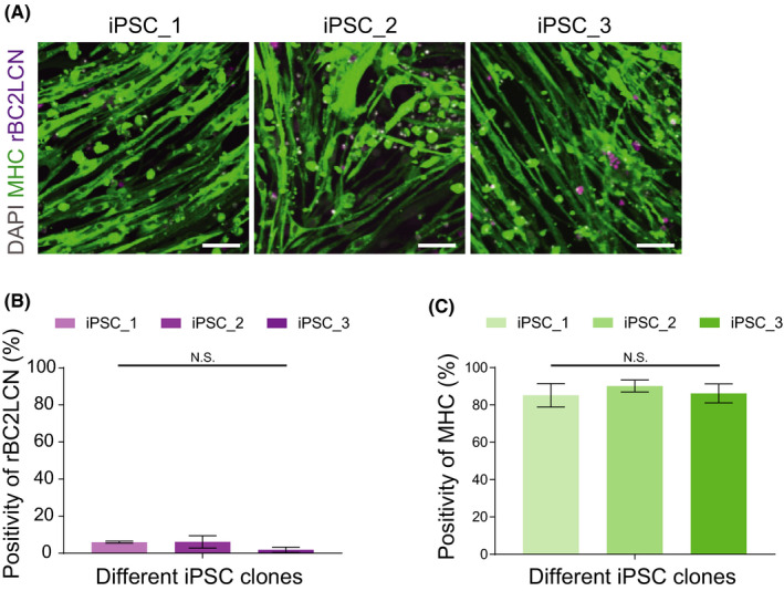
Human pluripotent stem cells have the potential to differentiate into various cell types including skeletal muscles (SkM), and they are applied to regenerative medicine or in vitro modelling for intractable diseases. A simple differentiation method is required for SkM cells to accelerate neuromuscular disease studies. Here, we established a simple method to convert human pluripotent stem cells into SkM cells by using temperature-sensitive Sendai virus (SeV) vector encoding myoblast determination protein 1 (SeV-Myod1), a myogenic master transcription factor. SeV-Myod1 treatment converted human embryonic stem cells (ESCs) into SkM cells, which expressed SkM markers including myosin heavy chain (MHC). We then removed the SeV vector by temporal treatment at a high temperature of 38℃, which also accelerated mesodermal differentiation, and found that SkM cells exhibited fibre-like morphology. Finally, after removal of the residual human ESCs by pluripotent stem cell-targeting delivery of cytotoxic compound, we generated SkM cells with 80% MHC positivity and responsiveness to electrical stimulation. This simple method for myogenic differentiation was applicable to human-induced pluripotent stem cells and will be beneficial for investigations of disease mechanisms and drug discovery in the future.

Keywords: Myod1; Sendai virus; differentiation method; disease modelling; high temperature treatment; human embryonic stem cells; human-induced pluripotent stem cells; skeletal muscle.


Conflict of interest statement

Jitsutaro Kawaguchi and Tsugumine Shu are employees of I'rom Group Co., Ltd. The remaining authors declare no competing interests.

Figures

FIGURE 1
FIGURE 1
Infection of SeV‐Myod1vector converted human pluripotent stem cells into skeletal muscle cells. (A) Construction of SeV‐Myod1 vector and schema of experimental design. Differentiation efficiency was analysed on day 8 to optimize the SeV‐Myod1 concentration and evaluate the cellular toxicity. (B) Cell damage (left panel) and cell survival (right panel) were analysed at each described MOI condition or at the condition of adding staurosporine and Triton‐X as positive control for cell death. (Independent experiments n = 3, mean ± SD, ****p < 0.0001). (C) Immunofluorescence staining for myosin heavy chain (MHC, green) and DAPI staining (white) on day 8. Representative images are shown. Scale bar =100 μm. (D) MHC positivity (% against DAPI) was quantified from (C). Quantified positivity was shown in separate graphs for once infection (left panel) and twice infection (right panel) of SeV‐Myod1. (Independent experiments n = 3, mean ± SD, **** p < 0.0001) MHC: myosin heavy chain, (E) Total cell number was counted from (C). (Independent experiments n = 3, mean ± SD, ***p < 0.001, ****p < 0.0001)
FIGURE 2
FIGURE 2
Transient cultivation at high temperature removed infected SeV‐Myod1. (A) Schema of SeV‐Myod1 vector infection and culture condition. The cells were incubated at 37ºC, 38ºC, 39ºC or 40ºC from day 3 to day 8, and the removal of SeV‐Myod1 vector was evaluated. (B) SeV RNA was analysed on day 8 and compared among the different temperature conditions. (Independent experiments n = 3, mean ± SD, **p < 0.01, ***p < 0.001). (C) The mRNA level of exogenous Myod1 delivered by SeV‐Myod1 was analysed on day 8 and compared among the different temperature conditions. (Independent experiments n = 3, mean ± SD, ***p < 0.001). (D) Differentiating cells were immunostained for SeV antibody (red). Nuclei were stained with DAPI (white). Scale bar = 100 μm. (E) Positivity of Sendai virus vector against total DAPI (%) was quantified and compared among the different temperature conditions. (Independent experiments, n = 3, mean ± SD, **p < 0.01, ***p < 0.001, NS: not significant)
FIGURE 3
FIGURE 3
Transient heat shock at temperature of 38ºC accelerated myogenic differentiation. (A) Cells were infected twice with SeV‐Myod1 at MOI 64 and then incubated at 37ºC, 38ºC, 39ºC or 40ºC from day 3 to day 8, as shown in the schema in Figure 2A. Myogenic differentiation efficiency was analysed by immunostaining for myosin heavy chain (MHC, green). Nuclei were stained with DAPI (white) to count the total cell number. Scale bar = 100 μm. (B) MHC positivity (%) against total DAPI was quantified and compared among the different temperature conditions. (Independent experiments n = 3, mean ± SD, ***p < 0.001, NS: not significant). (C) MHC‐positive cell area per MHC‐positive cell was quantified and compared among the different temperature conditions. (Independent experiments n = 3, mean ±SD, *p < 0.05, ** p < 0.01, ***p < 0.001). (D) MHC expression levels were analysed and compared among the different temperature conditions. (Independent experiments n = 2, mean ± SD)
FIGURE 4
FIGURE 4
Removal of residual pluripotent stem cells by rBC2LCN‐PE38 enhanced the purity of differentiated skeletal muscle cells. (A) Schema of experimental design. Pluripotent cell killer compound (rBC2LCN‐PE38) was added from day 5 to day 8, and myogenic cell differentiation efficiency was evaluated on day 14. rBC2KCN, a human pluripotent stem cell‐specific lectin, targets the surface of human pluripotent stem cells and is utilized for delivering fluorescent dye or PE38 to human pluripotent stem cells. (B) Presence of pluripotent stem cells on day 5. MHC immunostaining (green) for myogenic cells, rBC2LCN‐fluorescent dye staining (magenta) for pluripotent‐state cell surface representing podocalyxin. Nuclei were stained with DAPI (white). Scale bar = 100 μm. (C) Stacked bar charts of the percentage of MHC‐positive cells and human ESCs stained by rBC2LCN‐635. There was almost no overlap between MHC‐ and rBC2LCN‐positive cells on day 5. (D) Residual human ESCs on day 14 were stained with MHC antibody or rBC2LCN‐635 dye and compared between groups without (0 ng/ml) or with (50 ng/ml) rBC2LCN‐PE38. Scale bar = 100 μm. (E) Numbers of rBC2LCN‐fluorescent dye‐stained pluripotent stem cells were compared after treatment without or with rBC2LCN‐PE38 (50 ng/ml). (Independent experiments, n = 3, mean ± SD, ****p < 0.0001). (F) MHC positivity (%) was compared after treatment without or with rBC2LCN‐PE38 (50 ng/ml). (Independent experiments, n = 3, mean ± SD, ****p < 0.0001)
FIGURE 5
FIGURE 5
SeV‐Myod1 and 38℃ treatment converted multiple induced pluripotent stem cells (iPSCs) into skeletal muscle (SkM) cells. (A) Immunostaining of SkM cells originating from three different iPSCs were immunostained by myosin heavy chain (MHC) antibody (green), rBC2LCN‐635 dye (magenta) and nucleus‐stained with DAPI (grey). Scale bars = 100 μm. (B) Positivities of rBC2LCN were calculated. Independent experiment, n = 3, mean ± S.D. (C) Positivities of MHC were calculated. Independent experiment, n = 3, mean ± S.D
FIGURE 6
FIGURE 6
SkM cells responded to electrical stimulations. (A) Differentiated SkM cells were cultured in a 96‐well plate. To evoke the Ca2+ response, SkM cells were subjected to 3 Hz electrical field stimulation (EFS) and were monitored by analysing the altered fluorescence, indicating the altered dynamics of cytosolic Ca2+ concentration, quantified as ΔF/F. (B) ΔF/F were monitored after EFS at 10 V/2 Hz, 10 V/10 Hz, 10 V/50 Hz and 20 V/50 Hz sequentially at 90‐second intervals

References

    1. Thomson JA. Embryonic stem cell lines derived from human blastocysts. Science. 1998;282:1145‐1147. - PubMed
    1. Chal JJ, Pourquié O. Making muscle: skeletal myogenesis in vivo and in vitro. Development. 2017;144(12):2104‐2122. 10.1242/dev.151035 - DOI - PubMed
    1. Davis RL, Weintraub H, Lassar AB. Expression of a single transfected cDNA converts fibroblasts to myoblasts. Cell. 1987;51:987‐1000. - PubMed
    1. Constantinides PG, Jones PA, Gevers W. Functional striated muscle cells from non‐myoblast precursors following 5‐azacytidine treatment. Nature. 1977;267:364‐366. - PubMed
    1. Tanaka A, Woltjen K, Miyake K, et al. Efficient and reproducible myogenic differentiation from human iPS cells: prospects for modeling Miyoshi Myopathy in vitro. PLoS One. 2013;8:e61540. - PMC - PubMed

Publication types

MeSH terms