. 2021 Aug 25:12:670772.
doi: 10.3389/fpls.2021.670772. eCollection 2021.

Tomato Divinyl Ether-Biosynthesis Pathway Is Implicated in Modulating of Root-Knot Nematode Meloidogyne javanica's Parasitic Ability

Affiliations

Tomato Divinyl Ether-Biosynthesis Pathway Is Implicated in Modulating of Root-Knot Nematode Meloidogyne javanica's Parasitic Ability

Payal Sanadhya et al. Front Plant Sci. .

Abstract

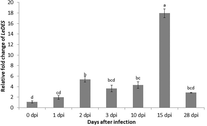
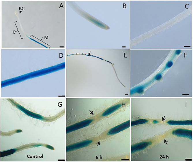
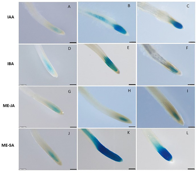
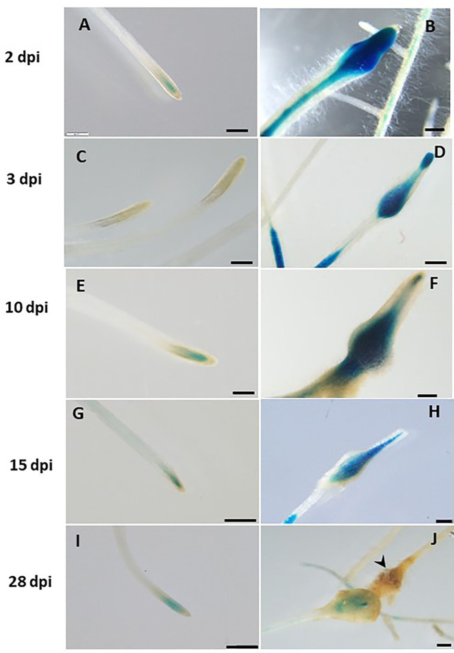
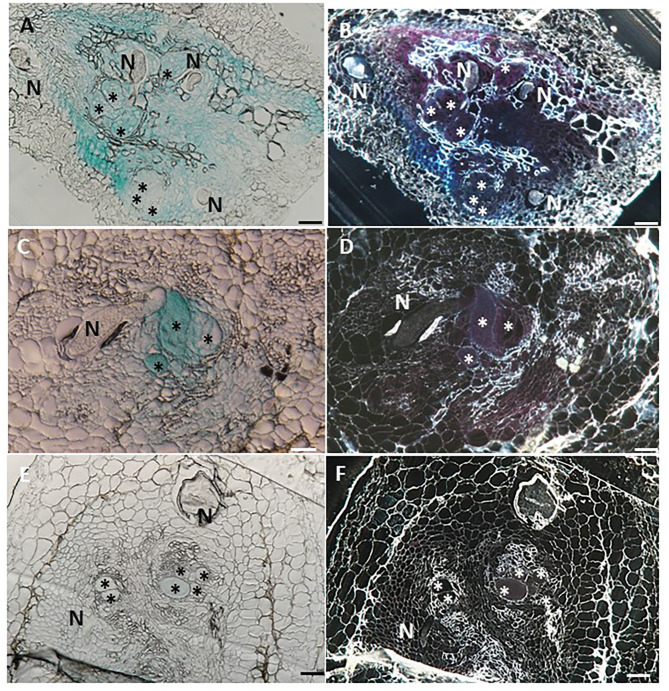
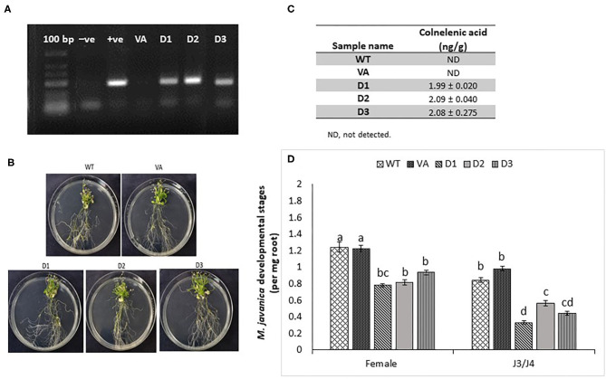
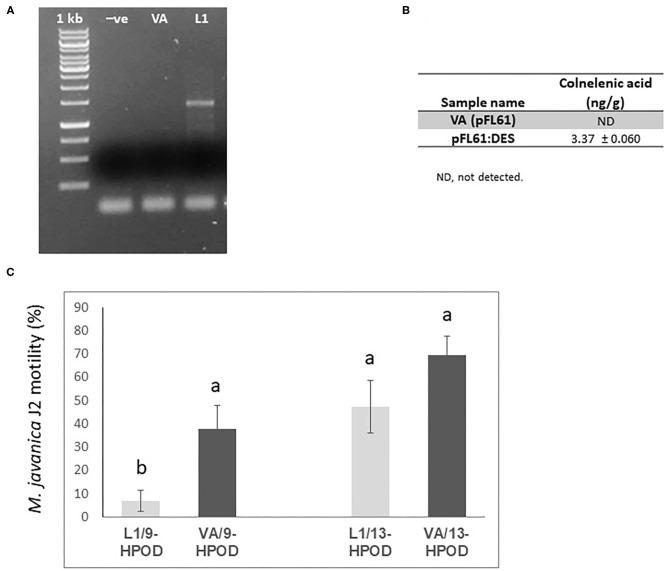
The role of the 9-lipoxygenase (9-LOX)-derived oxylipins in plant defense is mainly known in solanaceous plants. In this work, we identify the functional role of the tomato divinyl ether synthase (LeDES) branch, which exclusively converts 9-hydroperoxides to the 9-divinyl ethers (DVEs) colneleic acid (CA) and colnelenic acid (CnA), during infection by the root-knot nematode Meloidogyne javanica. Analysis of LeDES expression in roots indicated a concurrent response to nematode infection, demonstrating a sharp increase in expression during the molting of third/fourth-stage juveniles, 15 days after inoculation. Spatiotemporal expression analysis using an LeDES promoter:GUS tomato line showed high GUS activity associated with the developing gall; however the GUS signal became more constricted as infection progressed to the mature nematode feeding sites, and eventually disappeared. Wounding did not activate the LeDES promoter, but auxins and methyl salicylate triggered LeDES expression, indicating a hormone-mediated function of DVEs. Heterologous expression of LeDES in Arabidopsis thaliana rendered the plants more resistant to nematode infection and resulted in a significant reduction in third/fourth-stage juveniles and adult females as compared to a vector control and the wild type. To further evaluate the nematotoxic activity of the DVEs CA and CnA, recombinant yeast that catalyzes the formation of CA and CnA from 9-hydroperoxides was generated. Transgenic yeast accumulating CnA was tested for its impact on M. javanica juveniles, indicating a decrease in second-stage juvenile motility. Taken together, our results suggest an important role for LeDES as a determinant in the defense response during M. javanica parasitism, and indicate two functional modes: directly via DVE motility inhibition effect and through signal molecule-mediated defense reactions to nematodes that depend on methyl salicylate.

Keywords: Meloidogyne javanica; divinyl ether synthase; hormone signaling; innate immunity; oxylipins; plant defense signaling.


Conflict of interest statement

The authors declare that the research was conducted in the absence of any commercial or financial relationships that could be construed as a potential conflict of interest.

Figures

Figure 1
Figure 1
Expression pattern of LeDES in tomato roots during the infection time course. To investigate the transcript levels of LeDES upon nematode infection, total RNA was isolated from each sample at 1, 2, 3, 10, 15, and 28 dpi or without infection; RNA was subjected to qRT-PCR and normalized to β-tubulin as the reference gene. Two biological replicates were taken and three independent qRT-PCRs were performed per sample, resulting in a total of six replicates for statistical analyses. The graph shows the mean and SD of the amount of LeDES transcript relative to non-inoculated samples. Error bars correspond to SD n = 2 while different letters above the bars denote a significant difference (P ≤ 0.05, ANOVA) between different treatments as analyzed by Tukey–Kramer multiple comparison test.
Figure 2
Figure 2
Studying basal and wound induced LeDES GUS expression profile. Typical expression pattern of pLeDES::GUS in non-inoculated tomato root lines. (A) Basal GUS activity in roots. (B–D) Magnified image of root cap (B), elongation zone (C), and maturation zone (D). (E,F) Promoter–GUS activity observed in lateral root primordials in non-inoculated roots. RC, root cap; E, elongation zone; M, maturation zone. Bars: (A,E) 1 mm; (B–D,F) 250 μm. Expression analysis of pLeDES::GUS tomato root lines in response to wounding (G–I). Histochemical GUS staining of pLeDES::GUS in control roots (G) and in roots 6 h (H) and 24 h (I) after wounding. Bars: (G–I) 1 mm. Arrows indicate site of mechanical wounding.
Figure 3
Figure 3
GUS staining in transgenic tomato roots carrying pLeDES::GUS following exogenous phytohormone application. One-week-old roots were subjected to GB as a control (A,D,G,J) or to GB containing IAA [1 and 5 μM; (B,C)], IBA [1 and 10 μM; (E,F)], Me-JA [0.01 and 0.1 mM; (H,I)], or Me-SA [1 and 5 mM; (K,L)], for 16 h. GUS staining was monitored histochemically in root tips. Figures are representative of at least three independent experiments. Bars: 250 μm.
Figure 4
Figure 4
Expression-pattern analysis of pLeDES::GUS root lines during nematode infection. Non-inoculated control root demonstrated consistent GUS staining in the apical meristem (cell-division zone) (A,C,E,G,I). Increased GUS staining was detected in the infected swelling site located in the elongation zone on 2 dpi (B), 3 dpi (D), and 10 dpi, premature developing gall (F). Strong GUS staining was observed in the vasculature and vessels associated with the developed gall at 15 dpi (H). At 28 dpi (J), GUS staining intensity decreased and became localized specifically to the cells surrounding the developed nematode. Bars: (A,C–E) 250 μm; (B,F) 100 μm; (G–J) 500 μm.
Figure 5
Figure 5
Histological GUS localization within nematode feeding site. For histological GUS localization, galls were fixed and embedded in Technovit 7100 and 3-μm-thick cross sections were analyzed using a light microscope equipped with a Nikon digital camera. All giant cells were mature and nematodes developed to the J4 stage. Histological analysis of roots expressing pLeDES::GUS on 15 dpi clearly shows GUS expression restricted to the vascular systems bordering the feeding sites, the giant cells and cells surrounding the developing female body (A,B). At 28 dpi, GUS signal is observed mostly in the mature feeding site (C,D) and (E,F). GUS staining is observed as blue color in whole mounts, and as a red precipitate in the dark field micrographs of the sections. N, female nematode body; *giant cells. Bars: 100 μm.
Figure 6
Figure 6
Response of Arabidopsis plants overexpressing tomato LeDES to nematode infection. (A) RT-PCR confirmation of LeDES expression with DES-specific primers. Lane 1, negative control; lane 2, cloned LeDES template (positive control); Lane 3, independent transgenic line transformed with empty vector; lanes 4–6, independently generated transgenic lines for LeDES (B) Arabidopsis plants expressing LeDES 30 dpi; root systems of all plants show normal root with no phenotypic growth changes. (C) Quantification of colnelenic acid produced in transgenic Arabidopsis root homogenates as measured by LC-MS. (D) Disease development in Meloidogyne-infected roots of transgenic Arabidopsis plants expressing LeDES compared to control lines. All plants were inoculated with 300 sterile pre-parasitic J2s and the infected roots were assessed for J3/J4 and mature female development at 28 dpi through observation under the dissecting microscope following staining with acid fuchsin dye. General Linear Mixed Model test to infection assay data was applied. Different letters above the columns indicate significant difference (P ≤ 0.05) among different Arabidopsis lines analyzed by Tukey post-hoc range test.
Figure 7
Figure 7
Effect of recombinant yeast expressing LeDES on M. javaniva J2 viability. (A) RT-PCR confirmation of LeDES expression in yeast. Lane 1, negative control; lane 2, transgenic yeast cells with empty vector; lane 3, transgenic yeast containing LeDES (L1) (B) Quantification of colnelenic acid production in transgenic yeast by LC–MS. Values are means of duplicate measurements. (C) Yeast carrying LeDES display bionematostatic activity. J2s were incubated in SD medium containing yeast strain and 9-HPODE or 13-HPODE for 24 h, then 300 J2s were added to the reaction for 12 h then subjected for sieving followed by microscopic observation. Different letters above the columns indicate significant difference (P ≤ 0.05) among the different treatments analyzed by Tukey post-hoc range test.

References

    1. Alkharouf N. W., Klink V. P., Chouikha I. B., Beard H. S., MacDonald M. H., Meyer S., et al. . (2006). Timecourse microarray analyses reveal global changes in gene expression of susceptible Glycine max (soybean) roots during infection by Heterodera glycines (soybean cyst nematode). Planta 224, 838–852. 10.1007/s00425-006-0270-8 - DOI - PubMed
    1. Bar-Or C., Kapulnik Y., Koltai H. (2005). A broad characterization of the transcriptional profile of the compatible tomato response to the plant parasitic root knot nematode Meloidogyne javanica. Eur. J. Plant Pathol. 111, 181–192. 10.1007/s10658-004-2134-z - DOI
    1. Bird D., Mc K., Williamson V. M., Abad P., McCarter J., Danchin E. G. J., et al. . (2009). The genomes of root-knot nematodes. Annu. Rev. Phytopathol. 47, 333–351. 10.1146/annurev-phyto-080508-081839 - DOI - PubMed
    1. Cabrera J., Díaz-Manzano F. E., Sanchez M., Rosso M. N., Melillo T., Goh T., et al. . (2014). A role for LATERAL ORGAN BOUNDARIES-DOMAIN 16 during the interaction Arabidopsis-Meloidogyne spp. provides a molecular link between lateral root and root-knot nematode feeding site development. New Phytol. 203, 632–645. 10.1111/nph.12826 - DOI - PubMed
    1. Chinnapandi B., Bucki P., Braun Miyara S. (2017). SlWRKY45, nematode-responsive tomato WRKY gene, enhances susceptibility to the root knot nematode; M. javanica infection. Plant Signal. Behav. 12:e1356530. 10.1080/15592324.2017.1356530 - DOI - PMC - PubMed

LinkOut - more resources