

Concentrated growth factor regulates the macrophage-mediated immune response
- PMID: 34513006
- PMCID: PMC8421811
- DOI: 10.1093/rb/rbab049
Concentrated growth factor regulates the macrophage-mediated immune response
Abstract
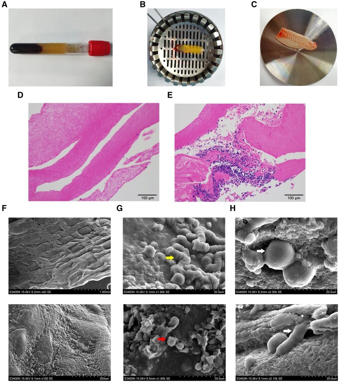
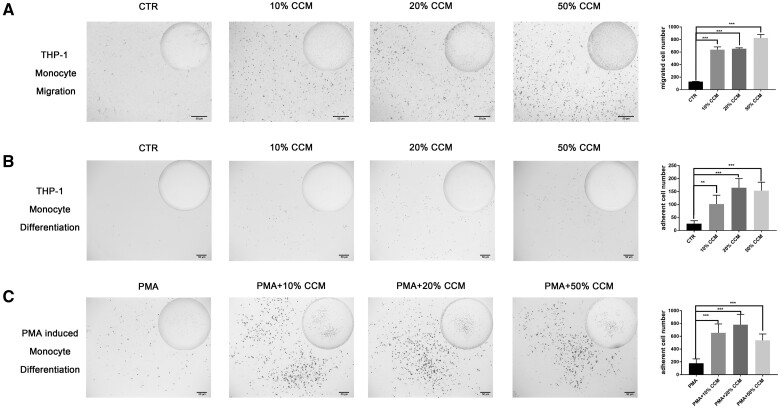
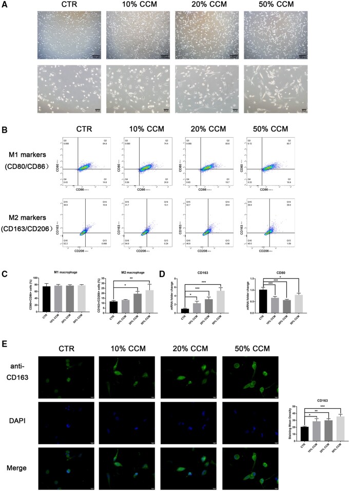
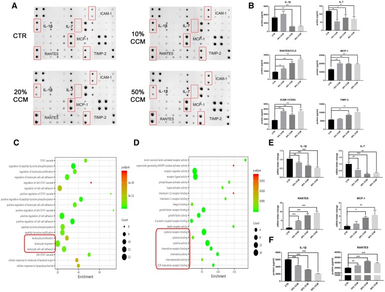
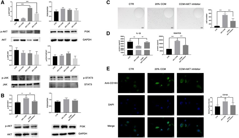
Concentrated growth factor (CGF) is a promising regenerative material that serves as a scaffold and adjunct growth factor for tissue engineering. The host immune response, particularly macrophage activity, plays a critical role in injury repair and tissue regeneration. However, the biological effect of CGF on the immune response is not clear. To enrich the theoretical groundwork for clinical application, the present study examined the immunoregulatory role of CGF in macrophage functional activities in vitro. The CGF scaffold appeared as a dense fibrin network with multiple embedded leukocytes and platelets, and it was biocompatible with macrophages. Concentrated bioactive factors in the CGF extract enhanced THP-1 monocyte recruitment and promoted the maturation of suspended monocytes into adherent macrophages. CGF extract also promoted THP-1 macrophage polarization toward the M2 phenotype with upregulated CD163 expression, as detected by cell morphology and surface marker expression. A cytokine antibody array showed that CGF extract exerted a regulatory effect on macrophage functional activities by reducing secretion of the inflammatory factor interleukin-1β while inducing expression of the chemokine regulated on activation, normal T cell expressed and secreted. Mechanistically, the AKT signaling pathway was activated, and an AKT inhibitor partially suppressed the immunomodulatory effect of CGF. Our findings reveal that CGF induces a favorable immune response mediated by macrophages, which represents a promising strategy for functional tissue regeneration.
Keywords: concentrated growth factor; immune response; macrophage; signaling pathway.
© The Author(s) 2021. Published by Oxford University Press.
Figures
Similar articles
-
In vitro and in vivo study of concentrated growth factor (CGF) mediating macrophage polarization in bone defect repair.Regen Ther. 2025 Apr 26;29:474-483. doi: 10.1016/j.reth.2025.04.013. eCollection 2025 Jun. Regen Ther. 2025. PMID: 40337617 Free PMC article.
-
Concentrated growth factor increases Schwann cell proliferation and neurotrophic factor secretion and promotes functional nerve recovery in vivo.Int J Mol Med. 2016 Feb;37(2):493-500. doi: 10.3892/ijmm.2015.2438. Epub 2015 Dec 18. Int J Mol Med. 2016. PMID: 26709397
-
The potential application of concentrated growth factor in pulp regeneration: an in vitro and in vivo study.Stem Cell Res Ther. 2019 May 20;10(1):134. doi: 10.1186/s13287-019-1247-4. Stem Cell Res Ther. 2019. PMID: 31109358 Free PMC article.
-
Considerations for Clinical Use of Concentrated Growth Factor in Maxillofacial Regenerative Medicine.J Craniofac Surg. 2021 Jun 1;32(4):1316-1321. doi: 10.1097/SCS.0000000000007182. J Craniofac Surg. 2021. PMID: 33055562 Review.
-
In vitro and in vivo effects of concentrated growth factor on cells and tissues.J Biomed Mater Res A. 2020 Jun;108(6):1338-1350. doi: 10.1002/jbm.a.36906. Epub 2020 Feb 28. J Biomed Mater Res A. 2020. PMID: 32090458 Review.
Cited by
-
The regulatory role of immune microenvironment-related cells and pathways in the pathogenesis of keloids.Front Immunol. 2025 Jul 11;16:1529564. doi: 10.3389/fimmu.2025.1529564. eCollection 2025. Front Immunol. 2025. PMID: 40718487 Free PMC article. Review.
-
Concentrated growth factor combined with iRoot BP Plus promotes inflamed pulp repair: an in vitro and in vivo study.BMC Oral Health. 2023 Apr 19;23(1):225. doi: 10.1186/s12903-023-02903-5. BMC Oral Health. 2023. PMID: 37076830 Free PMC article.
-
Advances in Research on Concentrated Growth Factor Applications for Androgenetic Alopecia Treatment: A Review.Med Sci Monit. 2025 Jul 3;31:e948054. doi: 10.12659/MSM.948054. Med Sci Monit. 2025. PMID: 40605258 Free PMC article. Review.
-
Efficacy of concentrated growth factor combined with coronally advanced flap in the treatment of gingival recession: a systematic review and meta-analysis.BMC Oral Health. 2025 Apr 9;25(1):508. doi: 10.1186/s12903-025-05890-x. BMC Oral Health. 2025. PMID: 40205617 Free PMC article.
-
Autologous Platelet-Rich Growth Factor Reduces M1 Macrophages and Modulates Inflammatory Microenvironments to Promote Sciatic Nerve Regeneration.Biomedicines. 2022 Aug 17;10(8):1991. doi: 10.3390/biomedicines10081991. Biomedicines. 2022. PMID: 36009539 Free PMC article.
References
-
- Anitua E, Alkhraisat MH, Orive G.. Perspectives and challenges in regenerative medicine using plasma rich in growth factors. J Control Release 2012;157:29–38. - PubMed
-
- Qiao J, An N, Ouyang X.. Quantification of growth factors in different platelet concentrates. Platelets 2017;28:774–8. - PubMed
-
- Dohan Ehrenfest DM, Rasmusson L, Albrektsson T.. Classification of platelet concentrates: from pure platelet-rich plasma (P-PRP) to leucocyte- and platelet-rich fibrin (L-PRF). Trends Biotechnol 2009;27:158–67. - PubMed
LinkOut - more resources
Full Text Sources
Research Materials
