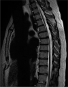
Indolent nonendemic central nervous system histoplasmosis presenting as an isolated intramedullary enhancing spinal cord lesion
- PMID: 34513158
- PMCID: PMC8422457
- DOI: 10.25259/SNI_345_2021
Indolent nonendemic central nervous system histoplasmosis presenting as an isolated intramedullary enhancing spinal cord lesion
Abstract
Background: Histoplasma capsulatum infection is largely seen in endemic regions; it results in symptomatic disease in <5% of those infected and is most often a self-limiting respiratory disease. Disseminated histoplasmosis is considered rare in the immunocompetent host. Central nervous system (CNS) dissemination can result in meningitis, encephalitis, and focal lesions in the brain and spinal cord, stroke, and hydrocephalus. An intramedullary spinal cord lesion as the only manifestation of CNS histoplasmosis has been rarely described.
Case description: We present an atypical case of a 44-year-old man from a nonendemic region, on adalimumab therapy for ulcerative colitis who developed an isolated intramedullary spinal cord lesion in the setting of disseminated histoplasmosis. His course was initially indolent with vague systemic symptoms that led to consideration of several other diagnoses including sarcoidosis and lymphoma. Biopsies of several positron emission tomography positive lymph nodes revealed granulomatous inflammation, but no firm diagnosis was achieved. He was ultimately diagnosed with histoplasmosis after an acute respiratory infection in the setting of anti-tumor necrosis factor therapy. With appropriate antifungal therapy, the spinal cord lesion regressed. The previous systemic biopsies were re-reviewed, and rare fungal elements consistent with H. capsulatum were identified. A presumptive diagnosis of CNS histoplasmosis was made in the absence of direct laboratory confirmation in the setting of rapid and complete resolution on antifungal therapy.
Conclusion: Disseminated histoplasmosis should be considered in granulomatous disease, even if the patient resides in a nonendemic region. Furthermore, clinicians should be mindful that CNS histoplasmosis may present in an atypical fashion.
Keywords: Abscess; Central nervous system; Histoplasmosis; Intramedullary; Spinal cord.
Copyright: © 2021 Surgical Neurology International.
Conflict of interest statement
There are no conflicts of interest.
Figures


References
-
- Bollyky PL, Czartoski TJ, Limaye A. Histoplasmosis presenting as an isolated spinal cord lesion. Arch Neurol. 2006;63:1802–3. - PubMed
-
- Hott JS, Horn E, Sonntag VK, Coons SW, Shetter A. Intramedullary histoplasmosis spinal cord abscess in a nonendemic region: Case report and review of the literature. J Spinal Disord Tech. 2003;16:212–5. - PubMed
-
- Livas IC, Nechay PS, Nauseef WM. Clinical evidence of spinal and cerebral histoplasmosis twenty years after renal transplantation. Clin Infect Dis. 1995;20:692–5. - PubMed
-
- Manning TC, Born D, Tredway TL. Spinal intramedullary histoplasmosis as the initial presentation of human immunodeficiency virus infection: Case report. Neurosurgery. 2006;59:E1146. discussion E1146. - PubMed
Publication types
LinkOut - more resources
Full Text Sources
