

Cauda equina arteriovenous fistula supplied by proximal radicular artery and concomitant sacral dural arteriovenous fistula: A case report and literature review
- PMID: 34513170
- PMCID: PMC8422537
- DOI: 10.25259/SNI_612_2021
Cauda equina arteriovenous fistula supplied by proximal radicular artery and concomitant sacral dural arteriovenous fistula: A case report and literature review
Abstract
Background: Cauda equina arteriovenous fistulas (AVFs) fed by the proximal radicular artery are exceedingly rare. Spinal dural arteriovenous fistulas (DAVFs) in the sacral region are rare and usually misdiagnosed. We report a case of a cauda equina AVF with concomitant sacral DAVF. We also review the coexistence of multiple types of spinal vascular malformations in a single patient.
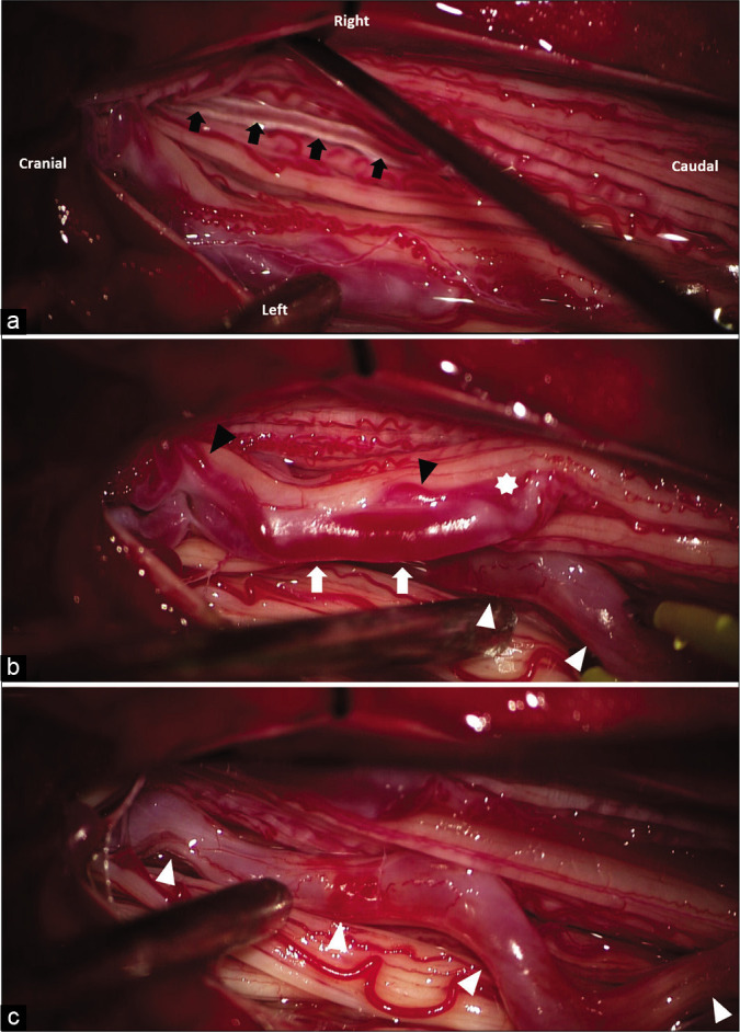
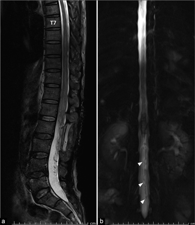
Case description: A 54-year-old man presented with progressive weakness of the lower extremities for 1 month. Magnetic resonance imaging (MRI) of the lumbosacral and thoracic spine showed spinal cord congestion, extending from the conus medullaris to the level of T7, and abnormal tortuous and dilated flow void, running from the level of L5 to T12 along anterior surface of the spinal cord. Spinal angiography demonstrated the fistula at the level of L2 below the conus medullaris. Based on intraoperative findings, the cauda equina AVF supplied by the proximal radicular artery with cranial drainage through the enlarged radicular vein was confirmed and successfully obliterated. Another enlarged arterialized radicular vein running parallel to another cauda equina nerve root is observed with unknown origin. After the operation, the patient showed mild improvement of his symptoms. Follow-up MRI and contrast-enhanced MR angiography revealed an another sacral DAVF vascularized by the lateral sacral artery.
Conclusion: The coexistence of different spinal vascular malformations in a same patient is extremely rare. Most authors of several studies hypothesized that venous hypertension and thrombosis due to the presence or treatment of the first spinal vascular lesion may produce a second DAVF.
Keywords: Cauda equina arteriovenous fistula; Filum terminale arteriovenous fistula; Multiple spinal vascular malformations; Radicular arteriovenous fistula; Sacral dural arteriovenous fistula.
Copyright: © 2021 Surgical Neurology International.
Conflict of interest statement
There are no conflicts of interest.
Figures


Similar articles
-
Sacral dural arteriovenous fistula of the filum terminale coexisting with partially thrombosed filum vein: A case report and literature review.Surg Neurol Int. 2022 Mar 4;13:78. doi: 10.25259/SNI_980_2021. eCollection 2022. Surg Neurol Int. 2022. PMID: 35399884 Free PMC article.
-
Anatomical considerations regarding a high-flow arteriovenous fistula below the conus medullaris in a patient with hereditary hemorrhagic telangiectasia: Case report.Interv Neuroradiol. 2023 Aug 24:15910199231196458. doi: 10.1177/15910199231196458. Online ahead of print. Interv Neuroradiol. 2023. PMID: 37621120
-
Cauda Equina and Filum Terminale Arteriovenous Fistulas: Anatomic and Radiographic Features.AJNR Am J Neuroradiol. 2020 Nov;41(11):2166-2170. doi: 10.3174/ajnr.A6813. Epub 2020 Oct 8. AJNR Am J Neuroradiol. 2020. PMID: 33033040 Free PMC article.
-
Concomitant Sacral Dural Arteriovenous Fistula and Conus Medullaris Arteriovenous Malformation with Respective Drainage Veins: Case Report and Literature Review.World Neurosurg. 2020 Sep;141:299-305. doi: 10.1016/j.wneu.2020.06.022. Epub 2020 Jun 11. World Neurosurg. 2020. PMID: 32535050 Review.
-
[Conus perimedullary arteriovenous fistula with multiple shunt points including the cauda equina: a case report].No Shinkei Geka. 2011 Apr;39(4):375-80. No Shinkei Geka. 2011. PMID: 21447852 Review. Japanese.
Cited by
-
Sacral dural arteriovenous fistula of the filum terminale coexisting with partially thrombosed filum vein: A case report and literature review.Surg Neurol Int. 2022 Mar 4;13:78. doi: 10.25259/SNI_980_2021. eCollection 2022. Surg Neurol Int. 2022. PMID: 35399884 Free PMC article.
-
Current Insights and Management Strategies for Lower Cervical Arteriovenous Fistulas: A Comprehensive Review.Asian J Neurosurg. 2025 May 5;20(3):462-477. doi: 10.1055/s-0045-1809046. eCollection 2025 Sep. Asian J Neurosurg. 2025. PMID: 40852074 Free PMC article. Review.
-
De novo formation of remote dural arteriovenous fistula following treated cavernous sinus dural arteriovenous fistula.World Neurosurg X. 2024 Mar 1;22:100307. doi: 10.1016/j.wnsx.2024.100307. eCollection 2024 Apr. World Neurosurg X. 2024. PMID: 38496348 Free PMC article.
References
-
- Anson JA, Spetzler RF. Classification of spinal arteriovenous malformations and implications for treatment. BNI Q. 1992;8:2–8.
-
- Dam-Hieu P, Mineo JF, Bostan A, Nonent M, Besson G. Concurrent spinal dural and intradural arteriovenous fistulas. Case report. J Neurosurg. 2001;95(Suppl 1):96–9. - PubMed
-
- Gioppo A, Faragò G, Giannitto C, Caputi L, Saladino A, Acerbi F, et al. Sacral dural arteriovenous fistulas: A diagnostic and therapeutic challenge-single-centre experience of 13 cases and review of the literature. J Neurointerv Surg. 2018;10:415–21. - PubMed
-
- Guédon A, Condette-Auliac S, Consoli A, di Maria F, Coskun O, Rodesch G. Primary conus medullaris arteriovenous shunt and secondary lumbo-sacral epidural arteriovenous fistula: One malformation can hide another. J Neuroradiol. 2021;48:16–20. - PubMed
Publication types
LinkOut - more resources
Full Text Sources