. 2021 Aug 26:11:715208.
doi: 10.3389/fcimb.2021.715208. eCollection 2021.

Dengue Virus Dysregulates Master Transcription Factors and PI3K/AKT/mTOR Signaling Pathway in Megakaryocytes

Affiliations

Dengue Virus Dysregulates Master Transcription Factors and PI3K/AKT/mTOR Signaling Pathway in Megakaryocytes

Anismrita Lahon et al. Front Cell Infect Microbiol. .

Abstract

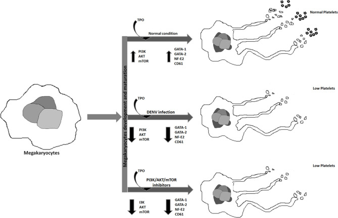
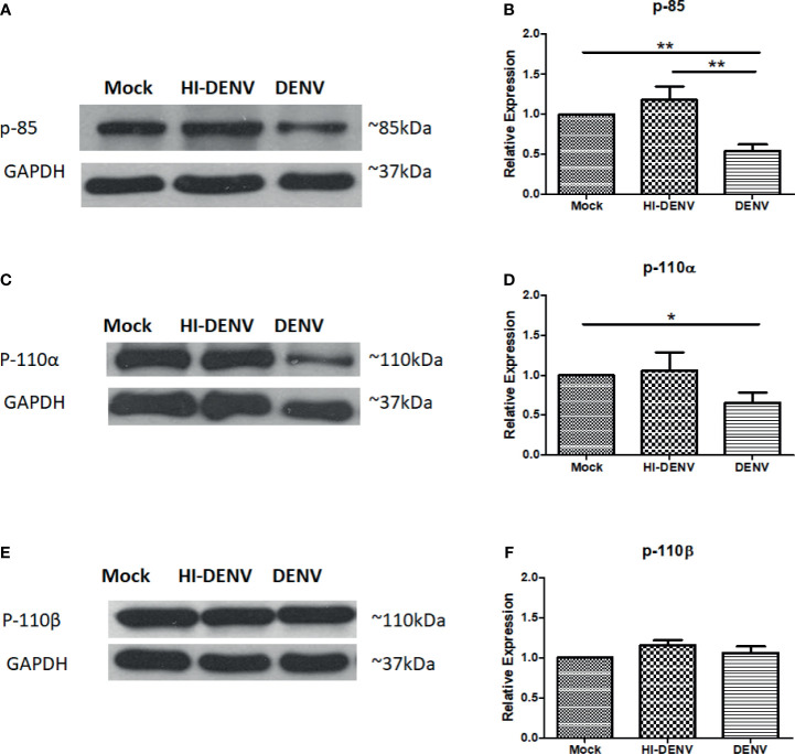
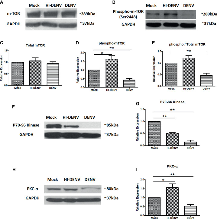
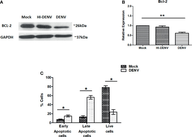
Dengue virus (DENV) infection can cause either self-limited dengue fever or hemorrhagic complications. Low platelet count is one of the manifestations of dengue fever. Megakaryocytes are the sole producers of platelets. However, the role of both host and viral factors in megakaryocyte development, maturation, and platelet production is largely unknown in DENV infection. PI3K/AKT/mTOR pathway plays a significant role in cell survival, maturation, and megakaryocyte development. We were interested to check whether pathogenic insult can impact this pathway. We observed decreased expression of most of the major key molecules associated with the PI3K/AKT/mTOR pathway in DENV infected MEG-01 cells. In this study, the involvement of PI3K/AKT/mTOR pathway in megakaryocyte development and maturation was confirmed with the use of specific inhibitors in infected MEG-01 cells. Our results showed that direct pharmacologic inhibition of this pathway greatly impacted megakaryopoiesis associated molecule CD61 and some essential transcription factors (GATA-1, GATA-2, and NF-E2). Additionally, we observed apoptosis in megakaryocytes due to DENV infection. Our results may suggest that DENV impairs PI3K/AKT/mTOR axis and molecules involved in the development and maturation of megakaryocytes. It is imperative to investigate the role of these molecules in the context of megakaryopoiesis during DENV infection to better understand the pathways and mechanisms, which in turn might provide insights into the development of antiviral strategies.

Keywords: AKT; GATA-2; MEG-01; NF-E2; dengue virus; mTOR; thrombopoietin.


Conflict of interest statement

The authors declare that the research was conducted in the absence of any commercial or financial relationships that could be construed as a potential conflict of interest.

Figures

Graphical Abstract
Graphical Abstract
Graphical abstract represents the effect of DENV infection and different inhibitors on megakaryopoiesis.
Figure 1
Figure 1
DENV infects MEG-01 cells and modulates the expression of AKT. (A) MEG-01 cells were infected with 1 MOI of DENV, and replication was determined from cell culture supernatants collected at day 1 post-infection through day 5 post-infection by using RT-PCR. (B) Replication was further confirmed by immunoblotting for DENV NS1 (~48 kDa) in 5th-day post infected cell lysate and (C) flow cytometric detection of DENV envelope protein using 4G2 antibody staining of infected cells (mean ± SE of infected cells = 23.4 ± 4.1). (D, E) Immunoblotting of pan-AKT and phosphorylated AKT (Ser473) in mock-infected, DENV inactivated virus treated, and DENV infected MEG-01 cell lysate at 5th day post-infection. (F–H) Relative expression of pan-AKT and phospho-AKT was calculated by using the densitometry method and presented as column graphs. The ratio of phospho-AKT and pan-AKT is also presented as column graphs. (I, J) Immunoblot of pan-AKT and phospho-AKT from the lysate of mock, inactivated virus treated, and DENV infected HEK293 cells and (K–M) presentation of relative expression of pan-AKT and phospho-AKT as column graphs of mock control, inactivated virus treated, and DENV infected HEK293 cells. (*pValue < 0.05; **pValue < 0.01).
Figure 2
Figure 2
Immunoblotting of PI3K regulatory and catalytic unit proteins. Representative immunoblots and relative expression of (A, B) p-85, (C, D) p110-α, and (E, F) p-110-β in mock-infected, DENV inactivated virus treated, and DENV infected MEG-01 cell lysate. (*pValue < 0.05; **pValue < 0.01).
Figure 3
Figure 3
Immunoblotting of mTOR and associated proteins. (A, B) Representative immunoblots of total mTOR and phospho-mTOR (Ser2448) in mock, DENV inactivated virus treated, and DENV infected MEG-01 cell lysate. Graphical representation of the expression of mTOR (C) total mTOR, (D) phospho-mTOR, and (E) ratio of phospho and total mTOR expression. Representative immunoblots and relative expression of (F, G) P70-S6 kinase and (H, I) PKC-α in mock-infected, DENV inactivated virus treated, and DENV infected MEG-01 cell lysate. (*pValue < 0.05; **pValue < 0.01).
Figure 4
Figure 4
DENV infection leads to apoptosis in MEG-01 cells. (A, B) Immunoblotting of Bcl-2 protein in mock-infected, DENV inactivated virus treated, and DENV infected MEG-01 cell lysate and represented as column graphs. (C) DENV infected and control MEG-01 cells were stained with FITC conjugated Annexin-V and Propidium Iodide and analyzed on flow cytometer. Graph representation of early apoptosis (only Annexin-V positive cells), late apoptosis (Annexin-V + PI positive cells), and live cells (double negative cells). (*pValue < 0.05; **pValue < 0.01).
Figure 5
Figure 5
Immunoblotting of development and maturation-specific transcription factors in MEG-01 cells. Immunoblot and graphical representation of the expression of GATA-1 (A, B), GATA-2 (C, D), and NF-E2 (E, F) in mock, DENV inactivated virus treated, and DENV infected MEG-01 cell lysate. Effect of different inhibitors of AKT (AKT-IV), PI3K (Ly294002), mTOR (Torin-1), and PKCα (HBBDE) on the expression of most affected transcription factors (G) and their graphical presentation (H, I) during DENV infection. (*pValue < 0.05; ***pvalue < 0.001).
Figure 6
Figure 6
Flow cytometry of CD61 in mock and DENV infected MEG-01 cells. MEG-01 cells were mock-treated or treated with inhibitors of AKT (AKT-IV), PI3K (Ly294002), and PKCα (HBBDE). (A) Histograms from different conditions represent the expression of CD61 and (B) column graph presentation of median fluorescence intensity (MFI) of CD61 in cells with the above-stated condition. (*pValue < 0.05; ***pvalue < 0.001).

References

    1. Arya R. P., Lahon A., Patel A. K. (2021). Dengue Virus Induced Interferon-β by Activating RNA Sensing Pathways in Megakaryocytes. Immunol. Lett. 236, 31–36. 10.1016/j.imlet.2021.06.001 - DOI - PubMed
    1. Backer J. M. (2010). The Regulation of Class IA PI 3-Kinases by Inter-Subunit Interactions. Curr. Top. Microbiol. Immunol. 346, 87–114. 10.1007/82_2010_52 - DOI - PMC - PubMed
    1. Basu A., Jain P., Gangodkar S. V., Shetty S., Ghosh K. (2008). Dengue 2 Virus Inhibits In Vitro Megakaryocytic Colony Formation and Induces Apoptosis in Thrombopoietin-Inducible Megakaryocytic Differentiation From Cord Blood CD34+ Cells. FEMS Immunol. Med. Microbiol. 53 (1), 46–51. 10.1111/j.1574-695X.2008.00399.x - DOI - PubMed
    1. Bollati M., Alvarez K., Assenberg R., Baronti C., Canard B., Cook S., et al. . (2010). Structure and Functionality in Flavivirus NS-Proteins: Perspectives for Drug Design. Antiviral Res. 87, 125–148. 10.1016/j.antiviral.2009.11.009 - DOI - PMC - PubMed
    1. Chen S., Hu M., Shen M., Wang S., Wang C., Chen F., et al. . (2018). IGF-1 Facilitates Thrombopoiesis Primarily Through Akt Activation. Blood 132, 210–222. 10.1182/blood-2018-01-825927 - DOI - PubMed

Publication types

MeSH terms

Substances