

Endodontic Management of a Two-rooted Mandibular First Premolar with Five Root Canals with Cone-beam Computed Tomography: A Case Report
- PMID: 34514072
- PMCID: PMC8417543
- DOI: 10.30476/DENTJODS.2020.83376.1049
Endodontic Management of a Two-rooted Mandibular First Premolar with Five Root Canals with Cone-beam Computed Tomography: A Case Report
Abstract
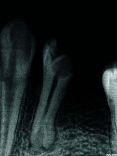
Proper knowledge of the anatomic structure of the root canal system is a vital prerequisite for successful root canal therapy. This report presents the endodontic management a two-rooted lower first premolar with five root canals. A similar case has not been reported to date. The use of cone beam computed tomography (CBCT) in rare and doubtful cases helps establish an accurate diagnosis and render successful endodontic treatment thereafter. This article helps broaden our knowledge about the possible anatomic diversities as to teeth with more roots and root canals than expected normally.
Keywords: Anatomic variations; Cone-beam computed tomography; Endodontic treatment; Mandibular first premolar; Tooth morphology.
Copyright: © Journal of Dentistry.
Conflict of interest statement
Conflict of Interest: None
Figures


References
-
- England MC Jr, Hartwell GR, Lance JR. Detection and treatment of multiple canals in mandibular premolars. J Endod. 1991; 17: 174–178. - PubMed
-
- Ingle JI. A standardized endodontic technique utilizing newly designed instruments and filling materials. Oral Surg Oral Med Oral Pathol. 1961; 14: 83–91. - PubMed
-
- Cleghorn BM, Christie WH, Dong CCS. The root and root canal morphology of the human mandibular first premolar: a literature review. J Endod. 2007; 33: 509–516. - PubMed
-
- Slowey RR. Root canal anatomy. Road map to successful endodontics. Dent Clin North Am. 1979; 23: 555–573. - PubMed
-
- Rotstein I, Ingle JI. Ingle’s Endodontics. 7th ed. PMPH USA: Raleigh, NC, USA; 2019. p. 29.
Publication types
LinkOut - more resources
Full Text Sources