Review
. 2021 Oct;4(10):2100104.
doi: 10.1002/adtp.202100104. Epub 2021 Aug 6.

Peptide-Based Inhibitors for SARS-CoV-2 and SARS-CoV

Affiliations
Review

Peptide-Based Inhibitors for SARS-CoV-2 and SARS-CoV

Disha Panchal et al. Adv Ther (Weinh). 2021 Oct.

Abstract

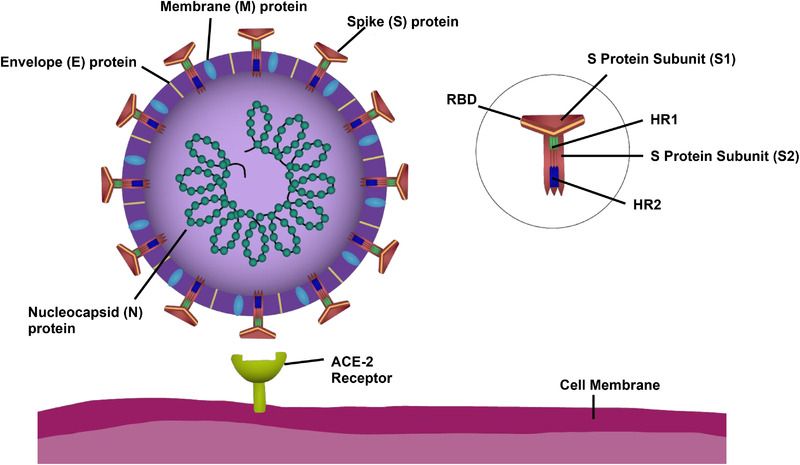
The COVID-19 (coronavirus disease) global pandemic, caused by the spread of the SARS-CoV-2 (severe acute respiratory syndrome coronavirus 2) virus, currently has limited treatment options which include vaccines, anti-virals, and repurposed therapeutics. With their high specificity, tunability, and biocompatibility, small molecules like peptides are positioned to act as key players in combating SARS-CoV-2, and can be readily modified to match viral mutation rate. A recent expansion of the understanding of the viral structure and entry mechanisms has led to the proliferation of therapeutic viral entry inhibitors. In this comprehensive review, inhibitors of SARS and SARS-CoV-2 are investigated and discussed based on therapeutic design, inhibitory mechanistic approaches, and common targets. Peptide therapeutics are highlighted, which have demonstrated in vitro or in vivo efficacy, discuss advantages of peptide therapeutics, and common strategies in identifying targets for viral inhibition.

Keywords: SARS‐CoV; SARS‐CoV‐2; SARS‐CoV‐2 mutants; coronavirus; peptide therapeutics.


Conflict of interest statement

V.A.K. has equity interests in start‐up companies attempting to translate peptides from peptide‐based technological platform.

Figures

Figure 1
Figure 1
Components of the SARS‐CoV‐2 virus and the host‐cell binding target ACE‐2 receptor. The envelope (E) protein, membrane (M) protein, nucleocapsid (N) protein, and Spike (S) protein are the key structural proteins of SARS‐CoV‐2. The structural proteins (N, E, M, and S) are highly conserved within the family Coronaviridae. The single positive strand nature of SARS‐CoV‐2 and its family members allows for rapid transcription of its RNA and infection of neighboring cells. The receptor binding domain (RBD) of the S protein is made of the S1 and S2 subunits. S2 is further divided into two heptad repeat regions, HR1 and HR2. S2 is essential for viral fusion and entry into the host cell.
Figure 2
Figure 2
Interaction of Spike RBD and ACE‐2. A) Bound complex between ACE‐2 (light blue) and Spike RBD (red). Tan shows PPI interface on ACE‐2. B) Close‐up view of the interaction interface. C–E) indicate and label crucial residues from Spike RBD which contribute to complex formation. PDB 7DMU.
Figure 3
Figure 3
Structure of the HR1‐HR2 trimeric fusion core. A) Cartoon representation showing HR1 in cyan and HR2 in dark blue. B) Top‐down view. C) Key interacting residues between HR2 (side chains shown in light blue) and HR1 (side chains shown in tan). PDB 6LXT.
Figure 4
Figure 4
Amino acid sequence alignment of the receptor‐binding domain and heptad repeat 1 (HR1) domain of both SARS‐CoV and SARS‐CoV‐2 virus. Conserved residues between both viruses are marked with asterisks (*), residues with similar properties are marked with a colon (:), while residues with only marginally similar properties are marked with a period (.). A) Amino acid sequence alignment of RBD of SARS‐CoV and SARS‐CoV‐2. The several residue changes in the SARS‐CoV‐2 RBD in comparison to SARS‐CoV allow for higher binding affinity between RBD and ACE‐2 at the RBD–ACE‐2 interface. B) Amino acid sequence alignment of HR1 domains of SARS‐CoV and SARS‐CoV‐2. The residue changes marked within the HR1 domain prompt study into differences in the interactions between HR1 and HR2 domains, which affect 6‐HB formation.
Figure 5
Figure 5
Structure of the SARS‐CoV‐2 Spike RBD. The left hand “closed” conformation does not bind ACE‐2. A conformational switch to the “open” structure can bind ACE‐2 and initiates viral infection. For the D614G strain, a single point mutation (residue in black, circled in yellow) causes Spike RBD to preferentially occupy the “open” conformation. PDB 6ZB4 and 7DK3.
Figure 6
Figure 6
Binding inhibitor mechanism for the SARS‐CoV‐2 virus. To the left, the RBD of the S1 subunit on the Spike protein of SARS‐CoV‐2 binds to the ACE‐2 receptor on the host‐cell, completing the first step of viral infection. To the right, a peptide inhibitor binding to the ACE‐2 receptor, preventing the Spike protein from binding by blocking the ACE‐2 receptor binding.
Figure 7
Figure 7
Fusion inhibitor mechanisms for the SARS‐CoV‐2 virus. A) Formation of a 6‐HB initiates viral fusion. B) The HR2 domain, comprised of 3 ɑ‐helices, present within the S2 subunit of SARS‐CoV‐2 interacts with the HR1 domain, ɑ‐helices, to form a six‐helical bundle (6‐HB). This process can be inhibited by the presence of three copies of a fusion peptide inhibitor.

References

    1. Listings of WHO's response to COVID‐19, World Health Organization 2020, https://www.who.int/news/item/29-06-2020-covidtimeline.
    1. Wu F., Zhao S., Yu B., Chen Y. M., Wang W., Song Z. G., Hu Y., Tao Z. W., Tian J. H., Pei Y. Y., Yuan M. L., Zhang Y. L., Dai F. H., Liu Y., Wang Q. M., Zheng J. J., Xu L., Holmes E. C., Zhang Y. Z., Nature 2020, 579, 265. - PMC - PubMed
    1. Zhu N., Zhang D., Wang W., Li X., Yang B., Song J., Zhao X., Huang B., Shi W., Lu R., Niu P., Zhan F., Ma X., Wang D., Xu W., Wu G., Gao G. F., Tan W., N. Engl. J. Med. 2020, 382, 727. - PMC - PubMed
    1. Drosten C., Günther S., Preiser W., van der Werf S., Brodt H. R., Becker S., Rabenau H., Panning M., Kolesnikova L., Fouchier R. A., Berger A., Burguière A. M., Cinatl J., Eickmann M., Escriou N., Grywna K., Kramme S., Manuguerra J. C., Müller S., Rickerts V., Stürmer M., Vieth S., Klenk H. D., Osterhaus A. D., Schmitz H., Doerr H. W., N. Engl. J. Med. 2003, 348, 1967. - PubMed
    1. Zhong N. S., Zheng B. J., Li Y. M., Poon; Xie Z. H., Chan K. H., Li P. H., Tan S. Y., Chang Q., Xie J. P., Liu X. Q., Xu J., Li D. X., Yuen K. Y., Peiris, Guan Y., Lancet 2003, 362, 1353. - PMC - PubMed