Review
. 2022 Feb;50 Suppl 1(Suppl 1):6-17.
doi: 10.1111/vcp.13031. Epub 2021 Sep 12.

Analysis of the gut microbiome in dogs and cats

Affiliations
Review

Analysis of the gut microbiome in dogs and cats

Jan S Suchodolski. Vet Clin Pathol. 2022 Feb.

Abstract

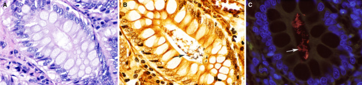
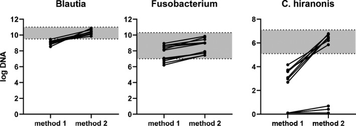
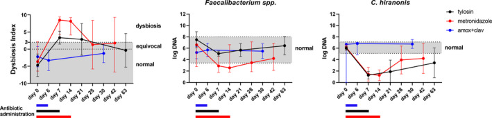
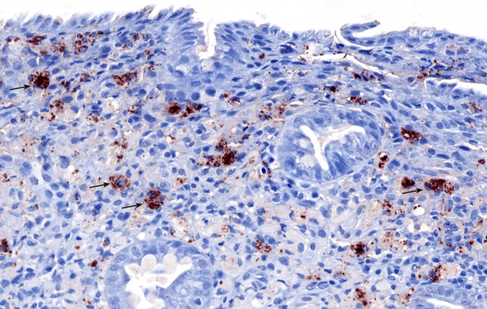
The gut microbiome is an important immune and metabolic organ. Intestinal bacteria produce various metabolites that influence the health of the intestine and other organ systems, including kidney, brain, and heart. Changes in the microbiome in diseased states are termed dysbiosis. The concept of dysbiosis is constantly evolving and includes changes in microbiome diversity and/or structure and functional changes (eg, altered production of bacterial metabolites). Molecular tools are now the standard for microbiome analysis. Sequencing of microbial genes provides information about the bacteria present and their functional potential but lacks standardization and analytical validation of methods and consistency in the reporting of results. This makes it difficult to compare results across studies or for individual clinical patients. The Dysbiosis Index (DI) is a validated quantitative PCR assay for canine fecal samples that measures the abundance of seven important bacterial taxa and summarizes the results as one single number. Reference intervals are established for dogs, and the DI can be used to assess the microbiome in clinical patients over time and in response to therapy (eg, fecal microbiota transplantation). In situ hybridization or immunohistochemistry allows the identification of mucosa-adherent and intracellular bacteria in animals with intestinal disease, especially granulomatous colitis. Future directions include the measurement of bacterial metabolites in feces or serum as markers for the appropriate function of the microbiome. This article summarizes different approaches to the analysis of gut microbiota and how they might be applicable to research studies and clinical practice in dogs and cats.

Keywords: Clostridium hiranonis; Dysbiosis Index; cats; dogs; fecal microbiota transplantation; metagenomics; microbiome.


Conflict of interest statement

The author is an employee of the Gastrointestinal Laboratory at Texas A&M University that offers microbiome and gastrointestinal function testing on a fee‐for‐service basis.

Figures

FIGURE 1
FIGURE 1
Photomicrograph of the colonic mucosa of a healthy dog. The bacteria within the crypts of healthy dogs are inconspicuous on routine hematoxylin and eosin stain (A). The Steiner silver stain (B) highlights abundant bacteria (arrow) within the crypts. Fluorescence in situ hybridization with EUB338 probe targeting all bacteria in the crypts. Labeled bacteria appear red (arrow). The autofluorescence of the intestinal mucosa appears green. DAPI (4′,6‐diamidino‐2‐phenylindole)‐stained nuclei of colonic mucosa appear blue. ×60 objective. Courtesy of Dr Paula Giaretta, DACVP, Universidade Federal de Minas Gerais, Brazil
FIGURE 2
FIGURE 2
Effect of the DNA extraction method on the abundance of fecal bacteria. Two different DNA extraction methods were compared for canine fecal samples, and the bacterial taxa were measured using identical quantitative PCR (qPCR) assays. Method 1 uses chemical lysis, whereas method 2 employs bead beating in addition to chemical lysis. Grey areas indicate the RIs for the targeted bacteria. Differences in methods will affect the measured the abundance in 16S rRNA gene sequencing and qPCR data. It is possible to establish RIs for specific taxa, but assays need to be analytically validated and performed with proper quality control to reproducibly assess the microbiota across studies and in clinical settings
FIGURE 3
FIGURE 3
The effect of different antibiotics on canine fecal microbiota. The data are summarized from three different studies: dogs receiving tylosin (n = 8), metronidazole (n = 16), and amoxicillin‐clavulanic acid (n = 6). Dots indicate median values, error bars indicate ranges, grey areas indicate the RIs. All samples were analyzed using the same method (ie, DNA extraction and quantitative PCR assays), and this allows for a better comparison of data across different studies. Furthermore, the data can be compared with existing RIs, allowing conclusions to be drawn as to the magnitude of changes (size effect) of an intervention within the microbiota (Dysbiosis Index [DI]) or on specific bacterial taxa (ie, short‐chain fatty acid producing Faecalibacterium spp. and bile acid‐converting C hiranonis). These data show that broad‐spectrum antibiotics affect the abundance of C hiranonis (below RI), while amoxicillin‐clavulanic acid has a limited effect on the DI and C hiranonis
FIGURE 4
FIGURE 4
Photomicrograph of an intestinal biopsy from a dog with granulomatous colitis shows strong immunolabeling for Escherichia coli in the cytoplasm of macrophages in the lamina propria (arrows). Red diaminobenzidine chromogen and hematoxylin counterstain, ×20 objective. Courtesy of Dr Patricia Ishii, DVM, Texas A&M University and Dr Paula Giaretta, DACVP, Universidade Federal de Minas Gerais, Brazil

References

    1. Swanson KS, Dowd SE, Suchodolski JS, et al. Phylogenetic and gene‐centric metagenomics of the canine intestinal microbiome reveals similarities with humans and mice. ISME J. 2011;5(4):639‐649. - PMC - PubMed
    1. Barry KA, Middelbos IS, Vester Boler BM, et al. Effects of dietary fiber on the feline gastrointestinal metagenome. J Proteome Res. 2012;11(12):5924‐5933. - PubMed
    1. Foster ML, Dowd SE, Stephenson C, Steiner JM, Suchodolski JS. Characterization of the fungal microbiome (mycobiome) in fecal samples from dogs. Vet Med Int. 2013;2013:1‐8. - PMC - PubMed
    1. Suchodolski JS, Morris EK, Allenspach K, et al. Prevalence and identification of fungal DNA in the small intestine of healthy dogs and dogs with chronic enteropathies. Vet Microbiol. 2008;132(3‐4):379‐388. - PubMed
    1. Ritchie LE, Steiner JM, Suchodolski JS. Assessment of microbial diversity along the feline intestinal tract using 16S rRNA gene analysis. FEMS Microbiol Ecol. 2008;66(3):590‐598. - PubMed