. 2022 Feb;101(2):166-176.
doi: 10.1177/00220345211037247. Epub 2021 Sep 11.

Periosteal Flaps Enhance Prefabricated Engineered Bone Reparative Potential

Affiliations

Periosteal Flaps Enhance Prefabricated Engineered Bone Reparative Potential

A G Abu-Shahba et al. J Dent Res. 2022 Feb.

Abstract

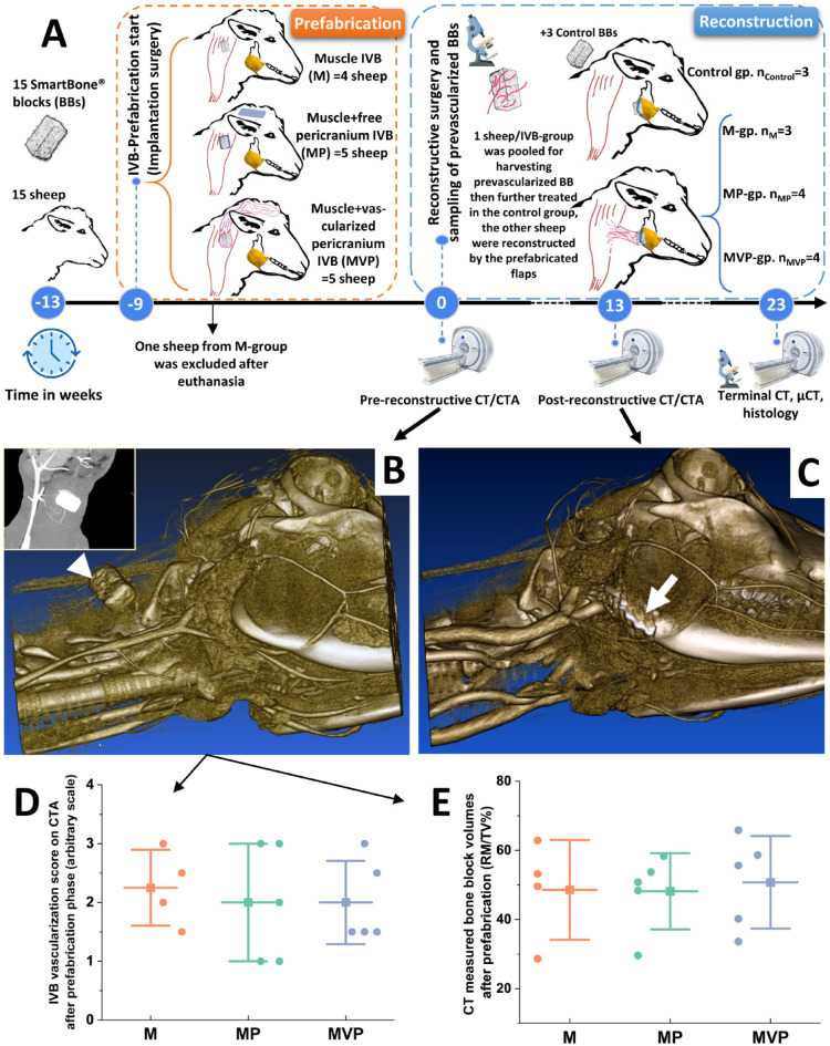
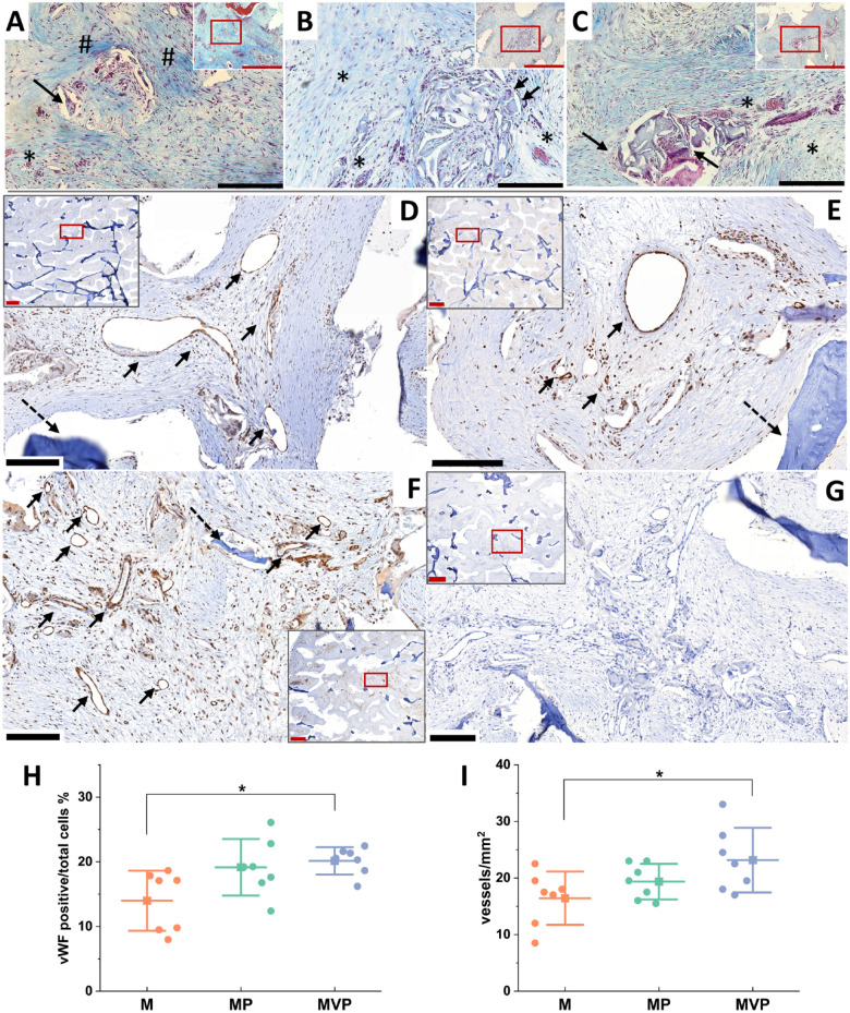
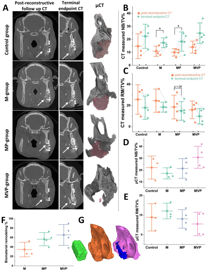
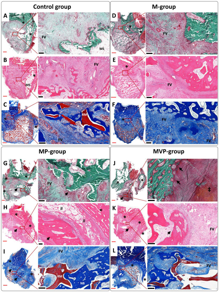
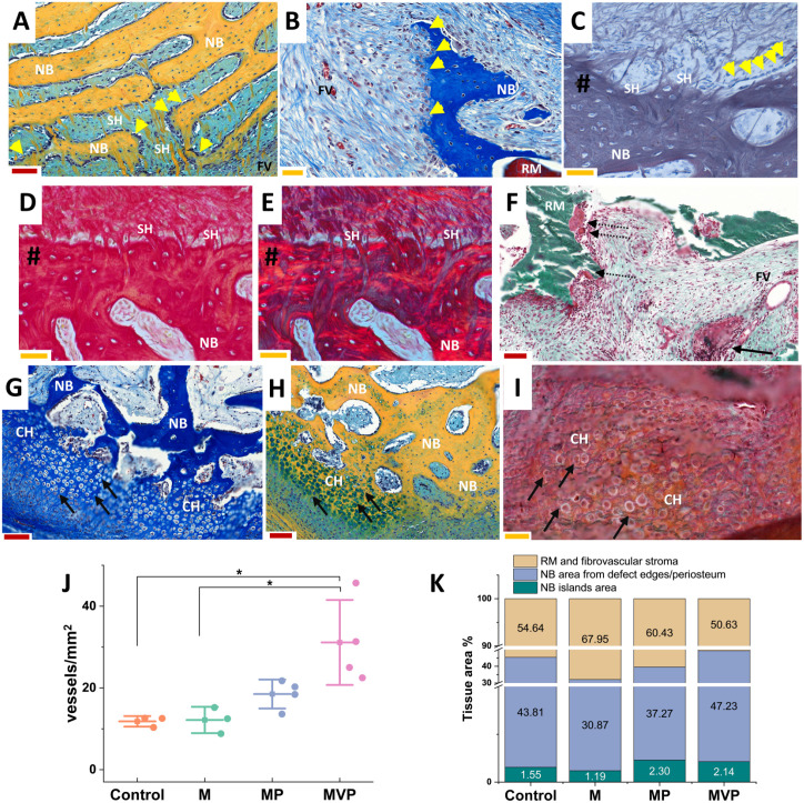
The clinical translation of bone tissue engineering for reconstructing large bone defects has not advanced without hurdles. The in vivo bioreactor (IVB) concept may therefore bridge between bone tissue engineering and reconstructive surgery by employing the patient body for prefabricating new prevascularized tissues. Ideally, IVB should minimize the need for exogenous growth factors/cells. Periosteal tissues are promising for IVB approaches to prefabricate tissue-engineered bone (TEB) flaps. However, the significance of preserving the periosteal vascular supply has not been adequately investigated. This study assessed muscle IVB with and without periosteal/pericranial grafts and flaps for prefabricating TEB flaps to reconstruct mandibular defects in sheep. The sheep (n = 14) were allocated into 4 groups: muscle IVB (M group; nM = 3), muscle + periosteal graft (MP group; nMP = 4), muscle + periosteal flap (MVP group; nMVP = 4), and control group (nControl = 3). In the first surgery, alloplastic bone blocks were implanted in the brachiocephalic muscle (M) with a periosteal graft (MP) or with a vascularized periosteal flap (MVP). After 9 wk, the prefabricated TEB flaps were transplanted to reconstruct a mandibular angle defect. In the control group, the defects were reconstructed by non-prevascularized bone blocks. Computed tomography (CT) scans were performed after 13 wk and after 23 wk at termination, followed by micro-CT (µCT) and histological analyses. Both CT and µCT analysis revealed enhanced new bone formation and decreased residual biomaterial volume in the MVP group compared with control and MP groups, while the M group showed less new bone formation and more residual biomaterial. The histological analysis showed that most of the newly formed bone emerged from defect edges, but larger areas of new bone islands were found in MP and MVP groups. The MVP group showed enhanced vascularization and higher biomaterial remodeling rates. The periosteal flaps boosted the reconstructive potential of the prefabricated TEB flaps. The regenerative potential of the periosteum was manifested after the transplantation into the mechanically stimulated bony defect microenvironment.

Keywords: bioreactors; flap prefabrication; mandibular reconstruction; periosteum; sheep; vascularization.


Conflict of interest statement

Declaration of Conflicting Interests: The authors declared no potential conflicts of interest with respect to the research, authorship, and/or publication of this article.

Figures

Figure 1.
Figure 1.
The study design and CT angiography results. The general outline of the study events is presented (A). At the end of the prefabrication phase, all sheep underwent pre-reconstructive computed tomography (CT) scans for assessing the prevascularized bone blocks (BBs) (white arrowhead in B). During the healing after the reconstructive transplantation surgery, the sheep underwent post-reconstructive CT scans for assessing the stability of the transplanted flaps into the mandibular angle defects (white arrow in C) and evaluating the ongoing new bone formation and biomaterial changes. These parameters were further reassessed by terminal-endpoint CT scans. The data of CT angiography (CTA) (exampled by the small window in B) were used for constructing 3-dimensional models (B, C). The analysis of the pre-reconstructive CTA revealed no differences among the employed in vivo bioreactors (IVBs) regarding the detected vasculature scaling around the BBs (D). The CT-measured BB volumes did not show major differences among the tested IVB conditions at the end of the prefabrication phase (E). The boxplots show mean (–■–), SD (whiskers), and averaged observation values (•). nM = 4. nMP = 5. nMVP = 5. M, intramuscular pouch; MP, a pericranial nonvascularized graft with the muscular pouch; MVP, pericranial vascularized flap with the muscular pouch.
Figure 2.
Figure 2.
Histological findings after the prefabrication phase. Photomicrographs of the Masson’s trichrome (MTC)–stained sections for the bone blocks (BBs) after the prefabrication phase in the tested in vivo bioreactors (IVBs): M (A), MP (B), and MVP (C). No ectopic bone formation was seen within the BBs. Active vascularization and degradation of the biomaterial were evident in all samples with associated macrophages and multinucleated giant cells (MNGCs) (black arrows in A–C). Biomaterial pores were infiltrated by well-vascularized fibrovascular stroma (*). Relatively less degradation and more fibrotic stroma were seen in M samples (# in A). Representations of the immunohistochemistry (IHC) for von Willebrand factor (vWF) and density of blood vessels in the prefabricated tissue-engineered bone (TEB) samples in different IVBs: M (D), MP (E), and MVP (F) after the prefabrication phase. More vascularization was seen in MVP samples, especially when compared to M samples. The black arrows show the detected blood vessels (D–F), and dashed arrows show residual biomaterial. The negative control for IHC is depicted (G). The quantified vWF positive/total cells (%) was higher in MVP samples compared with M samples as was the density of the blood vessels (vessels/mm2) (H, I). Red scale bars in section overview = 1,000 µm; in higher magnification (for red boxes), the black scale bars = 200 µm. The boxplots show mean (–■–), SD (whiskers), and averaged measurements from segments of the BB samples (•), *P < 0.05. M, intramuscular pouch; MP, a pericranial nonvascularized graft with the muscular pouch; MVP, pericranial vascularized flap with the muscular pouch.
Figure 3.
Figure 3.
Post-reconstructive radiographic analyses results. Representations of the computed tomography (CT) and micro-CT (µCT) analyses (A). Comparing the CT images from post-reconstructive follow-up scans (left column in A) to the terminal-endpoint CTs (middle column in A) showed progressive bone defect healing and biomaterial degradation in all groups. Most of the detected new bone formation progressed from the edges and lingual aspect of the defect (white arrows). The 3-dimensional (3D) models from the µCT scans were coronally cut at the same level of the shown CT images to depict the residual biomaterial (shaded red) and the formed new bone at a higher resolution (right column in A). The least residual biomaterial was evidently seen in the MVP group. The quantification for the CT-measured newly formed bone volume (NB/TV%) revealed a trend of increasing new bone volumes between the 2 CT time points within all groups with a corresponding decrease in the residual biomaterial volumes (RM/TV%) (B, C). The µCT analysis revealed an increased mean new bone formation (NB/TV%) and the least residual biomaterial volumes (RM/TV%) in the MVP group (D, E). The 3D-constructed models from pre-, post-reconstructive, and terminal CTs were analyzed for assessing biomaterials remodeling in relation to the prefabrication technique (F, G). Representative 3D models (G) depict bone block from tissue-engineered bone (TEB) flap before transplantation (green left model), TEB reconstructed mandibular defect (gold middle model), and newly formed bone (pink) and residual biomaterial (blue) at the terminal state model (the model to the right). The 3D model comparison revealed a higher mean remodeling percentage in MP and MVP groups compared to the M group (F). The boxplots show mean (–■–), SD (whiskers), and averaged observation values (•), *P < 0.05. nControl = 3. nM = 3. nMP = 4. nMVP = 4. M, intramuscular pouch; MP, a pericranial nonvascularized graft with the muscular pouch; MVP, pericranial vascularized flap with the muscular pouch.
Figure 4.
Figure 4.
Photomicrographs represent the histological findings of the terminal-endpoint samples. The undecalcified sections were stained by Masson Goldner trichrome (A, D, G, J), and the decalcified sections were stained by hematoxylin and eosin (B, E, H, K) and Masson’s trichrome (C, F, I, L). Most of the detected new bone was seen mainly in relation to the defect edges/periosteum (*), especially lingually, with the related marrow spaces (MSs). However, bone islands (black arrows) were frequently seen in MP and MVP groups (G–L). The ingrowing intramembranous new bone (NB) enveloped areas of the residual biomaterial (RM), which was generally infiltrated with a fibrovascular stroma (FV). Some areas of mixed endochondral and intramembranous ossification were seen (‡). Perivascular fatty infiltration (#) was a common finding in the muscular components of the prefabricated tissue-engineered bone (TEB) flaps. The red scale bars in whole-slide images = 2,000 µm; black scale bars in higher magnification (of red boxes) = 200 µm. M, intramuscular pouch; MP, a pericranial nonvascularized graft with the muscular pouch; MVP, pericranial vascularized flap with the muscular pouch.
Figure 5.
Figure 5.
Detailed histological findings and measurements. Photomicrographs (A–E) illustrate the characteristic finding of the radiating, speckled, cellular, perforating collagen fibers (SH) related to the newly formed bone (NB). The perforating fibers (SH) showed similarities with Sharpey’s fibers, as shown in decalcified sections stained with reticulin (C) and picrosirius red under brightfield (D) and polarized light (E) (# indicates bone surface). The newly formed bone showed related osteoblastic cells (yellow arrowheads, A–C). The biomaterial degradation foci (F) showed groups of macrophages and multinucleated giant cells (dashed arrows) with remnants of biomaterial (RM), and collections of lymphocytes and plasma cells (black arrow in F) were not infrequently seen close to a nearby vascular channel within the fibrovascular stroma (FV). Areas of endochondral-like ossification were seen (black arrows in G–I), where the newly formed bone was observed to replace areas of hypertrophied nested chondrocyte-like cells (CH). The represented sections were stained by Movat’s pentachrome (A, H), Masson’s trichrome (B, G), reticulin (C), picrosirius red (D, E), and Masson Goldner trichrome for undecalcified sections (F, I). An increased density of blood vessels (vessels/mm2) was observed in MVP group samples compared to the control and M groups (J). The mean tissue area of the newly formed bone with its related marrow spaces was the least in the M group, while it was the largest in the MVP group (K). Larger areas of the newly formed bone islands were seen in both MP and MVP groups (K). The red scale bars (A, F–H) = 100 µm, and yellow scale bars (B–E, I) = 50 µm. The boxplot (J) shows mean (–■–), SD (whiskers), and averaged observation values (•), *P < 0.05. The stacked column chart (K) shows the average measured areas in histological sections. nControl = 3. nM = 3. nMP = 4. nMVP = 4. M, intramuscular pouch; MP, a pericranial nonvascularized graft with the muscular pouch; MVP, pericranial vascularized flap with the muscular pouch.

Similar articles

Cited by

References

    1. Aaron JE. 2012. Periosteal Sharpey’s fibers: a novel bone matrix regulatory system? Front Endocrinol. 3:98. - PMC - PubMed
    1. Aaron JE, Skerry TM. 1994. Intramembranous trabecular generation in normal bone. Bone Miner. 25(3):211–230. - PubMed
    1. Akar B, Tatara AM, Sutradhar A, Hsiao H-Y, Miller M, Cheng M-H, Mikos AG, Brey EM. 2018. Large animal models of an in vivo bioreactor for engineering vascularized bone. Tissue Eng Part B Rev. 24(4):317–325. - PMC - PubMed
    1. Ayoub A, Challa SR, Abu-Serriah M, McMahon J, Moos K, Creanor S, Odell E. 2007. Use of a composite pedicled muscle flap and rhBMP-7 for mandibular reconstruction. Int J Oral Maxillofac Surg. 36(12):1183–1192. - PubMed
    1. Battaglia S, Ratti S, Manzoli L, Marchetti C, Cercenelli L, Marcelli E, Tarsitano A, Ruggeri A. 2020. Augmented reality-assisted periosteum pedicled flap harvesting for head and neck reconstruction: an anatomical and clinical viability study of a galeo-pericranial flap. J Clin Med. 9(7):2211. - PMC - PubMed

Publication types