Ultrasonographic Tissue Perfusion in Peri-implant Health and Disease
- PMID: 34515570
- PMCID: PMC8982012
- DOI: 10.1177/00220345211035684
Ultrasonographic Tissue Perfusion in Peri-implant Health and Disease
Abstract
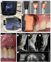
Color flow ultrasonography has played a crucial role in medicine for its ability to assess dynamic tissue perfusion and blood flow variations as an indicator of a pathologic condition. While this feature of ultrasound is routinely employed in various medical fields, its intraoral application for the assessment of tissue perfusion at diseased versus healthy dental implants has never been explored. We tested the hypothesis that quantified tissue perfusion of power Doppler ultrasonography correlates with the clinically assessed inflammation of dental implants. Specifically, we designed a discordant-matched case-control study in which patients with nonadjacent dental implants with different clinical diagnoses (healthy, peri-implant mucositis, or peri-implantitis) were scanned and analyzed with real-time ultrasonography. Forty-two posterior implants in 21 patients were included. Ultrasound scans were obtained at the implant regions of midbuccal, mesial/distal (averaged as interproximal), and transverse to compute the velocity- and power-weighted color pixel density from color velocity (CV) and color power (CP), respectively. Linear mixed effect models were then used to assess the relationship between the clinical diagnoses and ultrasound CV and CP. Overall, the results strongly suggested that ultrasound's quantified CV and CP directly correlate with the clinical diagnosis of dental implants at health, peri-implant mucositis, and peri-implantitis. This study showed for the first time that ultrasound color flow can be applicable in the diagnosis of peri-implant disease and can act as a valuable tool for evaluating the degree of clinical inflammation at implant sites.
Keywords: blood flow velocity; dental implant; evidence-based dentistry; peri-implant disease; peri-implantitis; ultrasonography.
Conflict of interest statement
Figures


Similar articles
-
Peri-Implant Health and Perfusion Parameters in Patients After Microvascular Jaw Reconstruction: A Clinical Cohort Study.Clin Implant Dent Relat Res. 2025 Feb;27(1):e70012. doi: 10.1111/cid.70012. Clin Implant Dent Relat Res. 2025. PMID: 39936507 Free PMC article.
-
Periodontal status of tooth adjacent to implant with peri-implantitis.J Dent. 2018 Mar;70:104-109. doi: 10.1016/j.jdent.2018.01.004. Epub 2018 Jan 8. J Dent. 2018. PMID: 29326047
-
Mucositis, peri-implantitis, implant success, and survival of implants in patients with treated generalized aggressive periodontitis: 3- to 16-year results of a prospective long-term cohort study.J Periodontol. 2012 Oct;83(10):1213-25. doi: 10.1902/jop.2012.110603. Epub 2012 Jan 20. J Periodontol. 2012. PMID: 22264211
-
Primary prevention of peri-implantitis: managing peri-implant mucositis.J Clin Periodontol. 2015 Apr;42 Suppl 16:S152-7. doi: 10.1111/jcpe.12369. J Clin Periodontol. 2015. PMID: 25626479
-
Peri-implant mucositis and peri-implantitis: clinical and histopathological characteristics and treatment.SADJ. 2012 Apr;67(3):122, 124-6. SADJ. 2012. PMID: 23198360 Review.
Cited by
-
Characterization of oral biomarkers during early healing at augmented dental implant sites.J Periodontal Res. 2025 Mar;60(3):206-214. doi: 10.1111/jre.13328. Epub 2024 Aug 1. J Periodontal Res. 2025. PMID: 39090529 Free PMC article. Clinical Trial.
-
Ultrasound identification of the cementoenamel junction and clinical correlation through ex vivo analysis.Sci Rep. 2024 Nov 13;14(1):27821. doi: 10.1038/s41598-024-79081-z. Sci Rep. 2024. PMID: 39537843 Free PMC article.
-
High-Frequency Ultrasound Characterization of Periodontal Soft Tissues Pre- and Post-Bacterial Inoculation.Ultrasound Med Biol. 2025 May;51(5):860-869. doi: 10.1016/j.ultrasmedbio.2025.01.014. Epub 2025 Feb 13. Ultrasound Med Biol. 2025. PMID: 39947944
-
Soft tissue elasticity at teeth and implant sites. A novel outcome measure of the soft tissue phenotype.J Periodontal Res. 2024 Dec;59(6):1130-1142. doi: 10.1111/jre.13296. Epub 2024 Jun 5. J Periodontal Res. 2024. PMID: 38837789 Free PMC article.
-
Intraoral perfusion assessment using endoscopic hyperspectral imaging (EHSI)- first description of a novel approach.Clin Oral Investig. 2025 Feb 5;29(2):115. doi: 10.1007/s00784-025-06197-5. Clin Oral Investig. 2025. PMID: 39907805 Free PMC article.
References
-
- Abrahamsson I, Soldini C. 2006. Probe penetration in periodontal and peri-implant tissues: an experimental study in the beagle dog. Clin Oral Implants Res. 17(6):601–605. - PubMed
-
- Ambrosini P, Cherene S, Miller N, Weissenbach M, Penaud J. 2002. A laser Doppler study of gingival blood flow variations following periosteal stimulation. J Clin Periodontol. 29(2):103–107. - PubMed
-
- Barootchi S, Ravida A, Tavelli L, Wang HL. 2020. Nonsurgical treatment for peri-implant mucositis: a systematic review and meta-analysis. Int J Oral Implantol (Berl). 13(2):123–139. - PubMed
-
- Berglundh T, Armitage G, Araujo MG, Avila-Ortiz G, Blanco J, Camargo PM, Chen S, Cochran D, Derks J, Figuero E, et al.. 2018. Peri-implant diseases and conditions: consensus report of workgroup 4 of the 2017 World Workshop on the Classification of Periodontal and Peri-implant Diseases and Conditions. J Periodontol. 89 Suppl 1:S313–S318. - PubMed
Publication types
MeSH terms
Substances
Grants and funding
LinkOut - more resources
Full Text Sources
Miscellaneous

