. 2021 Oct 25;203(22):e0041921.
doi: 10.1128/JB.00419-21. Epub 2021 Sep 13.

Fluorescence Imaging-Based Discovery of Membrane Domain-Associated Proteins in Mycobacterium smegmatis

Affiliations

Fluorescence Imaging-Based Discovery of Membrane Domain-Associated Proteins in Mycobacterium smegmatis

Corelle A Z Rokicki et al. J Bacteriol. .

Abstract

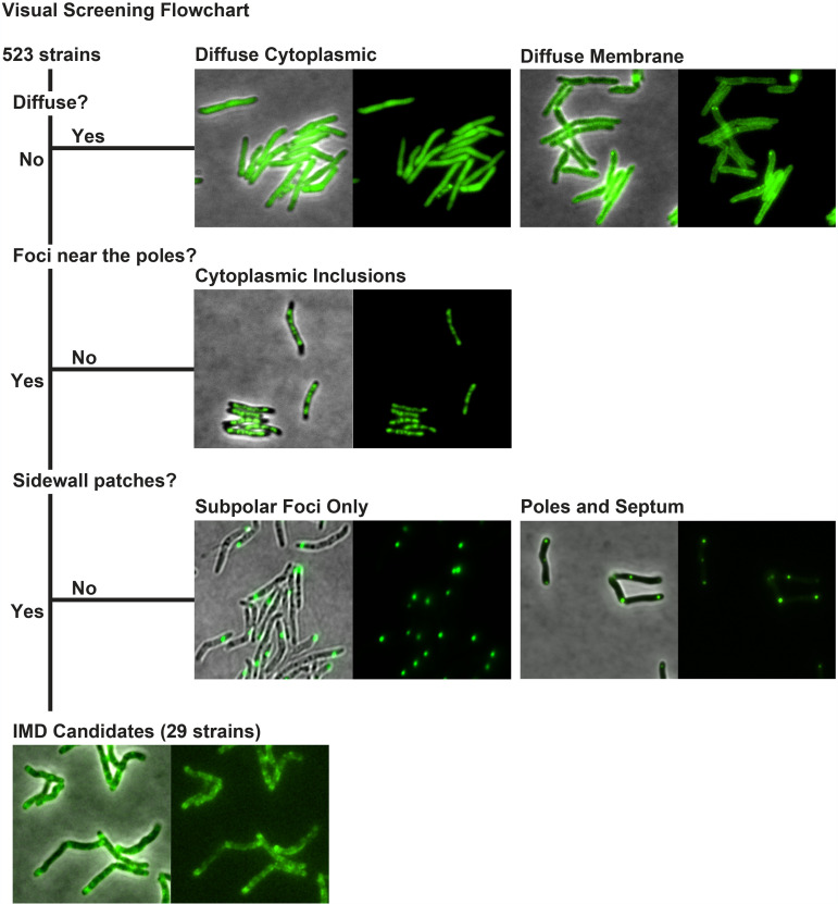
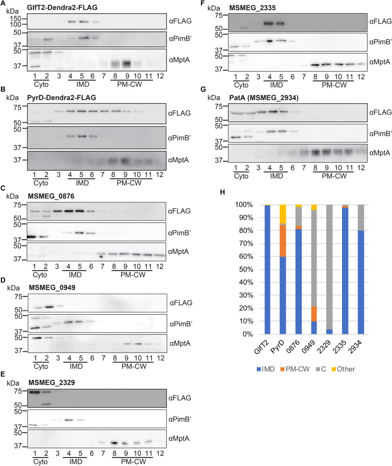
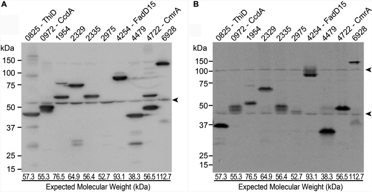
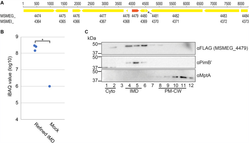
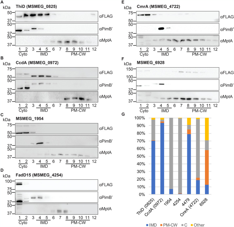
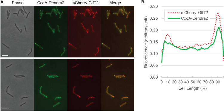
Mycobacteria spatially organize their plasma membrane, and many enzymes involved in envelope biosynthesis associate with a membrane compartment termed the intracellular membrane domain (IMD). The IMD is concentrated in the polar regions of growing cells and becomes less polarized under nongrowing conditions. Because mycobacteria elongate from the poles, the observed polar localization of the IMD during growth likely supports the localized biosynthesis of envelope components. While we have identified more than 300 IMD-associated proteins by proteomic analyses, only a few of these have been verified by independent experimental methods. Furthermore, some IMD-associated proteins may have escaped proteomic identification and remain to be identified. Here, we visually screened an arrayed library of 523 Mycobacterium smegmatis strains, each producing a Dendra2-FLAG-tagged recombinant protein. We identified 29 fusion proteins that showed polar fluorescence patterns characteristic of IMD proteins. Twenty of these had previously been suggested to localize to the IMD based on proteomic data. Of the nine remaining IMD candidate proteins, three were confirmed by biochemical methods to be associated with the IMD. Taken together, this new colocalization strategy is effective in verifying the IMD association of proteins found by proteomic analyses while facilitating the discovery of additional IMD-associated proteins. IMPORTANCE The intracellular membrane domain (IMD) is a membrane subcompartment found in Mycobacterium smegmatis cells. Proteomic analysis of purified IMD identified more than 300 proteins, including enzymes involved in cell envelope biosynthesis. However, proteomics on its own is unlikely to detect every IMD-associated protein because of technical and biological limitations. Here, we describe fluorescent protein colocalization as an alternative, independent approach. Using a combination of fluorescence microscopy, proteomics, and subcellular fractionation, we identified three new proteins associated with the IMD. Such a robust method to rigorously define IMD proteins will benefit future investigations to decipher the synthesis, maintenance, and functions of this membrane domain and help delineate a more general mechanism of subcellular protein localization in mycobacteria.

Keywords: Mycobacterial Systems Resource; fluorescence microscopy; membrane domain; membrane proteins; mycobacterium.


Figures

FIG 1
FIG 1
Visual screening of IMD protein candidates from 523 fluorescence images deposited in the Mycobacterial Systems Resource. Fluorescence images were visually screened for proteins that show fluorescence patterns that are similar to those of known IMD proteins. Excluding those fluorescence patterns that were diffuse, cytoplasmic inclusions, subpolar foci only, or polar foci and septum without sidewall patches, a total of 29 proteins were identified as the candidates of IMD proteins, showing subpolar foci and sidewall patches.
FIG 2
FIG 2
Validation of known IMD protein candidates by density gradient fractionation. (A to G) Among IMD protein candidates identified by the image screening, seven proteins, which were previously found in the IMD proteome, were examined by sucrose density gradient fractionation. Lysate was prepared from cells producing these proteins and was fractionated by sucrose density gradient sedimentation. An equal volume of each gradient fraction was separated by SDS-PAGE, and the FLAG epitope was detected by anti-FLAG immunoblotting. PimB′ (41.1 kDa), IMD marker; MptA (54.3 kDa), PM-CW marker. The apparent molecular weight of MptA on SDS-PAGE gel is about 40 kDa (32, 35). (H) Quantification of anti-FLAG immunoblot bands shown in panels A to G. C, cytoplasm. See Materials and Methods for details of the quantitative analysis.
FIG 3
FIG 3
Immunoblot analysis of newly discovered IMD protein candidates. (A) Crude cell lysates of the eight new IMD protein candidates (as well as MSMEG_2329 and MSMEG_2335, analyzed in Fig. 2) were analyzed by SDS-PAGE, and FLAG epitope was detected by immunoblotting. Expected molecular weight of each protein is indicated at the bottom of the lane. (B) Crude cell lysates were analyzed by PAGE under a nondenaturing condition, and Dendra2 fluorescence was detected in gel by a fluorescence imager. Arrowheads indicate nonspecific proteins.
FIG 4
FIG 4
MSMEG_4479 is identified in the IMD proteome. (A) The genome region surrounding MSMEG_4479. The nucleotide sequence in the region is identical between the two commonly used genome sequences (TIGR, NC_008596, MSMEG_ locus tag prefix; IGMCB, NC_018289, MSMEI_ locus tag prefix). Note that MSMEG_4479 does not have a corresponding MSMEI_ locus number while all other genes surrounding the region have a locus number with both locus tag prefixes. (B) Enrichment of MSMEG_4479 in the IMD, revealed by the reanalysis of previously published proteome data (refined IMD versus mock; see reference 13) using the TIGR genome sequence. iBAQ values were used to compare the protein abundance. Both IMD and mock preparations were analyzed in technical triplicate, but MSMEG_4479 was detected from the mock preparation in only one analysis. *, P < 0.05 by t test (two-tailed). (C) Subcellular localization of MSMEG_4479 was analyzed by density gradient fractionation. See the legend to Fig. 2 for details. Representative data of a biological duplicate are shown.
FIG 5
FIG 5
Subcellular fractionation of newly discovered IMD protein candidates. (A to F) Six proteins, which showed Dendra2 fluorescence patterns suggestive of IMD localization but were not previously identified in the IMD proteome, were analyzed by density gradient fractionation. See the legend to Fig. 2 for details. Representative data of two independent experiments are shown (see panel G for details). (G) Immunoblot bands of each protein, as shown in panels A to F and Fig. 4C, were quantified across density gradient fractions and calculated for the percent subcellular localization as described in Materials and Methods. For the new IMD proteins (ThiD, CcdA, and MSMEG_4479), cell culture, cell lysis, density gradient fractionation, and immunoblotting were repeated twice by independent researchers. Average percent localizations of the biological duplicates are shown. For other proteins, immunoblotting was repeated twice by independent researchers, and average percent localization of the technical duplicates is shown.
FIG 6
FIG 6
Colocalization of CcdA-Dendra2-FLAG and HA-mCherry-GlfT2, visualized by fluorescence microscopy. (A) HA-mCherry-GlfT2 is produced from the endogenous locus and previously confirmed as an IMD marker (13). Two different fields are shown as representative images. Bar, 4 μm. (B) Fluorescence intensity profile along the long axis of the cell. Cell length is shown as percent, and cells were aligned so that a more intense pole is positioned to the right (100%). Average intensity profiles of 14 cells are shown.

References

    1. Daffé M, Marrakchi H. 2019. Unraveling the structure of the mycobacterial envelope. Microbiol Spectr 10.1128/microbiolspec.GPP3-0027-2018. - DOI - PMC - PubMed
    1. Jackson M. 2014. The mycobacterial cell envelope-lipids. Cold Spring Harb Perspect Med 4:a021105. 10.1101/cshperspect.a021105. - DOI - PMC - PubMed
    1. Kaur D, Guerin ME, Skovierová H, Brennan PJ, Jackson M. 2009. Chapter 2: biogenesis of the cell wall and other glycoconjugates of Mycobacterium tuberculosis. Adv Appl Microbiol 69:23–78. 10.1016/S0065-2164(09)69002-X. - DOI - PMC - PubMed
    1. Dulberger CL, Rubin EJ, Boutte CC. 2020. The mycobacterial cell envelope–a moving target. Nat Rev Microbiol 18:47–59. 10.1038/s41579-019-0273-7. - DOI - PubMed
    1. Rahlwes KC, Sparks IL, Morita YS. 2019. Cell walls and membranes of Actinobacteria. Subcell Biochem 92:417–469. 10.1007/978-3-030-18768-2_13. - DOI - PubMed

Publication types

MeSH terms

Substances

LinkOut - more resources