. 2021 Sep 10;7(37):eabj2101.
doi: 10.1126/sciadv.abj2101. Epub 2021 Sep 10.

Neutrophil extracellular traps enhance macrophage killing of bacterial pathogens

Affiliations

Neutrophil extracellular traps enhance macrophage killing of bacterial pathogens

Andrew J Monteith et al. Sci Adv. .

Abstract

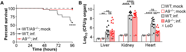
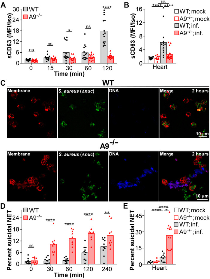
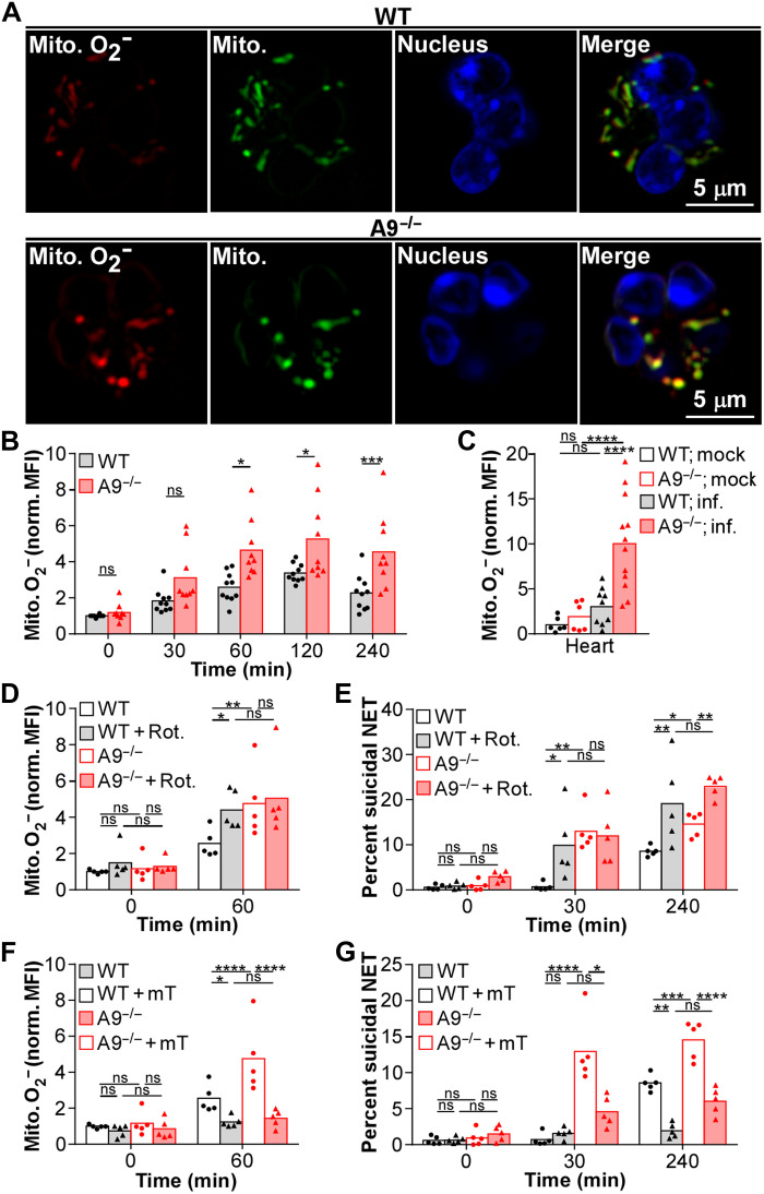
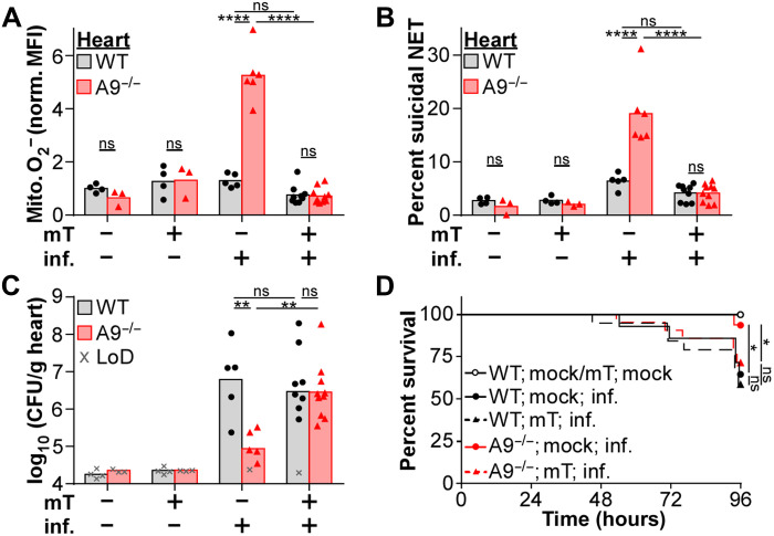
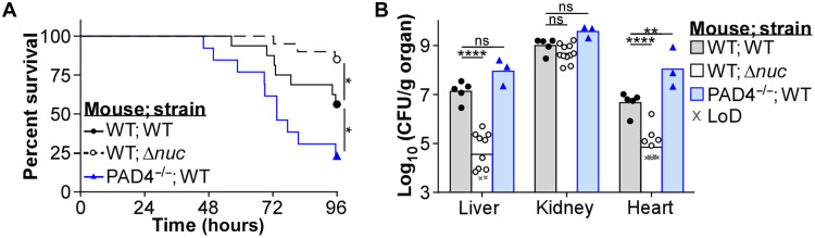
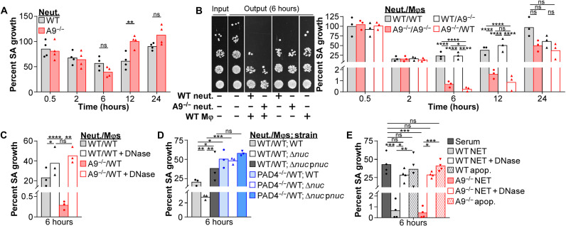
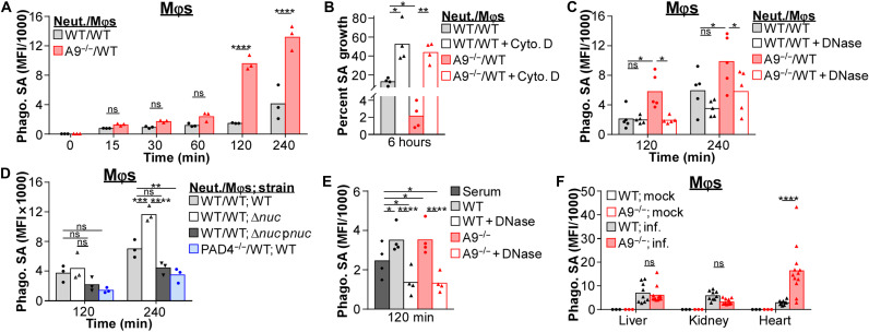
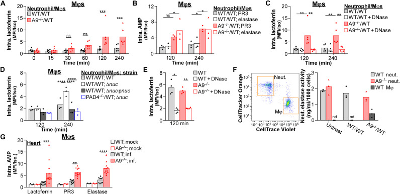
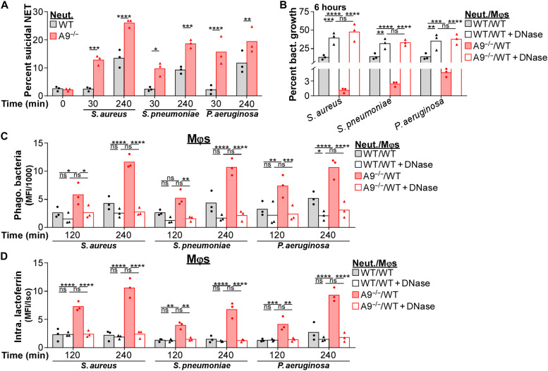
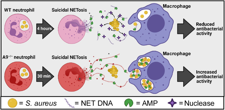
Neutrophils and macrophages are critical to the innate immune response, but cooperative mechanisms used by these cells to combat extracellular pathogens are not well understood. This study reveals that S100A9-deficient neutrophils produce higher levels of mitochondrial superoxide in response to Staphylococcus aureus and, as a result, form neutrophil extracellular traps (suicidal NETosis). Increased suicidal NETosis does not improve neutrophil killing of S. aureus in isolation but augments macrophage killing. NET formation enhances antibacterial activity by increasing phagocytosis by macrophages and by transferring neutrophil-specific antimicrobial peptides to them. Similar results were observed in response to other phylogenetically distinct bacterial pathogens including Streptococcus pneumoniae and Pseudomonas aeruginosa, implicating this as an immune defense mechanism that broadly enhances antibacterial activity. These results demonstrate that achieving maximal bactericidal activity through NET formation requires macrophages and that accelerated and more robust suicidal NETosis makes neutrophils adept at increasing antibacterial activity, especially when A9 deficient.


Figures

Fig. 1.
Fig. 1.. A9−/− mice are more protected from systemic MRSA infection.
Mice were systemically infected (inf.) with USA300 [colony-forming units (CFU) = 2 × 10⁷]. (A) During the infection, mouse survival was monitored. Each point represents the percentage of living mice (mock, n = 14, WT; inf., n = 31, A9−/−; inf., n = 29). (B) At 4 dpi, organs were homogenized and CFU was counted using spot plating [limit of detection (LoD)]. Each point represents a single mouse (mock, n = 11) (WT; inf., n = 13) (A9−/−; inf., n = 16). (A) Log-rank (Mantel-Cox) test or (B) two-way analysis of variance (ANOVA) with Tukey multiple comparisons test (**P ≤ 0.01 and ****P ≤ 0.0001; ns, not significant).
Fig. 2.
Fig. 2.. A9−/− neutrophils have accelerated and more robust suicidal NETosis in response to S. aureus.
(A) Neutrophils were cultured with S. aureus [multiplicity of infection (MOI) = 10] and primary degranulation [surface CD63 (sCD63)] by neutrophils (Ly6G+CD11b+) was quantified by flow cytometry. Median fluorescence intensity (MFI) normalized to an isotype (Iso) control. Each point represents neutrophils isolated from a single mouse (n = 14). (B) Mice were systemically infected (inf.) with S. aureus (CFU = 2 × 10⁷). At 4 dpi, organs were homogenized and primary degranulation by neutrophils was quantified by flow cytometry. MFI normalized to an isotype control. Each point represents a single mouse (mock, n = 11) (WT; inf., n = 13) (A9−/−; inf., n = 16). (C and D) Neutrophils were cultured with S. aureus (MOI = 10). (C) Representative images of neutrophils (red) stimulated for 2 hours with a nuclease-deficient (Δnuc) strain of S. aureus (green) are provided. Extracellular DNA was stained using Helix NP Blue. (D) The percentage of neutrophils undergoing suicidal NETosis (dead: extracellular dsDNA+MPO+H3Cit+) in response to S. aureus was quantified by flow cytometry. Each point represents neutrophils isolated from a single mouse (n = 9). (E) Mice were systemically infected with S. aureus (CFU = 2 × 10⁷). At 4 dpi, organs were homogenized and the percentage of neutrophils undergoing suicidal NETosis was quantified by flow cytometry. Each point represents a single mouse (mock, n = 6) (WT; inf., n = 9) (A9−/−; inf., n = 12). Two-way ANOVA with (A and D) Sidak’s or (B and E) Tukey multiple comparisons test (*P ≤ 0.05, **P ≤ 0.01, and ****P ≤ 0.0001).
Fig. 3.
Fig. 3.. Increased mitochondrial O2 heightens suicidal NETosis in A9−/− neutrophils responding to S. aureus.
(A and B) Neutrophils were cultured with S. aureus (MOI = 10). (A) Neutrophils were stimulated with S. aureus for 1 hour, and representative images of mitochondrial (Mito.) O2 (red; MitoSOX) and biomass (green; MitoTracker) are provided. Nuclear DNA was stained (blue; Hoechst). (B) Neutrophils (Ly6G+CD11b+) were cultured with S. aureus, and production of mitochondrial O2 was quantified by flow cytometry. MitoSOX MFI was normalized by MitoTracker MFI. Each point represents neutrophils isolated from a single mouse (n = 9). (C) Mice were systemically infected (inf.) with S. aureus (CFU = 2 × 10⁷). At 4 dpi, organs were homogenized and production of mitochondrial O2 in neutrophils was quantified in the heart by flow cytometry. MitoSOX MFI was normalized by MitoTracker MFI. Each point represents a single mouse (mock, n = 6) (WT; inf., n = 9) (A9−/−; inf., n = 12). (D to G) Neutrophils pretreated with (D and E) rotenone (Rot.; 0.5 μM) for 15 min or (F and G) MitoTEMPO (mT; 0.5 μM) for 2 hours were cultured with S. aureus (MOI = 10). Neutrophils were stimulated with S. aureus, and (D and F) the production of mitochondrial O2 and (E and G) the percentage of neutrophils undergoing suicidal NETosis (Dead: extracellular dsDNA+MPO+H3Cit+) were quantified by flow cytometry. (D and F) MitoSOX MFI was normalized by MitoTracker MFI. Each point represents neutrophils isolate from a single mouse (n = 5). Two-way ANOVA with (B) Sidak’s or (C to G) Tukey multiple comparisons test (*P ≤ 0.05, **P ≤ 0.01, ***P ≤ 0.001, and ****P ≤ 0.0001).
Fig. 4.
Fig. 4.. MitoTEMPO treatment of A9−/− mice reduces suicidal NETosis coinciding with enhanced lethality and heart colonization.
Mice were treated with MitoTEMPO (mT; 0.7 mg/kg) by intraperitoneal injection 24 hours before and every 24 hours during systemic infection (inf.) with S. aureus (A to C, CFU = 2 × 10⁷; D, CFU = 5 × 10⁷). (A to C) At 4 dpi, organs were homogenized, and (A) the production of mitochondrial O2 by neutrophils (Ly6G+CD11b+) and (B) the percentage of neutrophils undergoing suicidal NETosis (Dead: extracellular dsDNA+MPO+H3Cit+) were quantified by flow cytometry, and (C) CFU was counted using spot plating (LoD). (A) MitoSOX MFI was normalized by MitoTracker MFI. Each point represents a single mouse (mock; mock/mT; WT, n = 4) (mock; mock/mT; A9−/−, n = 3) (inf.; mock; WT, n = 5) (inf.; mock; A9−/−, n = 6) (inf.; mT; WT, n = 9) (inf.; mT; A9−/−, n = 10). (D) During the infection, mouse survival was monitored. Each point represents the percentage of living mice (mock; mock; WT/A9−/−, n = 7) (mock; mT; WT/A9−/−, n = 8) (inf.; mock; WT, n = 13) (inf.; mock; A9−/−, n = 15) (inf.; mT; WT, n = 19) (inf.; mT; A9−/−, n = 21). (A to C) Two-way ANOVA with Tukey multiple comparisons test or (D) log-rank (Mantel-Cox) test (*P ≤ 0.05, **P ≤ 0.01, and ****P ≤ 0.0001).
Fig. 5.
Fig. 5.. NET formation contributes to the protection of mice during systemic infection with S. aureus.
Mice were systemically infected with S. aureus (CFU = 2 × 10⁷). (A) During the infection, mouse survival was monitored. Each point represents the percentage of living mice (WT; WT, n = 16) (WT; Δnuc, n = 20) (PAD4−/−; WT, n = 13). (B) At 4 dpi, organs were homogenized and CFU was counted using spot plating (LoD). Each point represents a single mouse (WT; WT, n = 5) (WT; Δnuc, n = 11) (PAD4−/−; WT, n = 3). (A) Log-rank (Mantel-Cox) test or (B) two-way ANOVA with Tukey multiple comparisons test (*P ≤ 0.05, **P ≤ 0.01, and ****P ≤ 0.0001).
Fig. 6.
Fig. 6.. NET formation from A9−/− neutrophils better restrict S. aureus growth in the presence of Mφs.
Immune cells were cultured with S. aureus (MOI = 1). (A) Neutrophil (Neut.) restriction of S. aureus growth was quantified by CFU spot plating. Percent growth of S. aureus (SA) calculated relative to S. aureus growth in the absence of neutrophils. Each point represents the mean result (biological triplicate) of neutrophils isolated from a single mouse (n = 4). (B to D) Neutrophil-Mφ coculture (ratio = 1:1) restriction of S. aureus growth was quantified by CFU spot plating. Percent growth of S. aureus calculated relative to S. aureus growth in the absence of immune cells. (B) Representative spot plating from a single experiment is provided. (C) Immune cells cultured with S. aureus in the presence of deoxyribonuclease (DNase) (8 U/ml). Each point represents the mean result (biological triplicate) of immune cells isolated from a single mouse (n = 3). (E) Mφ restriction of S. aureus growth was quantified by CFU spot plating. NETs or apoptotic debris (apop.) used for S. aureus opsonization were isolated from neutrophils stimulated with phorbol 12-myristate 13-acetate (PMA) (100 nM) for 4 hours or an anti-FAS antibody (100 ng/ml) for 16 hours. Percent growth of S. aureus calculated relative to S. aureus growth in the absence of immune cells. Mφs cultured with S. aureus in the presence of DNase (8 U/ml). Each point represents the mean result (biological triplicate) of Mφs isolated from a single mouse (n = 4). Two-way ANOVA with (A) Sidak’s or (B to E) Tukey multiple comparisons test (*P ≤ 0.05, **P ≤ 0.01, ***P ≤ 0.001, and ****P ≤ 0.0001).
Fig. 7.
Fig. 7.. NET formation enhances phagocytosis of S. aureus by Mφs.
(A to E) Immune cells were cultured with fluorescently labeled S. aureus (MOI = 10). (A) Phagocytosis of S. aureus by Mφs in coculture (ratio = 1:1) with neutrophils (Neut.) was quantified by flow cytometry. Background MFI was subtracted from each time point. Each point represents immune cells isolated from a single mouse (n = 3). (B) Mφs were pretreated with cytochalasin D (Cyto. D; 10 μg/ml) for 1 hour before adding neutrophils and S. aureus. Percent growth of S. aureus was calculated relative to S. aureus growth in the absence of immune cells. Each point represents the mean result (biological triplicate) of immune cells isolated from a single mouse (n = 4). (C and D) Phagocytosis of S. aureus by Mφs in coculture with neutrophils was quantified by flow cytometry. Background MFI was subtracted from each time point. (C) Immune cells cultured with S. aureus in the presence of DNase (8 U/ml). Each point represents immune cells isolated from a single mouse (C, n = 5; D, n = 3). (E) Phagocytosis of S. aureus by Mφs was quantified by flow cytometry. Background MFI was subtracted from each time point. NETs used for S. aureus opsonization were isolated from neutrophils stimulated with PMA (100 nM) for 4 hours and cultured with Mφs in the presence of DNase (8 U/ml). Each point represents immune cells isolated from a single mouse (n = 4). (F) Mice were systemically infected with a fluorescent strain of S. aureus (pSarA_sfGFP; CFU = 2 × 10⁷). At 4 dpi, organs were homogenized and S. aureus levels within Mφs (CD11b+F4/80+Ly6G) in the heart were quantified by flow cytometry. Background MFI from uninfected mice was subtracted from infected. Each point represents a single mouse (mock, n = 3) (WT; inf., n = 9) (A9−/−; inf., n = 12). Two-way ANOVA with (A) Sidak’s or (B to F) Tukey multiple comparisons test (*P ≤ 0.05, **P ≤ 0.01, ***P ≤ 0.001, and ****P ≤ 0.0001).
Fig. 8.
Fig. 8.. NET formation transfers neutrophil-specific AMPs to Mφs.
(A to F) Immune cells were cultured with S. aureus (MOI = 10). (A to D) The intracellular (Intra.) abundance of lactoferrin, PR3, and elastase within Mφs in coculture (ratio = 1) with neutrophils (Neut.) following stimulation with S. aureus were quantified by flow cytometry. MFI was normalized to an isotype control. (C) Immune cells were cultured with S. aureus in the presence of DNase (8 U/ml). Each point represents immune cells isolated from a single mouse (A, n = 6; B and D, n = 3; C, n = 5). (E) Intracellular abundance of lactoferrin within Mφs following stimulation with S. aureus was quantified by flow cytometry. MFI was normalized to an isotype control. NETs used for S. aureus opsonization were isolated from neutrophils stimulated with PMA (100 nM) for 4 hours and cultured with Mφs in the presence of DNase (8 U/ml). Each point represents immune cells isolated from a single mouse (n = 4). (F) Cocultures were stimulated with S. aureus, and after 2 hours, neutrophils and Mφs were isolated by fluorescent sorting. Lysates from isolated cells were quantified for neutrophil elastase activity and normalized by cell number (nd, no activity detected). Each point represents the mean result (biological duplicate) of immune cells isolated from a single mouse (n = 3). (G) Mice were systemically infected with S. aureus (CFU = 2 × 10⁷). At 4 dpi, organs were homogenized and intracellular lactoferrin, PR3, and elastase levels within Mφs (CD11b+F4/80+Ly6G) in the heart were quantified by flow cytometry. MFI was normalized by an isotype control. Each point represents a single mouse (mock, n = 3) (WT; inf., n = 9) (A9−/−; inf., n = 12). Two-way ANOVA with (A) Sidak’s or (B to E and G) Tukey multiple comparisons test (*P ≤ 0.05, **P ≤ 0.01, ***P ≤ 0.001, and ****P ≤ 0.0001).
Fig. 9.
Fig. 9.. NET formation better restricts growth of multiple bacterial pathogens in the presence of Mφs.
Immune cells were cultured with S. aureus, S. pneumoniae, and P. aeruginosa (A, C, and D, MOI = 10; B, MOI = 1). (A) Neutrophils (Neut.) were stimulated with bacteria and the percentage of neutrophils (Ly6G+CD11b+) undergoing suicidal NETosis (Dead: extracellular dsDNA+MPO+H3Cit+) was quantified by flow cytometry. Each point represents neutrophils isolated from a single mouse (n = 3). (B) Neutrophil-Mφ coculture (ratio = 1:1) restriction of bacterial growth was quantified by CFU spot plating. Percent growth of bacteria was calculated relative to bacterial growth in the absence of immune cells. Immune cells cultured with bacteria in the presence of DNase (8 U/ml). Each point represents the mean result (biological triplicate) of immune cells isolated from a single mouse (n = 3). (C and D) Cocultures were stimulated with fluorescently labeled bacteria and (C) phagocytosis of bacteria by Mφs, and (D) the level of intracellular lactoferrin within Mφs was quantified by flow cytometry. Immune cells cultured with bacteria in the presence of DNase (8 U/ml). (C) Background MFI was subtracted from each time point, and (D) MFI was normalized by an isotype control. Each point represents immune cells isolated from a single mouse (n = 3). Two-way ANOVA with (A) Sidak’s or (B to D) Tukey multiple comparisons test (*P ≤ 0.05, **P ≤ 0.01, ***P ≤ 0.001, and ****P ≤ 0.0001).
Fig. 10.
Fig. 10.. Proposed model for how accelerated NET formation by A9−/− neutrophils enhances Mφ antibacterial activity.
Upon engaging S. aureus, WT neutrophils undergo suicidal NETosis after 4 hours. However, S. aureus secretes a nuclease that degrades NETs and uncouples the cooperation between neutrophils and Mφs during infection. In contrast, A9−/− neutrophils in response to S. aureus undergo comparable levels of suicidal NETosis as WT after 30 min. This accelerated and more robust NET formation outpaces/overwhelms S. aureus, which allows for increased phagocytosis of S. aureus into Mφs, transferring of neutrophil-specific AMPs to Mφs, and increased antibacterial activity. Therefore, NET formation acts as a conduit for neutrophils and Mφs to combat bacterial pathogens cooperatively during infection that is particularly important within the immunological niche of the heart. Created with BioRender.com.

References

    1. Klein M., Wang A., Infective endocarditis. J. Intensive Care Med. 31, 151–163 (2016). - PubMed
    1. Anderson D. J., Moehring R. W., Sloane R., Schmader K. E., Weber D. J., Fowler V. G. Jr., Smathers E., Sexton D. J., Bloodstream infections in community hospitals in the 21st century: A multicenter cohort study. PLOS ONE 9, e91713 (2014). - PMC - PubMed
    1. Collins L. V., Kristian S. A., Weidenmaier C., Faigle M., Van Kessel K. P., Van Strijp J. A., Gotz F., Neumeister B., Peschel A., Staphylococcus aureus strains lacking D-alanine modifications of teichoic acids are highly susceptible to human neutrophil killing and are virulence attenuated in mice. J. Infect. Dis. 186, 214–219 (2002). - PubMed
    1. Green J. N., Kettle A. J., Winterbourn C. C., Protein chlorination in neutrophil phagosomes and correlation with bacterial killing. Free Radic. Biol. Med. 77, 49–56 (2014). - PubMed
    1. Hampton M. B., Winterbourn C. C., Modification of neutrophil oxidant production with diphenyleneiodonium and its effect on bacterial killing. Free Radic. Biol. Med. 18, 633–639 (1995). - PubMed

LinkOut - more resources