. 2021 Sep 10;373(6560):eabj5338.
doi: 10.1126/science.abj5338. Epub 2021 Sep 10.

Nucleolar maturation of the human small subunit processome

Affiliations

Nucleolar maturation of the human small subunit processome

Sameer Singh et al. Science. .

Abstract

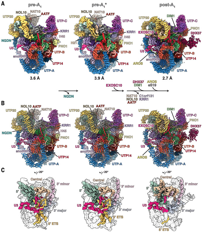
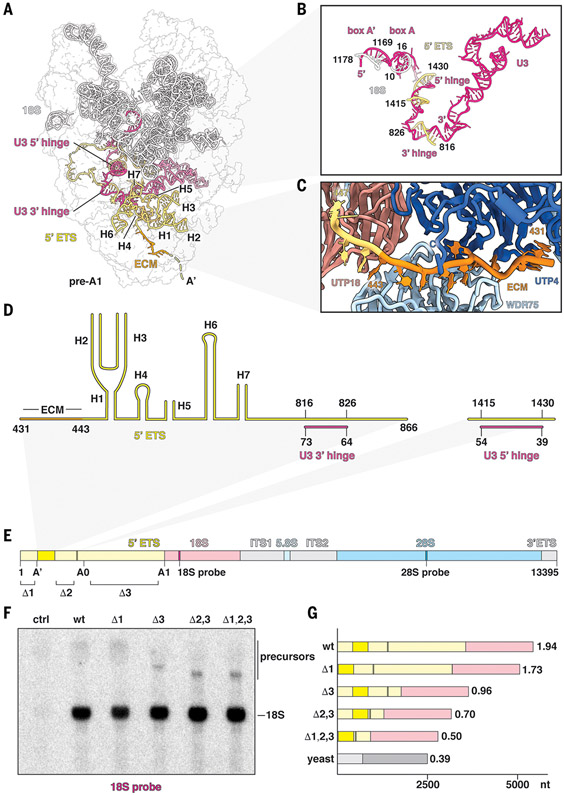
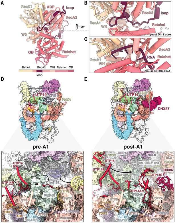
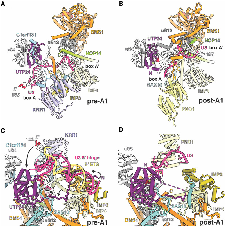
The human small subunit processome mediates early maturation of the small ribosomal subunit by coupling RNA folding to subsequent RNA cleavage and processing steps. We report the high-resolution cryo–electron microscopy structures of maturing human small subunit (SSU) processomes at resolutions of 2.7 to 3.9 angstroms. These structures reveal the molecular mechanisms that enable crucial progressions during SSU processome maturation. RNA folding states within these particles are communicated to and coordinated with key enzymes that drive irreversible steps such as targeted exosome-mediated RNA degradation, protein-guided site-specific endonucleolytic RNA cleavage, and tightly controlled RNA unwinding. These conserved mechanisms highlight the SSU processome’s impressive structural plasticity, which endows this 4.5-megadalton nucleolar assembly with the distinctive ability to mature the small ribosomal subunit from within.


Figures

Fig. 1.
Fig. 1.. Nucleolar maturation of human SSU processome.
(A) Cryo-EM density maps of nucleolar states pre-A1, pre-A1*, and post-A1 at 3.6, 3.9, and 2.7 Å, respectively. Modules, assembly factors, and RNA are labeled, and compositional changes are indicated at the bottom. (B) Atomic models of the three states displayed and labeled as in (A). (C) Structures of RNA elements in each state. 5′ domain, green; central domain, beige; 3′ major domain, gray; 3′ minor domain, light pink; 5′ ETS, yellow; U3 snoRNA, pink.
Fig. 2.
Fig. 2.. Recognition and processing of the human 5′ ETS.
(A) Architectural view of state pre-A1 with pre-18S (white), 5′ ETS (yellow), and U3 snoRNA (pink). The ECM (orange) and U3 hinges are indicated. (B) Detailed view of U3 snoRNA and its interactions with pre-18S and 5′ ETS RNAs. (C) Recognition of the ECM by UTP4 (dark blue) and WDR75 (light blue). (D) Secondary structure of the observed 5′ ETS in state pre-A1. (E) Schematic representation of a synthetic human rDNA locus containing distinct sequences for the small and large ribosomal subunits (18S and 28S probe, respectively). Ordered regions of the 5′ ETS are indicated in dark yellow, and segments for truncations are indicated with Δ1, Δ2, and Δ3. (F) Northern blot analysis of cells transfected without plasmid (control, ctrl), wild-type (wt), and truncated synthetic human rDNA loci (Δ1; Δ3; Δ2,3; and Δ1,2,3). Precursors and mature 18S rRNA originating from the synthetic template are indicated. (G) Schematic illustrating the ratios between 5′ ETS and 18S of wild-type and truncated rDNAs. Ratios between 5′ ETS and 18S are indicated at the end of each bar, with yeast (bottom bar) serving as reference. nt, nucleotides.
Fig. 3.
Fig. 3.. Structural remodeling facilitates licensing for exosome-mediated maturation.
(A) (Top) Architecture of state pre-A1. (Bottom) Zoomed view highlighting the NGDN (cyan, transparent surface) binding site near UTP20 (yellow), U3IP2 (purple), and ribosomal proteins eS4, eS24, and uS4. (B) (Top) Architecture of state pre-A1*. (Bottom) Zoomed view highlighting the loss of NGDN (cyan) and rearrangement of the U3IP2 N terminus. (C) (Top) Architecture of state post-A1. (Bottom) Zoomed view highlighting the presence of the EXOSC10 (pink, transparent surface) lasso with four peptide epitopes (I to IV) near the moved eS4.
Fig. 4.
Fig. 4.. Regulation of the DEAH-box helicase DHX37.
(A) Crystal structure of the yeast Dhr1 core enzyme with the RecA2-associated autoinhibitory loop. A schematic of the yeast Dhr1 domain organization is shown at the bottom. ADP, adenosine diphosphate; WH, winged helix domain; OB, oligonucleotide/oligosaccharide binding-fold domain. (B and C) Detailed views of the Dhr1 autoinhibitory loop in the substrate channel (B) compared with the substrate RNA in the mouse Dhx37 structure (PDB ID 6O16) (C). (D) (Top) Structure of human pre-A1 state highlighting UTP14 proximal proteins. (Bottom) Detailed view of UTP14 and the recruitment domain (UTP14R). Black arrows depict subsequent movements of UTP14R, WDR46, and PNO1 from pre-A1 to post-A1. (E) (Top) Structure of human post-A1 state highlighting UTP14 proximal proteins. (Bottom) Detailed view of UTP14 and its recruitment, activation, and stabilizing domains (UTP14R, UTP14Act, and UTP14S, respectively). Positioning of these domains depends on movement of UTP20 (UTP14R), processing of the 5′ ETS (UTP14Act), and movement of Pno1 (UTP14S). The black arrow depicts the putative path of UTP14R toward its binding site, as seen in the yeast state Dis-C. The putative AIM of TDIF2 is indicated.
Fig. 5.
Fig. 5.. Protein-guided mechanism of cleavage at site A1.
(A) Catalytic core of state pre-A1 with U3 boxes A and A′ (pink) and base-pairing 18S (white). C1orf131 (light blue) occludes the nuclease UTP24 (purple), and nucleotide 2 of the 18S rRNA (red) is positioned far away. (B) In state post-A1, C1orf131 has disappeared and KRR1 has been replaced by PNO1 (yellow). Rearrangements in U3 box A and A′ allow nucleotide 2 (red) of the 18S rRNA to be in close proximity to the UTP24 nuclease active site (magnesium ion, green circle). (C) In state pre-A1, the UTP24 N terminus (purple) is sequestered via interactions with the 5′ hinge (pink), 5′ ETS (yellow), and the IMP3 N terminus (olive). Black arrows highlight structural changes within the N terminus of UTP24, the 5′ end of 18S rRNA, and other proteins that are required for A1 cleavage. (D) Destabilization of the 5′ ETS and accompanying changes in the post-A1 state allow the rearranged UTP24 N terminus to stabilize incoming substrate 18S rRNA.
Fig. 6.
Fig. 6.. Human diseases of the SSU processome.
(A) SSU processome factors with disease-causing mutations are highlighted within state post-A1. MC, microcephaly; BWCNS, Bowen-Conradi syndrome; NAIC, North American Indian childhood cirrhosis; 5q-, 5q- syndrome; DBA, Diamond-Blackfan anemia; ACC, aplasia cutis congenita. (B) Detailed view of AROS (yellow, transparent surface) and its interacting proteins. DBA1-causing mutations in eS19 are colored red. (C) Detailed view of BMS1 (orange), uS12 (gray), and neighboring proteins. Disease-causing mutations in BMS1 (ACC) and uS12 (MC) are labeled and shown as red spheres. (D) A close-up view of the interface between eS19 (gray) and AROS (yellow, transparent surface). DBA1-causing mutations in eS19 are labeled and shown as red sticks.
Fig. 7.
Fig. 7.. Model for nucleolar maturation of the human ribosomal small subunit.
(A) Illustration depicting the assembly of human SSU processomes within the nucleolus, with fibrillar center (FC, red), dense fibrillar component (DFC, yellow), and granular component (GC, blue). The expanded human 5′ ETS (yellow) is shown interacting with IDRs of nucleolar proteins. Processing of pre-18S rRNA progresses via different states of the SSU processome (black box). (B to D) Detailed views of three states indicating the chronology of events with components present in states pre-A1 (B), pre-A1* (C), and post-A1 (D). Components leaving and joining the particles are indicated, and inhibitory mechanisms are highlighted.

References

    1. Bohnsack KE, Bohnsack MT, Uncovering the assembly pathway of human ribosomes and its emerging links to disease. EMBO J. 38, e100278 (2019). doi: 10.15252/embj.2018100278; pmid: 31268599 - DOI - PMC - PubMed
    1. Lafontaine DLJ, Riback JA, Bascetin R, Brangwynne CP, The nucleolus as a multiphase liquid condensate. Nat. Rev. Mol. Cell Biol 22, 165–182 (2021). doi: 10.1038/s41580-020-0272-6; pmid: 32873929 - DOI - PubMed
    1. Michot B, Bachellerie JP, Secondary structure of the 5′ external transcribed spacer of vertebrate pre-rRNA. Presence of phylogenetically conserved features. Eur. J. Biochem 195, 601–609 (1991). doi: 10.1111/j.1432-1033.1991.tb15743.x; pmid: 1999184 - DOI - PubMed
    1. Hunziker M et al., Conformational switches control early maturation of the eukaryotic small ribosomal subunit. eLife 8, e45185 (2019). doi: 10.7554/eLife.45185; pmid: 31206356 - DOI - PMC - PubMed
    1. Yao R-W et al., Nascent pre-rRNA sorting via phase separation drives the assembly of dense fibrillar components in the human nucleolus. Mol. Cell 76, 767–783.e11 (2019). doi: 10.1016/j.molcel.2019.08.014; pmid: 31540874 - DOI - PubMed

Publication types