Review
. 2022 Feb;57(1):16-47.
doi: 10.1080/10409238.2021.1961674. Epub 2021 Sep 13.

Regulation of protein function and degradation by heme, heme responsive motifs, and CO

Affiliations
Review

Regulation of protein function and degradation by heme, heme responsive motifs, and CO

Angela S Fleischhacker et al. Crit Rev Biochem Mol Biol. 2022 Feb.

Abstract

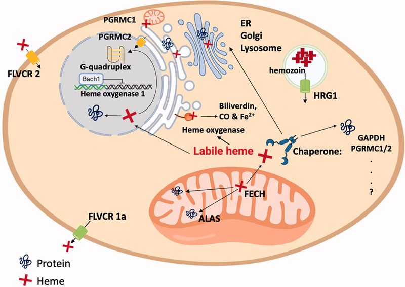
Heme is an essential biomolecule and cofactor involved in a myriad of biological processes. In this review, we focus on how heme binding to heme regulatory motifs (HRMs), catalytic sites, and gas signaling molecules as well as how changes in the heme redox state regulate protein structure, function, and degradation. We also relate these heme-dependent changes to the affected metabolic processes. We center our discussion on two HRM-containing proteins: human heme oxygenase-2, a protein that binds and degrades heme (releasing Fe2+ and CO) in its catalytic core and binds Fe3+-heme at HRMs located within an unstructured region of the enzyme, and the transcriptional regulator Rev-erbβ, a protein that binds Fe3+-heme at an HRM and is involved in CO sensing. We will discuss these and other proteins as they relate to cellular heme composition, homeostasis, and trafficking. In addition, we will discuss the HRM-containing family of proteins and how the stability and activity of these proteins are regulated in a dependent manner through the HRMs. Then, after reviewing CO-mediated protein regulation of heme proteins, we turn our attention to the involvement of heme, HRMs, and CO in circadian rhythms. In sum, we stress the importance of understanding the various roles of heme and the distribution of the different heme pools as they relate to the heme redox state, CO, and heme binding affinities.

Keywords: CO; Heme; circadian rhythm; enzymology; gas sensing/signaling; heme oxygenase; heme responsive motif; iron homeostasis; nuclear receptor; redox.


Conflict of interest statement

Disclosure statement

No potential conflict of interest was reported by the author(s).

Figures

Figure 1.
Figure 1.
Comparison of the heme oxygenases. (A) The heme degradation pathway involves the conversion of heme to biliverdin by heme oxygenase in a reaction that is dependent on cytochrome P450 reductase and requires seven electron equivalents, provided by NADPH, and O2. In addition to producing biliverdin, CO and Fe2+ iron are released. Biliverdin is subsequently converted to bilirubin by biliverdin reductase to complete the heme degradation pathway. (B) A linear representation of HO2 and HO1 for comparison of the features of the two proteins. High sequence similarity and structural homology exists in the catalytic core regions of the proteins. The catalytic core regions include a conserved histidine residue (His45 in human HO2, His25 in human HO1) that binds heme in preparation for catalytic turnover. Both proteins associate with the ER membrane via a C-terminal domain that is connected to the catalytic core via an intrinsically disordered region. Low sequence similarity exists in the intrinsically disordered region; HO2 contains two HRMs in this region (centered at Cys265-Pro266 and Cys282-Pro283) while a PEST domain has been identified in the same region of HO1. In addition, HO2 contains an extended N-terminal domain relative to HO1. (C) The two HRMs located in the intrinsically disordered region of HO2 act as a thiol/disulfide redox switch. Cys265 and Cys282 form a disulfide bond under oxidizing conditions that precludes heme binding from this region (left). Upon reduction of the disulfide bond (right), the thiols act as ligands to Fe3+-heme. The imidazole moiety of His256 provides an additional axial ligand to the iron of the Fe3+-heme coordinated by the thiol of Cys265.
Figure 2.
Figure 2.
Heme binding to Rev-erbβ. In the presence of a disulfide bond between Cys384 (of an HRM) and a nearby cysteine (Cys374), His568 and a neutral axial ligand coordinate Fe3+-heme independently of the HRMs (lower left). Upon reduction of the disulfide bond, Cys384 displaces the neutral axial ligand, increasing the heme-binding affinity of Rev-erbβ (upper left). In addition to a thiol/disulfide redox switch, an additional level of redox regulation in Rev-erbβ is the Fe3+/Fe2+ couple. Upon reduction of the heme to the Fe2+ state, Cys384 dissociates and is replaced by a neutral axial ligand to form the 6-coordinated heme (top right) or the 5-coordinated heme (bottom, center). The ligand switch is associated with a significant decrease in the heme-binding affinity of Rev-erbβ. However, Rev-Erbβ binds CO and NO with very high affinity (lower right). CO and NO increase the apparent redox potential of the Fe3+/Fe2+ couple by an EC mechanism, in which the reduction of Fe3+- to Fe2+-heme is tightly coupled to CO/NO binding, allowing Fe3+-Rev-erbβ to act as a gas sensor. A color version of the figure is available online.
Figure 3.
Figure 3.
Conceptual model of heme storage, transport and trafficking. Heme trafficking among different cellular compartment and their directions are indicated with black arrows. Putative heme transporters are shown with yellow or green barrels. Potential heme storage methods include G-quadruplex and hemozoin formation. Identified heme chaperones are listed. A color version of the figure is available online.
Figure 4.
Figure 4.
Interdependent iron and heme homeostasis regulation. Major components in heme and iron homeostasis maintenance and their interdependent regulation are shown in a simplified manner. Stimulating effects are shown in red arrows, repressing effects are shown in blue “T” shapes, and binding or transporting events are shown in black errors. A color version of the figure is available online.
Figure 5.
Figure 5.
Heme-HRM interactions alter protein-ligand interactions. Interactions between heme and the HRM lead to conformational changes that can promote or interfere with ligand binding. In the absence of heme, an HRM-containing protein (green) interacts with a ligand such as DNA, RNA, or another protein (blue; ligand A) while potentially not interacting with others (purple; ligand B). In the presence of heme, heme binding to the HRM is predicted to either sterically interfere with binding of ligand A by binding in the interface of the two biomolecules (upper) or by causing a conformational change in the HRM-containing protein (lower). The conformational change is induced by ligation of the heme via two protein residues, Cys of the HRM as well as a His from another region of the protein, and blocks the binding of ligand A. Additionally, either mode of heme binding can expose new binding sites on the HRM-containing protein to interact with ligand B, inducing a functional outcome such as protein degradation. A color version of the figure is available online.
Figure 6.
Figure 6.
CO-sensing scheme by heme binding proteins. The gas sensing proteins have the heme-containing sensor domain that is involved in binding the diatomic gas (in this case, CO) and a functional domain. Binding of a gaseous molecule to the heme Fe2+ complex results in a structural change in the sensor domain. The structural change is transduced to the functional domain resulting in functional outcomes, for example, either opening or closing of heme-bound ion channels, turning on or off transcriptional activity of a heme-bound transcription factor. In certain proteins that exists in the oxidized state, CO can also drive the reduction of the bound Fe3+-heme (indicated by the broken arrow). In the presence of suitable electron donors, reductive carbonylation of the oxidized protein takes place – this involves thermodynamically coupling the unfavorable heme reduction with the highly favorable association of CO with the Fe2+-heme. The heme coordination sphere undergoes a structural change during the redox switching and gas binding, resulting in a global structural change in the protein. A color version of the figure is available online.

References

    1. Airola MV, Du J, Dawson JH, Crane BR. 2010. Heme binding to the mammalian circadian clock protein period 2 is nonspecific. Biochemistry. 49(20):4327–4338. - PMC - PubMed
    1. Andrews NC, Schmidt PJ. 2007. Iron homeostasis. Annu Rev Physiol. 69:69–85. - PubMed
    1. Aono S 2003. Biochemical and biophysical properties of the CO-sensing transcriptional activator CooA. Acc Chem Res. 36(11):825–831. - PubMed
    1. Appel A, Bercaw J, Bocarsly A, Dobbek H, DuBois D, Dupuis M, Ferry J, Fujita E, Hille R, Kenis P, et al. 2013. Frontiers, opportunities, and challenges in biochemical and chemical catalysis of CO2 fixation. Chem Rev. 113(8):6621–6658. - PMC - PubMed
    1. Atamna H, Brahmbhatt M, Atamna W, Shanower GA, Dhahbi JM. 2015. ApoHRP-based assay to measure intracellular regulatory heme. Metallomics. 7(2):309–321. - PMC - PubMed

Publication types

LinkOut - more resources