Multicenter Study
. 2021 Nov;32(11):2920-2932.
doi: 10.1681/ASN.2021030382. Epub 2021 Sep 13.

The Clinical Application of Urine Soluble CD163 in ANCA-Associated Vasculitis

Collaborators, Affiliations
Multicenter Study

The Clinical Application of Urine Soluble CD163 in ANCA-Associated Vasculitis

Sarah M Moran et al. J Am Soc Nephrol. 2021 Nov.

Abstract

Background: Up to 70% of patients with ANCA-associated vasculitis (AAV) develop GN, with 26% progressing to ESKD. Diagnostic-grade and noninvasive tools to detect active renal inflammation are needed. Urinary soluble CD163 (usCD163) is a promising biomarker of active renal vasculitis, but a diagnostic-grade assay, assessment of its utility in prospective diagnosis of renal vasculitis flares, and evaluation of its utility in proteinuric states are needed.

Methods: We assessed a diagnostic-grade usCD163 assay in (1) a real-world cohort of 405 patients with AAV and 121 healthy and 488 non-AAV disease controls; (2) a prospective multicenter study of 84 patients with potential renal vasculitis flare; (3) a longitudinal multicenter cohort of 65 patients with podocytopathy; and (4) a cohort of 29 patients with AAV (with or without proteinuria) and ten controls.

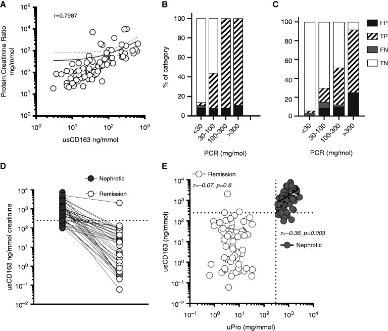
Results: We established a diagnostic reference range, with a cutoff of 250 ng/mmol for active renal vasculitis (area under the curve [AUC], 0.978). Using this cutoff, usCD163 was elevated in renal vasculitis flare (AUC, 0.95) but remained low in flare mimics, such as nonvasculitic AKI. usCD163's specificity declined in patients with AAV who had nephrotic-range proteinuria and in those with primary podocytopathy, with 62% of patients with nephrotic syndrome displaying a "positive" usCD163. In patients with AAV and significant proteinuria, usCD163 normalization to total urine protein rather than creatinine provided the greatest clinical utility for diagnosing active renal vasculitis.

Conclusions: usCD163 is elevated in renal vasculitis flare and remains low in flare mimics. Nonspecific protein leakage in nephrotic syndrome elevates usCD163 in the absence of glomerular macrophage infiltration, resulting in false-positive results; this can be corrected with urine protein normalization.

Keywords: ANCA; CD163; biomarker; crescentic glomerulonephritis; macrophage; urine.


Figures

None
Graphical abstract
Figure 1.
Figure 1.
Real-world clinical testing for usCD163 using a clinically accredited ELISA test (cohort 1). (A) Comparison of all usCD163 values in healthy control (HC) and patients with vasculitis in remission and with active RV. Both normalized (to urine creatinine, left axis) and non-normalized (right axis) values are presented. The dotted line reflects the optimal cut point for normalized values (250 ng/mmol) and the solid line reflects the optimal cut point for non-normalized values (1.3 ng/mL). Receiver-operator characteristic curve AUC values for (B) non-normalized and (C) normalized values, comparing active RV and remission vasculitis, are included. (D) Correlation of clinically accredited (Euroimmun) precoated ELISA kit with the R&D Duoset kit (as used in prior work).
Figure 2.
Figure 2.
Prospective evaluation of usCD163 testing in the clinical setting of possible RV flare (cohort 2). (A) Comparison of normalized usCD163 values in those subsequently adjudicated as having suffered an RV flare versus those that did not have a RV flare. ****P<0.0001, Mann–Whitney test. (B) Receiver-operator characteristic curve for normalized usCD163 comparing RV flare with no RV flare in this prospective cohort. (C) Normalized usCD163 values in patients subsequently evaluated as not having suffered an RV flare. (D) Normalized usCD163 values according to the pretest physician assessment of “possible” or “highly probable” RV flare. In each case, the dotted line represents the upper limit of normal (250 ng/mmol).
Figure 3.
Figure 3.
Assessment of the effect of proteinuria on usCD163 estimation. (A) Correlation of usCD163 with urine total protein concentration. (B) Effect of increasing amount of proteinuria on false-positive (FP) rate in cohort 2, a restricted clinical setting of clinical suspicion of RV flare, and in (C) cohort 1, a real-world cohort reflecting the general use of the test in clinical practice. (D) usCD163 values in paired active and remission nephrotic syndrome in patients with biopsy sample–proven podocytopathy. (E) Correlation of usCD163 values in patients with active nephrotic syndrome and in remission. FN, false negative; TN, true negative; TP, true positive
Figure 4.
Figure 4.
Assessment of methods for correction of usCD163 in the setting of heavy proteinuria (cohort 4). (A) Correlation of FE-CD163 with urine PCR (uPCR). Light gray data points reflect healthy controls. (B) FE-CD163 in patients with vasculitis in remission with, and without, heavy proteinuria. Healthy control (HC) shown for comparison. (C) usCD163, normalized to creatinine, in patients with active and remission RV (with and without proteinuria). The c-statistic refers to the area under the respective receiver-operating characteristic curve in attempting to differentiate active RV from both proteinuric and nonproteinuric patients in remission. (D–F) Various mechanisms for normalizing usCD163 in the setting of proteinuria: (D) normalized to total urine protein, (E) normalized using the albumin/sCD163 ratio of serum to urine, and (F) normalized to urine albumin. (G) The total protein-normalized cutoff of 2.5 ng/mg was applied to those cohort 1 patients with AAV and a urine protein value >0.5 g/L. *P<0.05, **P<0.01, ***P<0.001, ****P<0.0001, Kruskal–Wallis test, with Dunn multiple comparisons test. Sens, sensitivity; spec, specificity.

References

    1. Jennette JC, Olson JL, Schwartz MM, Silva FG, Thomas DB: Pauci-immune and antineutrophil cytoplasmic autoantibody–mediated crescentic glomerulonephritis and vasculitis. In: Heptinstall’s Pathology of the Kidney, edited by Olson JL, Jennette JC, Schwartz MM, Silva FG, Philadelphia, Lippincott Williams and Wilkins, 2007, pp 643–674
    1. Booth AD, Almond MK, Burns A, Ellis P, Gaskin G, Neild GH, et al. ; Pan-Thames Renal Research Group : Outcome of ANCA-associated renal vasculitis: a 5-year retrospective study. Am J Kidney Dis 41: 776–784, 2003 - PubMed
    1. Mohammad AJ, Segelmark M: A population-based study showing better renal prognosis for proteinase 3 antineutrophil cytoplasmic antibody (ANCA)-associated nephritis versus myeloperoxidase ANCA-associated nephritis. J Rheumatol 41: 1366–1373, 2014 - PubMed
    1. Lionaki S, Hogan SL, Jennette CE, Hu Y, Hamra JB, Jennette JC, et al. : The clinical course of ANCA small-vessel vasculitis on chronic dialysis. Kidney Int 76: 644–651, 2009 - PMC - PubMed
    1. Poggio ED, McClelland RL, Blank KN, Hansen S, Bansal S, Bomback AS, et al. ; Kidney Precision Medicine Project : Systematic review and meta-analysis of native kidney biopsy complications. Clin J Am Soc Nephrol 15: 1595–1602, 2020 - PMC - PubMed

Publication types

MeSH terms