Review
. 2022 Mar 1;14(3):a040923.
doi: 10.1101/cshperspect.a040923.

Axon Regeneration: A Subcellular Extension in Multiple Dimensions

Affiliations
Review

Axon Regeneration: A Subcellular Extension in Multiple Dimensions

Carla C Winter et al. Cold Spring Harb Perspect Biol. .

Abstract

Axons are a unique cellular structure that allows for the communication between neurons. Axon damage compromises neuronal communications and often leads to functional deficits. Thus, developing strategies that promote effective axon regeneration for functional restoration is highly desirable. One fruitful approach is to dissect the regenerative mechanisms used by some types of neurons in both mammalian and nonmammalian systems that exhibit spontaneous regenerative capacity. Additionally, numerous efforts have been devoted to deciphering the barriers that prevent successful axon regeneration in the most regeneration-refractory system-the adult mammalian central nervous system. As a result, several regeneration-promoting strategies have been developed, but significant limitations remain. This review is aimed to summarize historic progression and current understanding of this exciting yet incomplete endeavor.


Figures

Figure 1.
Figure 1.
Major milestones of axon regeneration research. In 1907, Tello (pupil of Ramón y Cajal) produced the first evidence that damaged central nervous system (CNS) axons can regenerate if placed near the permissive peripheral nervous system (PNS) environment. Tello anastomosed an optic nerve stump (1) to a sciatic nerve graft (2) and observed a scar (3) as well as purported RGC axons crossing the site of anastomosis (4) regenerating into the sciatic nerve (7). (5) Indicates a vein, (6) indicates neurilemma of the sciatic nerve (adapted from Tello 1907). Tello's work was complemented by Ramón y Cajal's histological descriptions of the degeneration and regeneration of the CNS, published in 1913 and translated in 1928 (adapted from Ramón y Cajal 1928). In 1981, David and Aguayo confirmed Tello's findings that CNS neurons can regenerate in the permissive PNS. (1) Diagram of the dorsal aspect of a rat spinal cord, with a peripheral nerve “bridge” between the medulla and thoracic spinal cord. Cross sections of the medulla and spinal cord show where the bridge was directly attached. (2) The origin of axons innervating the graft were determined with horseradish peroxidase (HRP) retrograde labeling and found to originate in the brainstem and spinal cord (figure adapted from David and Aguayo 1981). In 1984, Richardson and Issa used peripheral nerve grafts to investigate the neuron intrinsic mechanisms of CNS injury and regeneration; long spinal axons of dorsal root ganglia (DRG) were 100 times more likely to regenerate into peripheral nerve grafts in the dorsal column (following bilateral dorsal column lesions) if their peripheral axons were also cut (adapted from Richardson and Issa 1984). In 1999, Neumann and Woolf showed that altering intrinsic growth capacity of DRGs with a similar preconditioning lesion can promote regeneration of central axons in CNS tissue. Animals with a peripheral axotomy 1 to 2 weeks prior to (but not concomitant with) a dorsal column lesion show growth into and above the lesion, and that DRG explants show increased neurite outgrowth following preconditioning lesion (image shows explant results; adapted from Neumann and Woolf 1999). Focusing on neuron extrinsic factors inhibiting regeneration, in 1988 Caroni and Schwab identified myelin-derived membrane proteins with nonpermissive substrate properties; a monoclonal antibody, IN-1, neutralizes the nonpermissive substrate properties (adapted from Caroni and Schwab 1988a). A landmark study by Park et al. in 2008 highlighted the utility of altering the intrinsic state of neurons to promote axon regeneration–PTEN deletion (conditional knockout [CKO]) was shown to promote exceptionally robust axon regeneration in an optic nerve crush model (scale bar, 100 µm) (adapted from Park et al. 2008). Figure created with BioRender.com.
Figure 2.
Figure 2.
Models to study axon regeneration. (A) Invertebrates (e.g., Caenorhabditis elegans and Drosophila melanogaster) and zebrafish are well established models to study axon regeneration. They have short reproductive cycles to quickly screen gene function. The transparent cuticle and wings, of C. elegans and Drosophila can be used for laser-induced axon regeneration studies in vivo. The growth permissive bridge at the injury site distinguishes the zebrafish as a central nervous system (CNS) regeneration-competent vertebrate. (B) Other vertebrates or mammals like the mouse, Mus musculus, have a regeneration competent peripheral nervous system (PNS), but incompetent CNS. In the mammalian system, (1) the dorsal root ganglion (DRG) injury is used to model regeneration-competent PNS injury, or (2) to study the “preconditioning” effect for central branch injury. (3) Sciatic nerve injury (SNI) is another PNS injury model that keeps the neural tube intact to facilitate regeneration. In the optic nerve crush (ONC), viral vectors are used to manipulate retinal ganglion cell (RGC) gene expression and/or anterograde axon labeling (4) to assess axon regeneration. Generally, this is measured from the injury site (5) and divided into short (>2 mm) and long (<2 mm) distance regeneration. Spinal cord injury (SCI) models used are (6) the contusion model to mimic clinical relevance, or more consistent models like (7) lateral or (8) full transection to study specific aspects of axon regeneration and the environmental contribution. Figure created with BioRender.com.
Figure 3.
Figure 3.
Soma, axonal, and environmental events following axon injury. (1) The initial response to injury of any axon is an immediate influx of calcium into the tip of the severed axon, which triggers activation of calpains and subsequent Ca2+-dependent membrane fusion to reseal the ruptured end of the axon. (2) Calcium also leads to activation of local protein synthesis (e.g., of STAT3 and importin-β, which are retrogradely transported to initiate regenerative programs at the soma) and protein activity such as DLK-1. (3) Additionally, an injury-induced back-propagating calcium wave induces changes in gene expression, such as the (4) epigenetic changes seen in regenerative peripheral nervous system (PNS) neurons (e.g., dorsal root ganglion [DRG]) via activation of protein kinase Cµ (PKCµ), resulting in nuclear export of HDAC5 (histone deacetylase 5). HDAC5's absence in the nucleus enables a transition from repressive DNA histone methylation to permissive acetylation within the nucleus, which (5) allows transcription factors such as ATF3 to bind to accessible sites. (6) In contrast, nonregenerative axons of the adult central nervous system (CNS) do not induce similar epigenetic changes so that DNA remains in a methylated, repressive state. (7) The postinjury microenvironment plays a critical role in dictating regeneration competence. Activated microglia clear apoptotic cells and debris from the damaged extracellular matrix, while releasing inflammatory mediators to recruit leukocytes from the periphery. Neutrophils secrete damaging proteolytic enzymes. Recruited macrophages to the injury site become indistinguishable from activated microglia, performing phagocytosis and proinflammatory roles. NG2-expressing oligodendrocyte precursor cells (OPCs) also contribute to inhibition of axon regeneration. Finally, astrocytes contribute to the formation of the glial scar in the CNS but not PNS to form a chemical and physical barrier to axon regeneration. (PIC) Peripheral immune cell, (CK) cytokine, (Ast.) astrocyte, (PGs) proteoglycans, (GPs) glycoproteins, (GIMs) growth inhibitory molecules, (FC) foam cell, (MΦ) macrophage, (µG) microglia, (ROS) reactive oxygen species, (PMN) polymorphonuclear cell (neutrophil). Figure created with BioRender.com.
Figure 4.
Figure 4.
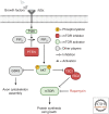
The PI3K/AKT/mTOR pathway. The PI3K/AKT/mTOR pathway is activated through the binding of growth factors to the receptor tyrosine kinase (RTK) complex, which in turn activates PI3K. The activated form of PI3K converts PIP2 to PIP3, which then mediates the phosphorylation of AKT. AKT acts on a wide range of substrates involved in the regulation of important cellular function. This includes blocking negative regulators of mTOR, such as TSC1 or TSC2. Active mTOR is associated with high-protein synthesis, cell proliferation, and cell growth. Phosphatase and tensin homolog (PTEN) acts as a tumor suppressor that negatively regulates this pathway by removing the 3-phosphate from PIP3. Loss of PTEN leads to overactivation of the mTOR pathway and has been shown to promote exceptionally robust axon regeneration following central nervous system (CNS) injury (Park et al. 2008; Liu et al. 2010). This effect can be abolished with the application of rapamycin, a known inhibitor of mTOR, indicating that the pro-regenerative effects of this treatment are mTOR-dependent (Park et al. 2008). mTOR-independent signaling mechanisms, however, appear to also be important for axon regeneration. This is underscored by the finding that genetic deletion of other negative regulators of mTOR, such as TSC2, only partially mimic the effects of PTEN deletion in the CNS (Park et al. 2008); therefore, PTEN deletion likely works via additional effectors (such as glycogen synthase kinase 3 [GSK3]) to promote axon regeneration. (RTK) Receptor tyrosine kinase, (PI3K) phosphoinositide 3-kinase, (PIP2) phosphatidylinositol 4,5-bisphosphate, (PIP3) phosphatidylinositol 3,4,5-triphosphate, (TSC) tuberous sclerosis complex, (mTOR) mammalian target of rapamycin. Figure created with BioRender.com.

References

    1. Abe N, Borson SH, Gambello MJ, Wang F, Cavalli V. 2010. Mammalian target of rapamycin (mTOR) activation increases axonal growth capacity of injured peripheral nerves. J Biol Chem 285: 28034–28043. 10.1074/jbc.M110.125336 - DOI - PMC - PubMed
    1. Al-Ali H, Beckerman SR, Bixby JL, Lemmon VP. 2017. In vitro models of axon regeneration. Exp Neurol 287: 423–434. 10.1016/j.expneurol.2016.01.020 - DOI - PMC - PubMed
    1. Anderson MA, Burda JE, Ren Y, Ao Y, O'Shea TM, Kawaguchi R, Coppola G, Khakh BS, Deming TJ, Sofroniew MV. 2016. Astrocyte scar formation AIDS central nervous system axon regeneration. Nature 532: 195–200. 10.1038/nature17623 - DOI - PMC - PubMed
    1. Anderson MA, O'Shea TM, Burda JE, Ao Y, Barlatey SL, Bernstein AM, Kim JH, James ND, Rogers A, Kato B, et al. 2018. Required growth facilitators propel axon regeneration across complete spinal cord injury. Nature 561: 396–400. 10.1038/s41586-018-0467-6 - DOI - PMC - PubMed
    1. Apara A, Galvao J, Wang Y, Blackmore M, Trillo A, Iwao K, Brown DP, Fernandes KA, Huang A, Nguyen T, et al. 2017. KLF9 and JNK3 interact to suppress axon regeneration in the adult CNS. J Neurosci 37: 9632–9644. 10.1523/JNEUROSCI.0643-16.2017 - DOI - PMC - PubMed

Publication types