. 2021 Sep 13;11(1):18119.
doi: 10.1038/s41598-021-97429-7.

The taxonomy of two uncultivated fungal mammalian pathogens is revealed through phylogeny and population genetic analyses

Affiliations

The taxonomy of two uncultivated fungal mammalian pathogens is revealed through phylogeny and population genetic analyses

Raquel Vilela et al. Sci Rep. .

Abstract

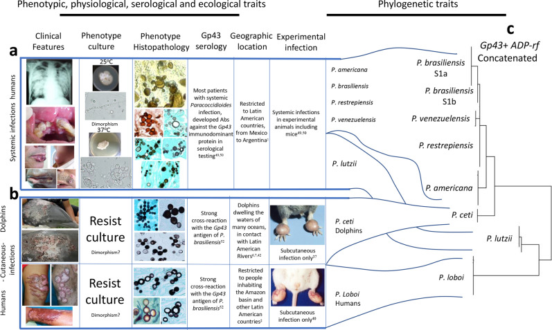
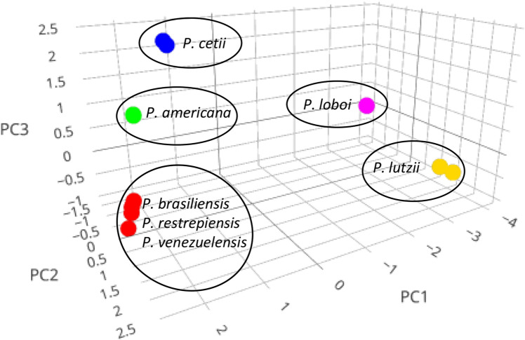
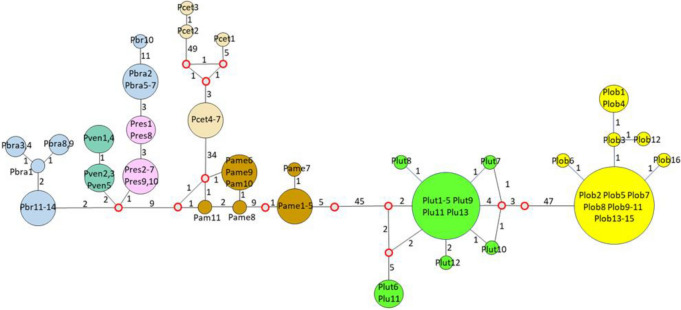
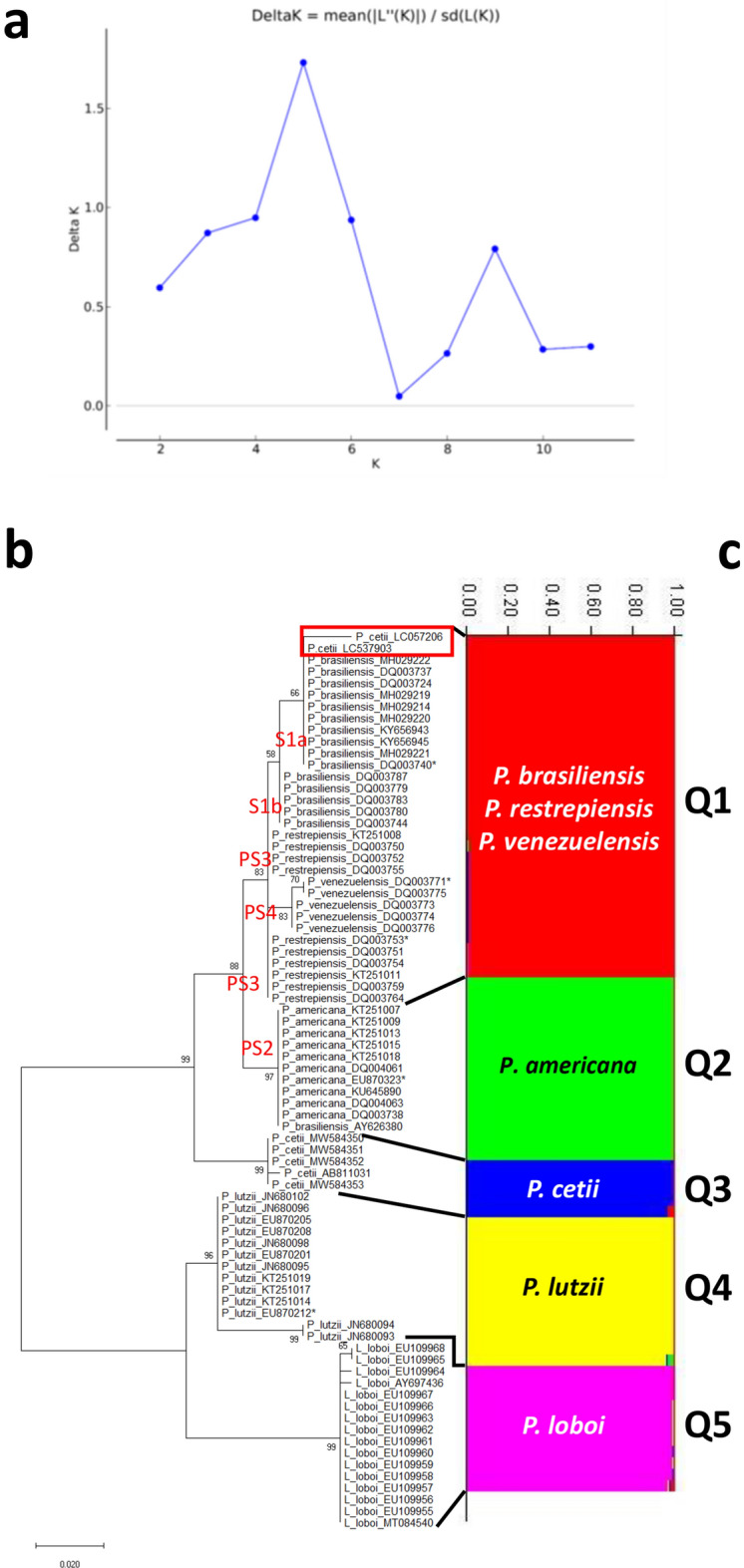
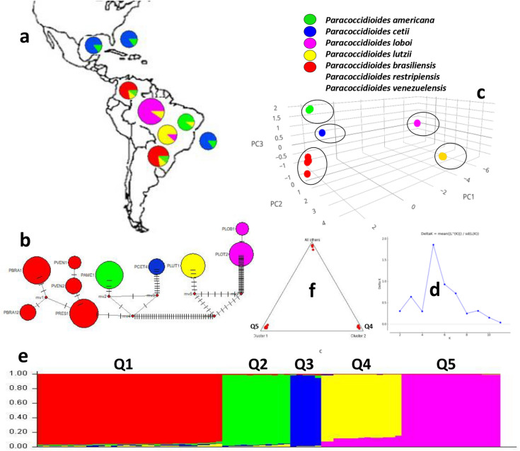
Ever since the uncultivated South American fungal pathogen Lacazia loboi was first described 90 years ago, its etiology and evolutionary traits have been at the center of endless controversies. This pathogen infects the skin of humans and as long believed, dolphin skin. However, recent DNA analyses of infected dolphins placed its DNA sequences within Paracoccidioides species. This came as a surprise and suggested the human and dolphin pathogens may be different species. In this study, population genetic analyses of DNA from four infected dolphins grouped this pathogen in a monophyletic cluster sister to P. americana and to the other Paracoccidioides species. Based on the results we have emended the taxonomy of the dolphin pathogen as Paracoccidioides cetii and P. loboi the one infecting human. Our data warn that phylogenetic analysis of available taxa without the inclusion of unusual members may provide incomplete information for the accurate classification of anomalous species.


Conflict of interest statement

The authors declare no competing interests.

Figures

Figure 1
Figure 1
Clinical features, phenotypic, physiological, ecological, and other traits, group Paracoccidioides species into two clusters (a,b). Panel (c) display the phylogenetic analysis of 59 Gp43 and ADP-rf Paracoccidioides species concatenated partial DNA sequences (including four USA dolphin). The phenotypic features shared by P. americana, P. brasiliensis (sensu stricto), P. lutzii, P. restrepiensis and P. venezuelensis is shown in panel “a”. Panel “b” comprises common phenotypic features shared by P. cetii and P. loboi. The phylogenetic traits (Panel c) using Gp43 and ADP-rf concatenated data in phylogenetic analysis, placed members of both clusters (a,b) on inverted phylogenetic groups. For instance, P. lutzii grouped with P. loboi whereas P. cetii clustered with the other Paracoccidioides species. However, without the inclusion of P. cetii DNA sequences, P. loboi would appear as an independent genus, as described by others,,,. (Clinical picture in panel b were courtesy of Drs. J. St. Leger, G. D. Bossart, L. Ajello, and P. Rosa).
Figure 2
Figure 2
Graphical output of the Gp43 DNA sequences PCA cluster analysis (total variation 90.3%, or 83.6% for PC1 (71.0%) and PC2 (12.6%)). The scatterplot shows the PC1, PC2 and PC3 results grouping Paracoccidioides species in five independent clusters including P. americana (green), P. cetii (blue), P. loboi (pink), and P. lutzii (yellow). Paracoccidioides brasiliensis (sensu stricto), P. restrepiensis, and P. venezuelensis appeared together in one of the clusters (red).
Figure 3
Figure 3
Neighbor-joining analysis of the Gp43 partial DNA sequences revealed several haplotypes. The size of the spheres is proportional to the number of individuals in each circle (numbers are shown inside the spheres). Median vectors (red open rings) represent missing or extinct species. Numbers between haplotypes indicate mutational steps. Large number of mutations were observed between Paracoccidioides lutzii (Plut-green) and P. loboi (Plob-yellow) (n = 54), between these 2 haplotypes and P. americana (Pame-brown) and P. cetii (Pcet-beige) (n = 52). Thirty-four mutations separate P. americana from P. cetii. In contrast, few mutations were observed between these four haplotypes and the remaining Paracoccidioides species (Pbra = P. brasiliensis [blue], Pres = P. restrepiensis [pink], Pven = P. venzuelensis [green]) (n = 11). Three Japanese dolphin haplotypes (Pcet = P. cetii [beige 1 to 3]) showed three missing or extinct haplotypes between them and the four USA dolphins (Pcet = P. cetii 4–7-beige) in this study.
Figure 4
Figure 4
Estimation of population using LnP(D) derived ΔK for K, from 1 to 10 (K = 5), using Gp43 DNA sequences (Panel a). Evolutionary analyses inferred by Maximum Likelihood of the Gp43 DNA sequences displays five monophyletic clusters (b) corresponding also to five STRUCTURE populations (Q1 to Q5) (c). STRUCTURE clustered P. brasiliensis (1Sa, 1Sb), P. restrepiensis (PS3), P. venezuelensis (PS4), in population Q1 (red), whereas the remaining species (P. americana [PS2, green], P. cetii [blue], P. lutzii [yellow] and P. loboi [pink]) grouped as independent populations (Q2 to Q5). The numbers on the y-axis show the subgroup membership and the x-axis the different accessions (Q) (Panel c). The distribution of accessions into different populations is indicate by color and shape.
Figure 5
Figure 5
Evolutionary analyses inferred by Maximum Likelihood of the ADP-rf, CHS4, KEX, and ITS, DNA sequences respectively, using homologous DNA sequences from well-known dimorphic Onygenales as outgroup (Table S1). In these trees, P. lutzii (yellow bars) consistently grouped as monophyletic clusters sister to P. loboi (pink bars). Except for Kex DNA sequences, grouping P. cetii (red rectangle) with P. brasiliensis (sensu lato) (bluish bar), the other DNA sequences in these analyses (ADP-rf, CHS4, and ITS) grouped P. cetii (blue bars) in monophyletic clusters. Paracoccidioides americana (green bars) clustered as a monophyletic group using ADP-rf and CHS4 DNA sequences and the other Paracoccidioides species (red bars) grouped in a single cluster. Using ITS DNA sequences, a poorly supported cluster was tentatively labelled as P. americana (green bar).
Figure 6
Figure 6
The concatenate Gp43 and ADP-rf data analyses was used to perform haplotypes analysis, to locate their geographical distributions (a,b), to estimate Principal Component Analysis (PCA) (c) and to investigate STRUCTURE population distribution (df) of several Paracoccidoides species including P. cetii from dolphins and P. loboi from humans. The geographical distribution of the five haplotypes (b) is shown in Panel (a). Five clusters are observed in haplotype analysis (b) each corresponding to P. loboi (PLOT-pink), P. lutzii (PLUT yellow), P. cetii (PCET-blue), P. americana (PAME-green). The other Paracoccidoides species formed single haplotypes (red). Small bars between haplotypes represent mutations and the median vectors (red dots) are missing or extinct haplotypes. The size of the spheres is proportional to the number of individuals in each circle. Five-populations were also found using PCA (c) and STRUCTURE (df) analyses. Panel d shows K = 5 value used to build the concatenated data. STRUCTURE analysis (e) showed P. americana (Q2-green), P. cetii (Q3-blue), P. loboi (Pink-Q5) P. lutzii (yellow-Q4), and the remaining Paracoccidioides species (Q1-red) clustering in independent barplots (e). The numbers on the y-axis show the subgroup membership and the x-axis the different accessions (Q). The distribution of accessions into different populations is indicate by color and shape. The triangle plot (f) is an analogous result obtained from STRUCTURE software outputs. The two cluster on the base of triangle corresponds to P. loboi (Q5) and P. lutzii (Q4) DNA sequences; the top harbor the remaining species in this study.

References

    1. Lôbo J. Um caso de blastomycose produzido por uma espécie nova encontrada em Recife. Rev. Med. Perenambuco. 1931;1:763–765.
    1. Lacaz CS, Baruzzi RG, Rosa MCB. Doença de Jorge Lôbo. IPSIS Gráfica e Editora; 1986. pp. 1–92.
    1. Baruzzi RG, Lacaz S, Souza PPA. História natural da doença de Jorge Lôbo. Occurrência entre os índios caiabi (Brasil central) Rev. Med. Trop. São Paulo. 1979;21:302–338. - PubMed
    1. Talhari S, Cunha MGS, Barros MLB, Gadelha AR. Doença de Jorge Lôbo. Estudo de 22 casos novos. Med. Cut. Ibero. Latin Amer. 1981;9:87–96. - PubMed
    1. Migaki G, Valerio MG, Irvine B, Garner FM. Lobo’s disease in an Atlantic bottle nosed dolphin. J. Am. Vet. Med. Assoc. 1971;159:578–582. - PubMed

Publication types