. 2021 Oct;6(10):1300-1308.
doi: 10.1038/s41564-021-00956-2. Epub 2021 Sep 13.

A regulatory cascade controls Staphylococcus aureus pathogenicity island activation

Affiliations

A regulatory cascade controls Staphylococcus aureus pathogenicity island activation

Andreas F Haag et al. Nat Microbiol. 2021 Oct.

Abstract

Staphylococcal pathogenicity islands (SaPIs) are a family of closely related mobile chromosomal islands that encode and disseminate the superantigen toxins, toxic shock syndrome toxin 1 and superantigen enterotoxin B (SEB). They are regulated by master repressors, which are counteracted by helper phage-encoded proteins, thereby inducing their excision, replication, packaging and intercell transfer. SaPIs are major components of the staphylococcal mobilome, occupying five chromosomal att sites, with many strains harbouring two or more. As regulatory interactions between co-resident SaPIs could have profound effects on the spread of superantigen pathobiology, we initiated the current study to search for such interactions. Using classical genetics, we found that, with one exception, their regulatory systems do not cross-react. The exception was SaPI3, which was originally considered defective because it could not be mobilized by any known helper phage. We show here that SaPI3 has an atypical regulatory module and is induced not by a phage but by many other SaPIs, including SaPI2, SaPIbov1 and SaPIn1, each encoding a conserved protein, Sis, which counteracts the SaPI3 repressor, generating an intracellular regulatory cascade: the co-resident SaPI, when conventionally induced by a helper phage, expresses its sis gene which, in turn, induces SaPI3, enabling it to spread. Using bioinformatics analysis, we have identified more than 30 closely related coancestral SEB-encoding SaPI3 relatives occupying the same att site and controlled by a conserved regulatory module, immA-immR-str'. This module is functionally analogous but unrelated to the typical SaPI regulatory module, stl-str. As SaPIs are phage satellites, SaPI3 and its relatives are SaPI satellites.


Conflict of interest statement

Competing interests: Authors declare no competing interests.

Figures

Figure 1
Figure 1. Helper SaPIbov1 promotes satellite SaPI3 induction and transfer.
Lysogenic S. aureus strains carrying phages 80α, 80α Δdut or ϕ11, and the indicated SaPI(s), were MC induced and titres of (a) SaPI3 seb::ermC and (b) SaPIbov1 tst::tetM determined by transduction into RN4220. Error bars indicate ±s.d. (n=4 (80α and 80α Δdut lysogens) or n=3 (Φ11 lysogens) biological replicates). Statistical analysis was performed using One-Way ANOVA on log10 transformed data followed by Sidak’s multiple comparisons test. p-values are as follows: * p<0.05, ** p<0.01, *** p<0.001, **** p<0.0001, ns not significant. Additional information is provided in the supplementary data file. Diamonds located at the baseline indicate replicates with no observed transductants. (c) Representative Southern blot analyses (n=3) of the strains analysed in panels (a) and (b). Samples were isolated 60, 90 or 120 min after induction with MC, separated on agarose gel and blotted with SaPI3-(top panel) or SaPIbov1- (bottom panel) specific probes. The upper band is ‘bulk’ DNA, including chromosomal, phage, and replicating SaPI; the lower band is SaPI linear monomers (L) released from SaPI-sized phage heads. (d) Representative Southern blot analyses (n=3) of SaPIbov1 and SaPI3 excision and replication following induction of the cloned 80α dut gene. The strains containing SaPIbov1 tst::tetM, SaPI3 seb::ermC or both islands were complemented with plasmid pJP2511 (empty plasmid) or its derivative expressing the 80α Dut protein (pJP2533). Samples were isolated 4 hours after induction with 5 μM CdCl2, which induces dut expression, and Southern blots were performed using SaPI3- and SaPIbov1-specific probes. The upper band is ‘bulk’ DNA. In these experiments, because no helper phage is present, the excised SaPI DNA appears as covalently closed circular molecules (CCC) rather than the linear monomers that are seen following helper-phage-mediated induction and packaging.
Figure 2
Figure 2. Impact of SaPI3 induction on helper SaPIs replication and transfer.
S. aureus lysogenic for 80α containing the indicated SaPIs were MC induced and the SaPI3 seb::ermC (erythromycin resistance marker) (a) or helper SaPI (tetracycline resistance marker) (b) titres were determined. Error bars indicate ±s.d. (n=3 biological replicates). Statistical analysis was performed using One-Way ANOVA on log 10 transformed data followed by Sidak’s multiple comparisons test. * p<0.05, ** p<0.01, *** p<0.001, **** p<0.0001, ns not significant. Additional information is provided in the supplementary data file. (c) Representative Southern blot analyses (n=3) analysis of mini-lysates from the defined strains taken 2 h after MC induction using probes specific for either SaPI3 (top panel), or the tetM marker of the helper SaPIs SaPIbov1, SaPI2, SaPIn1 or a SaPIbov2-specific probe (bottom panel).
Figure 3
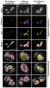
Figure 3. Sis and ImmR colocalise in vivo.
S. aureus RN4220 expressing either (a) SaPIbov1 Sis-mCherry and SaPI3 ImmR-GFP, (b) mCherry and SaPI3 ImmR-GFP or (c) SaPIbov1 Sis-mCherry and GFP were induced with 5 μM CdCl2 for 16 h and samples prepared and subjected to super-resolution microscopy. Maximum projections (top panels, scale bars 5 μm) and representative 3D-reconstructions (bottom panels, scale bars 0.2 μm) for each sample are shown. Depicted colours are false coloured. Clear: cell wall stained with wheat germ agglutinin Alexa Fluor 467 (WGA); purple: mCherry; yellow: GFP. Representative images from 3 independent experiments are shown.
Figure 4
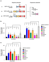
Figure 4. The helper SaPI Sis inducer alleviates SaPI3 ImmR repression.
(a) Schematic representation of the reporter and expression plasmids used. The β-lactamase reporter gene (blaZ) is under the control of SaPI3 ImmR, in presence or absence of ImmA. Expression of different cloned sis genes is under the control of a cadmium-inducible promoter (Pcad). (b) Strains containing either pJP2524 or pJP2525, as well as any of the different pCN51-derivative plasmids expressing the different Sis homologs, were assayed for β-lactamase activity 5 h after induction with 1 μM CdCl2. (c) Strains containing pJP2526 and any of the different pCN51-derivative plasmids expressing the different Sis homologs were assayed for β-lactamase activity 5 h after induction with 1 μM CdCl2. Error bars indicate ±s.d. (n=3 biological replicates). Statistical analysis was performed using One-Way ANOVA followed by Holm-Sidak’s multiple comparisons test. p-values are as follows: * p<0.05, ** p<0.01, *** p<0.001, **** p<0.0001, ns not significant. Additional information is provided in the supplementary data file.
Figure 5
Figure 5. ImmA is required for full activation of SaPI3.
(a) 80α lysogens carrying SaPIbov1 and the indicated SaPI3 islands were MC induced and the transfer of SaPI3 assessed in RN4220. (b) The SaPI3 immR or stir’ genes were cloned into pUT18c (pJP2531 or pJP2532, respectively) and the immA gene was cloned into pKNT25 (pJP2530). The pUT18c- and pKNT25-derivative plasmids were co-transformed into E. coli strain BTH101. Serial dilutions of an overnight culture were plated onto LB supplemented with kanamycin, ampicillin and 100 μM IPTG and 20 μg ml-1 X-gal. BTH101 transformed with pUT18c-zip and pKNT25-zip or pUT18c and pKNT25 served as positive or negative controls for protein-protein interactions, respectively. (c) RN4220 80α lysogens carrying the SaPI3 seb::tetM WT or the ΔimmA mutant were transformed with either an empty control plasmid (pCN51), a plasmid expressing the wild-type ImmA (pJP2520) or a plasmid expressing an ImmA version mutated in the protein’s active site (pJP2521, ImmA E51A/E52A). Cultures were grown to early exponential phase, MC treated and the expression of ImmA was induced with 1 μM CdCl2. To analyze SaPI3 transfer, the lysates were transduced in RN4220. Error bars indicate ±s.d. (n=3 biological replicates). Statistical analysis was performed using log10 transformed data and an unpaired, two-sided Student’s t-test (a) or One-Way ANOVA followed by Sidak’s multiple comparisons test (c). p-values are as follows: * p<0.05, ** p<0.01, *** p<0.001, **** p<0.0001, ns not significant. Additional information is provided in the supplementary data file.
Figure 6
Figure 6. Model of SaPI3 activation.
(a) SaPI3 is maintained in the chromosome by its ImmR repressor, which represses expression from both PimmR and Pstr’ promoters. (b) Helper SaPI Sis proteins (1) bind to ImmR and sequester it from its DNA target (2) allowing initial expression of SaPI3 replication genes from the str’ promoter as well as of ImmA (3) from the immR promoter. (c) ImmA can either bind to ImmR and remove it from its DNA target (4a) or can bind to the complex of ImmR and Sis (4b) leading to ImmR degradation (5a&b). Although this has not been experimentally tested, once induction of SaPI3 has started, the island can provide its own Sis (6) to sequester any ImmR produced and ensure replication.

References

    1. Lindsay JA, Ruzin A, Ross HF, Kurepina N, Novick RP. The gene for toxic shock toxin is carried by a family of mobile pathogenicity islands in Staphylococcus aureus . Mol Microbiol. 1998;29:527–543. - PubMed
    1. Gillet Y, et al. Association between Staphylococcus aureus strains carrying gene for Panton-Valentine leukocidin and highly lethal necrotising pneumonia in young immunocompetent patients. Lancet. 2002;359:753–759. - PubMed
    1. Penadés JR, Christie GE. The phage-inducible chromosomal islands: a family of highly evolved molecular parasites. Annu Rev Virol. 2015;2:181–201. - PubMed
    1. Novick RP. Pathogenicity islands and their role in staphylococcal biology. Microbiol Spectr. 2019;7 - PMC - PubMed
    1. Tormo MA, et al. Staphylococcus aureus pathogenicity island DNA is packaged in particles composed of phage proteins. J Bacteriol. 2008;190:2434–2440. - PMC - PubMed

Publication types

MeSH terms