Review
. 2021 Dec;76(12):3659-3686.
doi: 10.1111/all.15089. Epub 2021 Sep 27.

Advances and highlights in biomarkers of allergic diseases

Affiliations
Review

Advances and highlights in biomarkers of allergic diseases

Ismail Ogulur et al. Allergy. 2021 Dec.

Abstract

During the past years, there has been a global outbreak of allergic diseases, presenting a considerable medical and socioeconomical burden. A large fraction of allergic diseases is characterized by a type 2 immune response involving Th2 cells, type 2 innate lymphoid cells, eosinophils, mast cells, and M2 macrophages. Biomarkers are valuable parameters for precision medicine as they provide information on the disease endotypes, clusters, precision diagnoses, identification of therapeutic targets, and monitoring of treatment efficacies. The availability of powerful omics technologies, together with integrated data analysis and network-based approaches can help the identification of clinically useful biomarkers. These biomarkers need to be accurately quantified using robust and reproducible methods, such as reliable and point-of-care systems. Ideally, samples should be collected using quick, cost-efficient and noninvasive methods. In recent years, a plethora of research has been directed toward finding novel biomarkers of allergic diseases. Promising biomarkers of type 2 allergic diseases include sputum eosinophils, serum periostin and exhaled nitric oxide. Several other biomarkers, such as pro-inflammatory mediators, miRNAs, eicosanoid molecules, epithelial barrier integrity, and microbiota changes are useful for diagnosis and monitoring of allergic diseases and can be quantified in serum, body fluids and exhaled air. Herein, we review recent studies on biomarkers for the diagnosis and treatment of asthma, chronic urticaria, atopic dermatitis, allergic rhinitis, chronic rhinosinusitis, food allergies, anaphylaxis, drug hypersensitivity and allergen immunotherapy. In addition, we discuss COVID-19 and allergic diseases within the perspective of biomarkers and recommendations on the management of allergic and asthmatic patients during the COVID-19 pandemic.

Keywords: COVID-19; allergen immunotherapy; allergic diseases; biomarkers; precision medicine.


Conflict of interest statement

Ismail Ogulur, Yagiz Pat, Elena Barletta, Lacin Cevhertas, Ruben Fernandez‐Santamaria, Mengting Huang, Manal Bel Imam, Jana Koch, Siyuan Ma, Debbie J. Maurer, Yaqi Peng, Urszula Radzikowska, Arturo O. Rinaldi, Juan Rodriguez‐Coira, Pattraporn Satitsuksanoa, Stephan R. Schneider, Alexandra Wallimann, Damir Zhakparov, Reihane Ziadlou, Marie‐Charlotte Brüggen, Katja Baerenfaller, Luo Zhang, Mubeccel Akdis declare no conflict of interest. Yasutaka Mitamura and Willem van de Veen report grants from Novartis. Milena Sokolowska reports grants from Swiss National Science Foundation, GSK and Novartis. Cezmi Akdis reports grants from Allergopharma, Idorsia, Swiss National Science Foundation, Christine Kühne‐Center for Allergy Research and Education, European Commission's Horison's 2020 Framework Programme, Cure, Novartis Research Institutes, Basel, Astra Zeneca, Switzerland, Scibase, Stockholm, advisory role for Sanofi/Regeneron, Glaxo Smith‐Kline, Novartis.

Figures

FIGURE 1
FIGURE 1
Biomarkers and cells to identify endotypes of asthma. Type 2‐high asthma can be characterized by sputum and blood eosinophils, serum sIgE, FeNO and periostin levels. Moreover, epithelial cytokines (TSLP, IL‐25, IL‐33), Type 2 cytokines (IL‐4, IL‐5, IL‐13) and RAGE are also important biomarkers. Type 2‐low asthma is generally based on neutrophilic or paucigranulocytic patterns. HMGB1 and calprotectin levels can also be useful biomarkers for type 2‐low asthma. Type 2‐high asthma includes a series of immune cells such as eosinophils, Th2, ILC2, B cells and mast cells. Conversely, neutrophils, Th1 and Th17 cells are recognized as the leading mediators involved in type 2‐low asthma. FeNO, fractional exhaled nitric oxide; HMGB1, high‐mobility group box protein; IL, interleukin; ILC, innate lymphoid cells; RAGE, receptor for advanced glycation end products; sIgE, specific immunoglobulin E; Th, T helper cell; TSLP, thymic stromal lymphoprotein
FIGURE 2
FIGURE 2
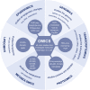
Overview of omics studies with definition and methods used. Definitions are written in italics below the titles, methods used are circled. ATAC‐seq, assay for transposase‐accessible chromatin using sequencing; CE‐MS, capillary electrophoresis coupled to mass spectrometry; ChiP, chromatin immunoprecipitation sequencing; GC‐MS, gas chromatography coupled to mass spectrometry; LC‐MS, liquid chromatography coupled to mass spectrometry; MALDI, matrix‐assisted laser desorption ionization; NGS, next‐generation sequencing; NMR, nuclear magnetic resonance; PacBio, real‐time single–molecule sequencing by Pacific Biosciences; PCR, polymerase chain reaction; seq, sequencing; SOLiD, sequencing by oligonucleotide ligation and detection
FIGURE 3
FIGURE 3
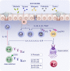
Schematic representation of allergic rhinitis (AR) molecular mechanisms and potential biomarkers. AR patients have a compromised epithelial barrier function. Exogenous agents in our environment can affect the nasal mucosa and increase barrier permeability. Primed DCs activate naïve T cells and induce their differentiation into Th2 cells. Additionally, activated epithelial cells secrete cytokines including IL‐25, IL‐33 and TSLP, activating ILC2s and Th2 cells, which in turn propagate a type 2 immune response. The presence of specific immune cells and their surface markers could serve as biomarkers for AR. CysLTR1 expression on ILC2s is upregulated and ST2 expression of pathogenic Th2 cells is increased in AR patients, while T cells are decreased. The production of type 2 cytokines, for example, IL‐4, IL‐5, IL‐9 and IL‐13, by pro‐inflammatory cells drives recruitment of eosinophils, activation of B cells leading to the local and systemic production of IgE, followed by mast cell degranulation. After class switching of B cells, their CD23 expression is increased. Furthermore, specific miRNA and protein levels such as periostin are changed in AR patients locally as well as in the blood and could be used as future biomarkers for diagnosis or to monitor treatment. CysLTR1, Cysteinyl Leukotriene Receptor 1; DC, dendritic cell; EOS, eosinophil; Ig, immunoglobulin; IL, interleukin; ILC, innate lymphoid cells; MC, mast cell; Path Th2, pathogenic memory Th2; TGF, transforming growth factor; Th, T helper cell; Tfh, T follicular helper cell; Tfr, T follicular regulatory cell; TSLP, thymic stromal lymphopoietin
FIGURE 4
FIGURE 4
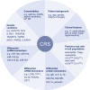
Features and biomarkers for CRS diagnosis, endotyping and treatment with a focus on recent findings. CRS severity is associated with lower age, female gender and a history of surgery. Comorbidities, such as asthma, AR or CF are relevant for categorization and treatment decisions of CRS. While clinical features such as the presence of nasal polyps are important factors to diagnose CRS and separate patients into CRSwNP and CRSsNP, other biomarkers, such as the presence of specific cell populations, protein, ncRNA or mRNA levels can help to distinguish further endotypes. For example, ECRSwNP is characterized by type 2 inflammation, including an increase in eosinophils, higher levels of total IgE and cytokines IL‐5, IL‐4 as well as IL‐13 and might therefore be responsive to treatment with available biologicals such as dupilumap. Furthermore, multiple genetic variations could aid in diagnosis of CRS or contribute to further understanding of the disease. AR, allergic rhinitis; CF, cystic fibrosis; COPD, chronic obstructive pulmonary disease; CRS, chronic rhinosinusitis; CRSwNP, CRS with nasal polyps; CRSsNP, CRS without nasal polyps; ECRSwVP, eosinophilic CRSwNP; mRNA, messenger RNA; ncRNA, noncoding RNA; PM, particulate matter; Treg, regulatory T cell
FIGURE 5
FIGURE 5
Mechanisms of allergic sensitization in food allergy. Allergic sensitization of food allergen occurs through damaged human skin or gastrointestinal tract. These food allergens can be obtained and processed by DCs. Impaired epithelial cells on the skin directly secrete cytokines such as IL‐25, IL‐33, and TSLP. Then, DCs migrate to draining lymph nodes where they present allergen‐derived peptides on MHC class II molecules to naive T cells. With the presence of several co‐stimulatory and cytokine signals, those T cells are differentiated to Th2 cells or TFH cells. Type 2 cytokines involving allergic inflammation including IL‐4, IL‐5, IL‐13, and IL‐9 are predominantly secreted by Th2 cells. Whereas, IL‐21, IL‐4, and IL‐13 are secreted by TFH cells and initiate the B‐cell class–switching to IgE, plasma cell differentiation and allergen‐specific IgE production. These processes can occur locally in the stomach and duodenum. The secreted allergen‐specific IgE antibodies from plasma cells bind to FcεRI molecules on mast cells and basophils. Subsequent exposure to allergen induces mast cell and basophil degranulation. DC, dendritic cell; FcεRI, Fc epsilon receptor I; Ig, immunoglobulin; IL, interleukin; MHC, major histocompatibility complex; TFH, T follicular helper cell; Th, T helper cell; TSLP, thymic stromal lymphoprotein
FIGURE 6
FIGURE 6
Biomarkers for the diagnosis, prognosis and management of food allergy. In allergen SPTs the skin is pricked and allergen extracts are inserted. The test is considered positive if local inflammation and histamine release occurs. Similarly, BAT and MAT measure the release of inflammatory mediators through degranulation of basophils and mast cells. Both allergen‐specific Th2 cells and their secreted cytokines (IL‐4, IL‐5 and IL‐13) are currently used as biomarkers to characterize the mechanism of food allergy and the effects of AIT in gaining allergen tolerance. Compared to T cells, B cells produce allergen‐specific IgE during the sensitization step. sIgE can be quantified in serum either through common ELISA assays or ImmunoCAP. In addition to IgE, the isotypes IgG and IgA are also involved in food allergies. The additional monitoring of Ig ratios (sIgE/total IgE and sIgG4/IgE) can be instrumental in following the course of development of food tolerance during AIT or OFC. Molecular biomarkers associated with food allergy were both genetic determinants and DNA methylation changes. Changes in protein and metabolite levels were recognized as diagnostic biomarkers in FAn, allowing the discrimination of non‐anaphylactic and anaphylactic events. The introduction of new omics technologies, studies of differences in gut microbiota and the use of high‐throughput approaches (genomics, epigenomics, transcriptomics, proteomics and metabolomics), as well as integration of the acquired data, are gaining importance in the food biomarkers research landscape. AIT, allergen immunotherapy; BAS, basophil; BAT; basophil activation test; ELISA, enzyme‐linked immunosorbent assay; Fan, food‐induced anaphylaxis; Ig: immunoglobulin; IL: interleukin; MAT: mast cell activation test; MC, mast cell; OFC, oral food challenge; SPT: skin prick test; Th: T helper cell
FIGURE 7
FIGURE 7
Biomarkers in drug hypersensitivity reactions. In immediate drug hypersensitivity reactions, the most validated biomarkers are the detection of drug‐sIgE by in vivo or in vitro tests, the analysis of activation markers on basophils after the stimulation with the culprit drug and the measurement of mediators and cytokines released by effector cells. The recent associations between specific gene variants or differential gene expression and immediate drug hypersensitivity reactions can be also be used as potential biomarkers. In non‐immediate drug hypersensitivity reactions, the assessment of drug‐specific cell proliferation, mainly by lymphocyte transformation test, the analysis of the specific cell subpopulations involved in the reaction, mainly by flow cytometry and immunohistochemistry, the measurement of cytokines, chemokines and cytotoxic markers, and the identification of specific cytokine producing cells by ELISpot are currently the most validated biomarkers. The analysis of specific gene alleles and post‐transcriptomic regulators could also be used as early biomarkers in non‐immediate drug hypersensitivity reactions. ELISpot, enzyme‐linked immune absorbent spot; sIgE, specific immunoglobulin E
FIGURE 8
FIGURE 8
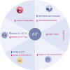
Effects of AIT on the immune response of allergic individuals. AIT in allergic individuals increases the number of Treg, ILC1 and intermediate‐type monocytes in circulation. At the same type, AIT reduces the number of eosinophils, non‐classical monocytes, Th2, Th2a, Th17, ILC2 and ILC3 in peripheral blood. AIT also reduces the mRNA levels of STAB1 in PBMCs. AIT, allergen immunotherapy; EOS, eosinophils; ILC, innate lymphoid cell; MO, monocytes; PBMC: peripheral blood mononuclear cell; STAB1, stabilin‐1; Th, T helper cell; T reg, regulatory T cell
FIGURE 9
FIGURE 9
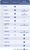
Detection of SARS‐CoV‐2 infection in allergic patients might be complex due to overlap of aeroallergy and COVID‐19 symptoms. Fever, cough, shortness of breath, body aches, and sore throat can indicate SARS‐CoV‐2 infection, whereas runny/stuffy nose, sneezing, and watery eyes point‐out more into respiratory diseases manifestation. Uncertainty regarding the source of observed manifestations should be always (tele)consulted with physicians and an extensive screening for SARS‐CoV‐2 should be assured to limit the spread of the virus. COVID‐19, coronavirus disease 2019; SARS‐CoV‐2, severe acute respiratory syndrome coronavirus 2

References

    1. Agache I, Akdis CA. Precision medicine and phenotypes, endotypes, genotypes, regiotypes, and theratypes of allergic diseases. J Clin Investig. 2019;129(4):1493‐1503. This extensive review discusses a precision medicine approach to optimize management of allergic diseases. - PMC - PubMed
    1. Aw Yong PY, Islam F, Harith HH, Israf DA, Tan JW, Tham CL. The potential use of honey as a remedy for allergic diseases: a mini review. Front Pharmacol. 2020;11:599080. - PMC - PubMed
    1. Yao Y, Chen CL, Yu D, Liu Z. Roles of follicular helper and regulatory T cells in allergic diseases and allergen immunotherapy. Allergy. 2021;76(2):456‐470. - PubMed
    1. Bousquet J, Grattan CE, Akdis CA, et al. Highlights and recent developments in allergic diseases in EAACI journals (2019). Clin Transl Allergy. 2020;10(1):56. - PMC - PubMed
    1. Agache I, Akdis CA. Endotypes of allergic diseases and asthma: an important step in building blocks for the future of precision medicine. Allergol Int. 2016;65(3):243‐252. - PubMed