Observational Study
. 2022 Feb;302(2):460-469.
doi: 10.1148/radiol.2021210986. Epub 2021 Sep 14.

Chest Radiograph Scoring Alone or Combined with Other Risk Scores for Predicting Outcomes in COVID-19

Affiliations
Observational Study

Chest Radiograph Scoring Alone or Combined with Other Risk Scores for Predicting Outcomes in COVID-19

Iain Au-Yong et al. Radiology. 2022 Feb.

Erratum in

Abstract

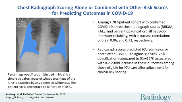
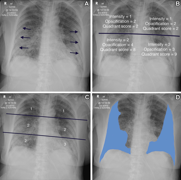
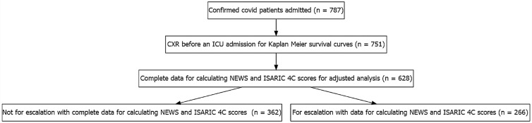
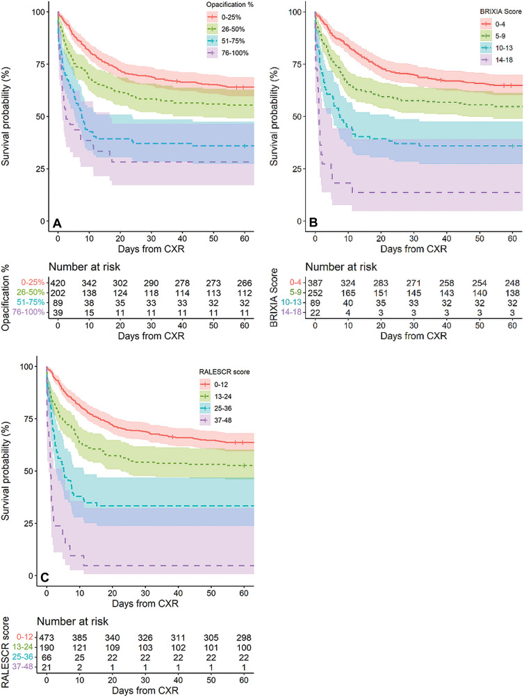
Background Radiographic severity may help predict patient deterioration and outcomes from COVID-19 pneumonia. Purpose To assess the reliability and reproducibility of three chest radiograph reporting systems (radiographic assessment of lung edema [RALE], Brixia, and percentage opacification) in patients with proven SARS-CoV-2 infection and examine the ability of these scores to predict adverse outcomes both alone and in conjunction with two clinical scoring systems, National Early Warning Score 2 (NEWS2) and International Severe Acute Respiratory and Emerging Infection Consortium: Coronavirus Clinical Characterization Consortium (ISARIC-4C) mortality. Materials and Methods This retrospective cohort study used routinely collected clinical data of patients with polymerase chain reaction-positive SARS-CoV-2 infection admitted to a single center from February 2020 through July 2020. Initial chest radiographs were scored for RALE, Brixia, and percentage opacification by one of three radiologists. Intra- and interreader agreement were assessed with intraclass correlation coefficients. The rate of admission to the intensive care unit (ICU) or death up to 60 days after scored chest radiograph was estimated. NEWS2 and ISARIC-4C mortality at hospital admission were calculated. Daily risk for admission to ICU or death was modeled with Cox proportional hazards models that incorporated the chest radiograph scores adjusted for NEWS2 or ISARIC-4C mortality. Results Admission chest radiographs of 50 patients (mean age, 74 years ± 16 [standard deviation]; 28 men) were scored by all three radiologists, with good interreader reliability for all scores, as follows: intraclass correlation coefficients were 0.87 for RALE (95% CI: 0.80, 0.92), 0.86 for Brixia (95% CI: 0.76, 0.92), and 0.72 for percentage opacification (95% CI: 0.48, 0.85). Of 751 patients with a chest radiograph, those with greater than 75% opacification had a median time to ICU admission or death of just 1-2 days. Among 628 patients for whom data were available (median age, 76 years [interquartile range, 61-84 years]; 344 men), opacification of 51%-75% increased risk for ICU admission or death by twofold (hazard ratio, 2.2; 95% CI: 1.6, 2.8), and opacification greater than 75% increased ICU risk by fourfold (hazard ratio, 4.0; 95% CI: 3.4, 4.7) compared with opacification of 0%-25%, when adjusted for NEWS2 score. Conclusion Brixia, radiographic assessment of lung edema, and percentage opacification scores all reliably helped predict adverse outcomes in SARS-CoV-2 infection. © RSNA, 2021 Online supplemental material is available for this article. See also the editorial by Little in this issue.


Conflict of interest statement

Disclosures of Conflicts of Interest: I.A.Y. Expert witness work for the General Medical Council; leadership of fiduciary role in the Royal College of Radiologists Part 2b Committee. Y.H. No relevant relationships. E.G. No relevant relationships. A.F. No relevant relationships. J.R.M. No relevant relationships. M.G. Restricted grant for work in a different field from Gilead; statistician member of the Trial Steering Committee for a clinical trial. A.R. No relevant relationships. I.J. No relevant relationships. M.S. No relevant relationships. S.B. No relevant relationships. S.C. No relevant relationships. S.H.P. No relevant relationships. J.W. No relevant relationships. C.J.C. No relevant relationships. T.C. disclosed no relevant relationships.

Figures

None
Graphical abstract
Determination of the various scoring systems. (A) Anteroposterior
radiograph in 78-year-old woman shows the classic changes of COVID-19
pneumonitis, which consist of opacification in a peripheral and basal
distribution (arrows). (B) Radiograph shows calculation of the radiographic
assessment of lung edema (RALE) score. The radiograph is divided into four
quadrants. Each quadrant is assigned an intensity score and an opacification
score. These are multiplied together for each quadrant, and all four scores
are added together. The patient has a RALE score of 21. (C) Radiograph shows
calculation of the Brixia score. The lungs are divided into six zones, and
the degree of opacification is scored as follows: interstitial opacities,
interstitial and alveolar opacities (interstitial predominate), and
interstitial and alveolar opacities (alveolar predominate), scored as 1, 2,
and 3, respectively. The patient has a Brixia score of 11 (1 + 2 + 2 + 1 + 2
+ 3). The highest possible Brixia score is 18. (D) Radiograph shows
percentage opacification, a simple visual estimate of the total percentage
of lung parenchymal opacification.
Figure 1:
Determination of the various scoring systems. (A) Anteroposterior radiograph in 78-year-old woman shows the classic changes of COVID-19 pneumonitis, which consist of opacification in a peripheral and basal distribution (arrows). (B) Radiograph shows calculation of the radiographic assessment of lung edema (RALE) score. The radiograph is divided into four quadrants. Each quadrant is assigned an intensity score and an opacification score. These are multiplied together for each quadrant, and all four scores are added together. The patient has a RALE score of 21. (C) Radiograph shows calculation of the Brixia score. The lungs are divided into six zones, and the degree of opacification is scored as follows: interstitial opacities, interstitial and alveolar opacities (interstitial predominate), and interstitial and alveolar opacities (alveolar predominate), scored as 1, 2, and 3, respectively. The patient has a Brixia score of 11 (1 + 2 + 2 + 1 + 2 + 3). The highest possible Brixia score is 18. (D) Radiograph shows percentage opacification, a simple visual estimate of the total percentage of lung parenchymal opacification.
Flowchart of patients admitted with reviewed chest radiograph (CXR)
from February to July 2020 at Nottingham University Hospitals. ICU =
intensive care unit, ISARIC-4C = International Severe Acute Respiratory and
Emerging Infection Consortium: Coronavirus Clinical Characterization
Consortium, NEWS2 = National Early Warning Score 2.
Figure 2:
Flowchart of patients admitted with reviewed chest radiograph (CXR) from February to July 2020 at Nottingham University Hospitals. ICU = intensive care unit, ISARIC-4C = International Severe Acute Respiratory and Emerging Infection Consortium: Coronavirus Clinical Characterization Consortium, NEWS2 = National Early Warning Score 2.
Kaplan-Meier survival curves during 60 days after chest radiographic
examination (CXR) stratified by quartiles of (A) percentage opacification,
(B) Brixia, and (C) radiographic assessment of lung edema (RALE) scores
(RALESCR).
Figure 3:
Kaplan-Meier survival curves during 60 days after chest radiographic examination (CXR) stratified by quartiles of (A) percentage opacification, (B) Brixia, and (C) radiographic assessment of lung edema (RALE) scores (RALESCR).

Comment in

References

    1. Dramé M , Tabue Teguo M , Proye E , et al. . Should RT-PCR be considered a gold standard in the diagnosis of COVID-19? J Med Virol 2020. ; 92 ( 11 ): 2312 – 2313 . - PMC - PubMed
    1. Wong HYF , Lam HYS , Fong AH-TT , et al. . Frequency and distribution of chest radiographic findings in patients positive for COVID-19 . Radiology 2020. ; 296 ( 2 ): E72 – E78 . - PMC - PubMed
    1. Cozzi A , Schiaffino S , Arpaia F , et al. . Chest x-ray in the COVID-19 pandemic: radiologists’ real-world reader performance . Eur J Radiol 2020. ; 132 109272 . - PMC - PubMed
    1. Ng MY , Wan EYF , Wong HYF , et al. . Development and validation of risk prediction models for COVID-19 positivity in a hospital setting . Int J Infect Dis 2020. ; 101 ( 74 ): 82 . - PMC - PubMed
    1. Lechien JR , Chiesa-Estomba CM , Place S , et al. . Clinical and epidemiological characteristics of 1420 European patients with mild-to-moderate coronavirus disease 2019 . J Intern Med 2020. ; 288 ( 3 ): 335 – 344 . - PMC - PubMed

Publication types