. 2022 Feb;49(3):861-870.
doi: 10.1007/s00259-021-05563-1. Epub 2021 Sep 14.

ImmunoPET of trophoblast cell-surface antigen 2 (Trop-2) expression in pancreatic cancer

Affiliations

ImmunoPET of trophoblast cell-surface antigen 2 (Trop-2) expression in pancreatic cancer

Weiyu Chen et al. Eur J Nucl Med Mol Imaging. 2022 Feb.

Abstract

Purpose: Without a standard test for pancreatic carcinomas, this highly lethal disease is normally diagnosed at its advanced stage, leading to a low survival rate of patients. Trophoblast cell-surface antigen 2 (Trop-2), a transmembrane glycoprotein, is associated with cell proliferation and highly expressed in most of solid epithelial tumors, including pancreatic cancer. A non-invasive method of imaging Trop-2 would greatly benefit clinical diagnosis and monitoring of pancreatic cancer. In the current study, 89Zr-labeled anti-Trop-2 antibody (AF650) was recruited for the systemic evaluation of Trop-2 as an immunoPET target for pancreatic cancer imaging.

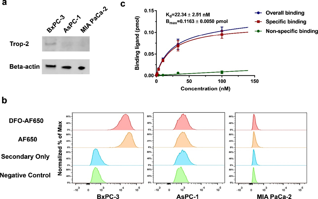
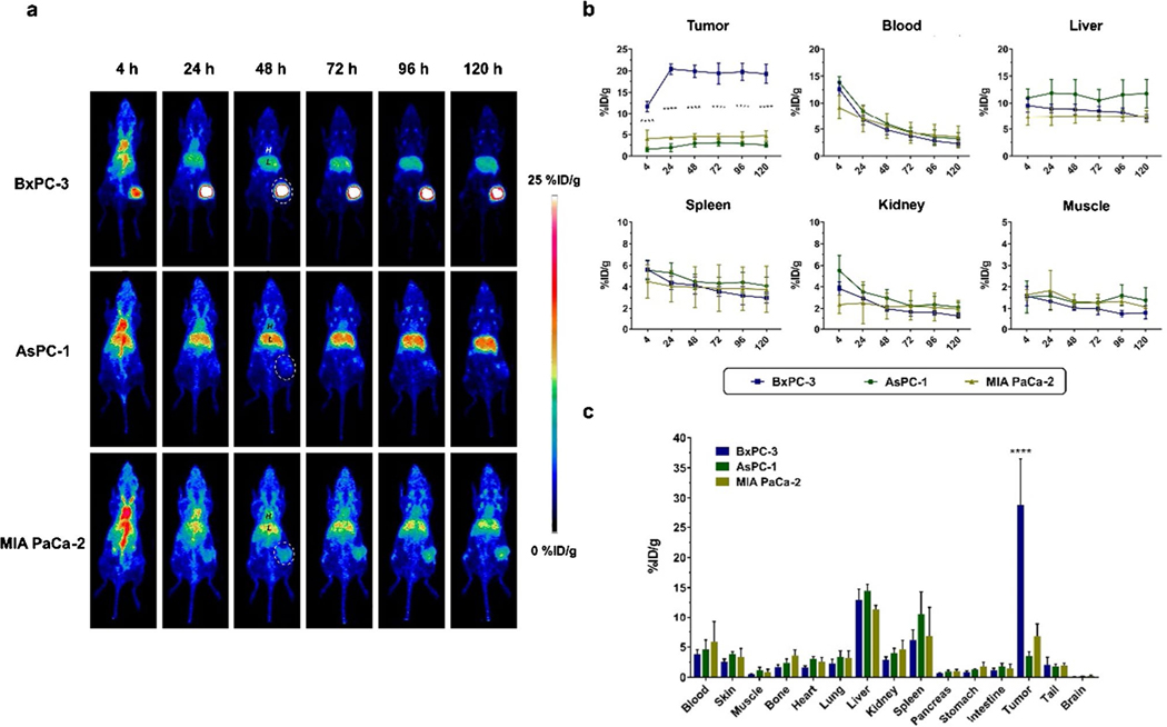
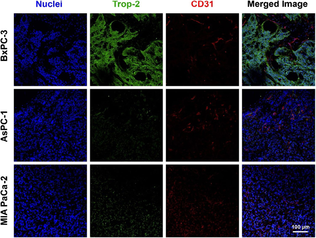
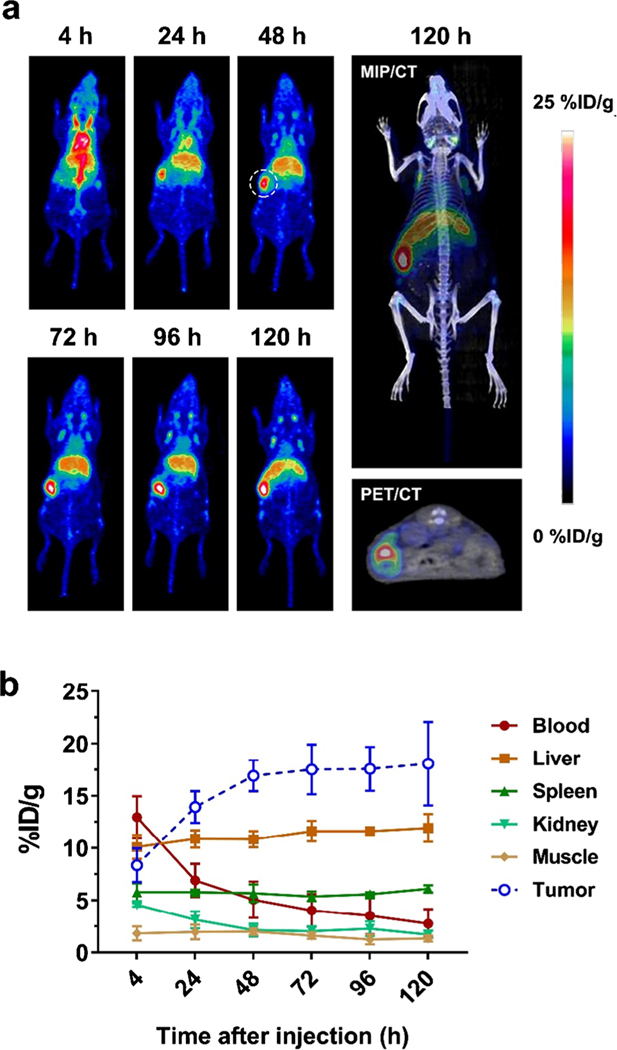
Methods: AF650 was conjugated with desferrioxamine (DFO) and then radiolabeled with 89Zr. Trop-2 expression levels were determined in three pancreatic cancer cell lines (BxPC-3, MIA PaCa-2, and AsPC-1) via western blot, flow cytometry, saturation binding assay, and immunofluorescence staining. The targeting capacity of 89Zr-DFO-AF650 was evaluated in mouse models with subcutaneous xenograft of pancreatic cancers via PET imaging and bio-distribution studies. In addition, a Trop-2-positive orthotopic cancer model was recruited for further validating the targeting specificity of 89Zr-DFO-AF650.

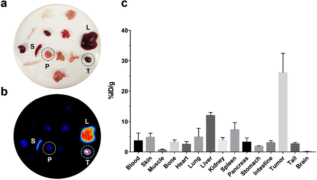
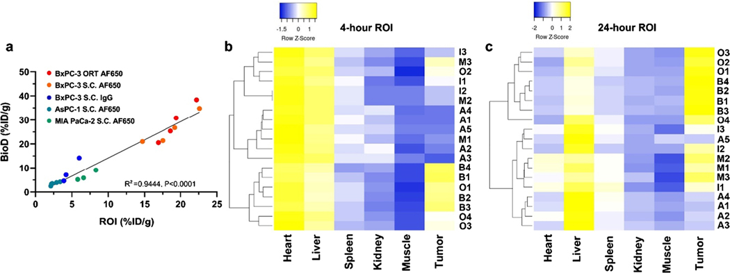
Results: BxPC-3 cells expressed high levels of Trop-2, while AsPC-1 and MIA PaCa-2 cells expressed low levels of Trop-2. Additionally, 89Zr-DFO-AF650 exhibited high specificity to Trop-2 in BxPC-3 cells (Kd = 22.34 ± 2.509 nM). In subcutaneous xenograft models, about 28.8 ± 7.63%ID/g tracer accumulated in the BxPC-3 tumors at 120 h post injection, which was much higher than those reaching MIA PaCa-2 (6.76 ± 2.08%ID/g) and AsPC-1 (3.51 ± 0.69%ID/g) tumors (n = 4). More importantly, 89Zr-DFO-AF650 could efficiently distinguish primary tumors in the orthotopic BxPC-3 cancer model, showing high correlation between PET imaging and bio-distribution and sensitivity.

Conclusions: 89Zr-DFO-AF650 can be effectively used to detect pancreatic cancer via Trop-2-mediated immunoPET in vivo, clearly revealing the great potential of Trop-2-based non-invasive imaging in pancreatic cancer detection and treatment monitoring.

Keywords: Molecular imaging; Monoclonal antibody (mAb); Pancreatic cancer; Positron emission tomography (PET); Trophoblast cell-surface antigen 2 (Trop-2); Zr-89.


Conflict of interest statement

Conflict of interest Weibo Cai is a scientific advisor, stockholder, and grantee of Focus-X Therapeutics, Inc. All other authors declare that they have no conflict of interest.

Figures

Fig. 1
Fig. 1
The binding affinity of AF650 to Trop-2. a The expression level of Trop-2 among three pancreatic cancer cell lines. b The Trop-2 expression among three cancer cell lines via DFO-AF650 or AF650 mediated flow cytometry. c The Trop-2 saturation binding assays of 89Zr-DFO-AF650 on BxPC-3 cells; unlabeled DFO-AF650 was recruited for determining the nonspecific binding (n = 3)
Fig. 2
Fig. 2
ImmunoPET of 89Zr-DFO-AF650 among subcutaneous pancreatic cancer models. a The serial PET maximum intensity projection (MIP) images of 89Zr-DFO-AF650 at 4, 24, 48, 72, 96, and 120 h post injection (n ≥ 3); tumors are indicated by white dash circles, H, blood pool; L, liver. b The curves of PET region of interest (ROI) including the tumor, heart, liver, spleen, kidney, and muscle at different time points. c The bio-distribution of 89Zr-DFO-AF650 among different organs harvested at day 5 (n ≥ 3). **** shows P < 0.0001
Fig. 3
Fig. 3
Trop-2 expression within tumor tissues from pancreatic cancer cells. The immunofluorescence staining of BxPC-3, AsPC-1, and MIA PaCa-2 tumor sections with DAPI (blue), Trop-2 (green), and CD31 (red) antibodies. Scale bar = 100 μm
Fig. 4
Fig. 4
Evaluation of 89Zr-DFO-AF650 in detecting BxPC-3 cancer cells in an orthotopic tumor model via ImmunoPET. a The MIP PET images of 89Zr-DFO-AF650 in mice bearing BxPC-3 orthotopic tumors at different time points, and the representative MIP/CT and PET/CT images at day 5 (n = 4); the tumor site is highlighted by the dashed line. b The time-activity curves of PET ROI among organs from 4 to 120 h after initial tracer administration (n = 4)
Fig. 5
Fig. 5
The high specificity of 89Zr-DFO-AF650 in detecting the orthotopic BxPC-3 tumor. The (a) optical and (b) MIP PET images of tissues harvested from mice injected with 89Zr-DFO-AF650 at day 5 (n = 4); c the bio-distribution of 89Zr-DFO-AF650 tracer among organs collected at day 5 (= 4)
Fig. 6
Fig. 6
Evaluation of 89Zr-DFO-AF650 imaging metrics for Trop-2 immunoPET among pancreatic cancer models. a The correlation between ROI and BioD (%ID/g); ORT, orthotopic model; S.C., subcutaneous model. bc The Z-normalized heat map of 89Zr-DFO-AF650 accumulated values in various organs via specified ROI (%ID/g) at 4 and 24 h post injection; groups including A: AF650 (AsPC-1, S.C.), B: AF650 (BxPC-3, S.C.), I: IgG (BxPC-3, S.C.), M: AF650 (MIA PaCa-2, S.C.), O: AF650 (BxPC-3, ORT)

References

    1. Siegel RL, Miller KD, Jemal A. Cancer statistics, 2020. CA Cancer J Clin. 2020;70:7–30. - PubMed
    1. Vincent A, Herman J, Schulick R, Hruban RH, Goggins M. Pancreatic cancer. Lancet. 2011;378:607–20. - PMC - PubMed
    1. Chen W, Ni D, Rosenkrans ZT, Cao T, Cai W. Smart H2S-triggered/therapeutic system (SHTS)-based nanomedicine. Adv Sci. 2019;6:1901724. - PMC - PubMed
    1. Sun T, Jiang D, Rosenkrans ZT, Ehlerding EB, Ni D, Qi C, et al. A melanin-based natural antioxidant defense nanosystem for theranostic application in acute kidney injury. Adv Funct Mater. 2019;29:1904833. - PMC - PubMed
    1. Wang YXJ, Huang H, Zheng CJ, Xiao BH, Chevallier O, Wang W. Diffusion-weighted MRI of the liver: challenges and some solutions for the quantification of apparent diffusion coefficient and intravoxel incoherent motion. Am J Nucl Med Mol Imaging. 2021;11:107–42. - PMC - PubMed