. 2021 Sep 14;52(1):117.
doi: 10.1186/s13567-021-00987-x.

Acid resistance system CadBA is implicated in acid tolerance and biofilm formation and is identified as a new virulence factor of Edwardsiella tarda

Affiliations

Acid resistance system CadBA is implicated in acid tolerance and biofilm formation and is identified as a new virulence factor of Edwardsiella tarda

Chunmei Du et al. Vet Res. .

Abstract

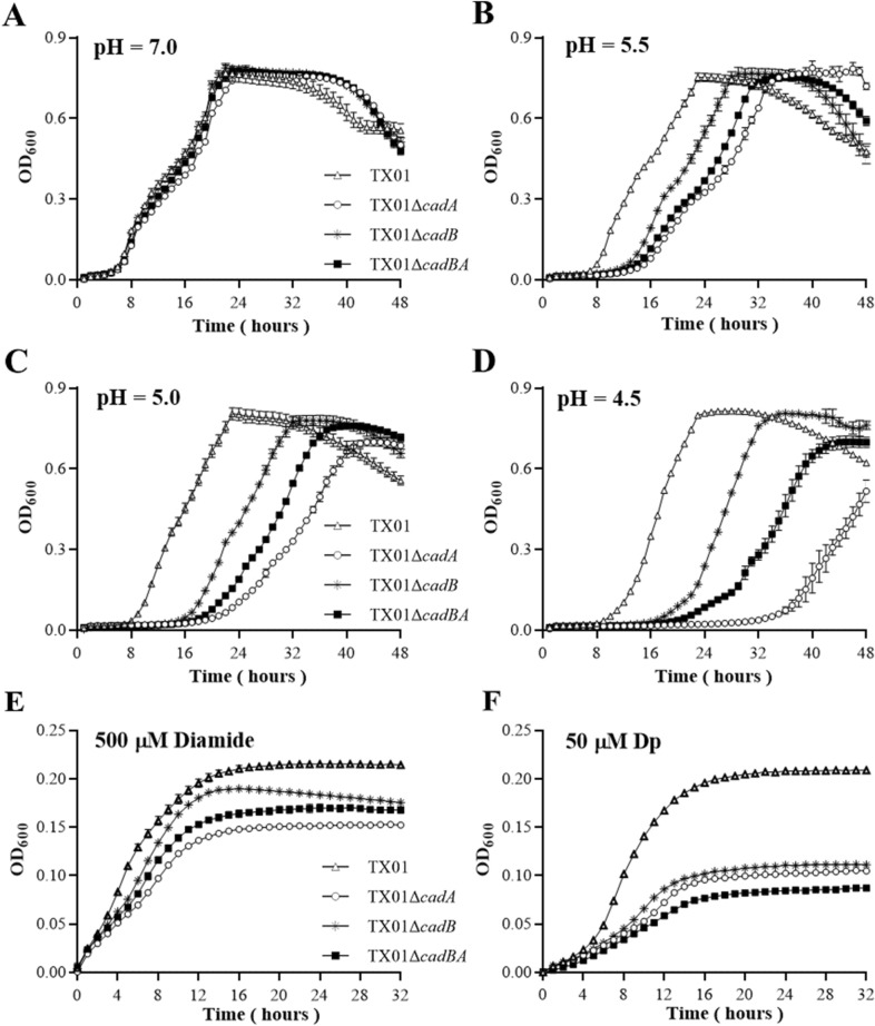
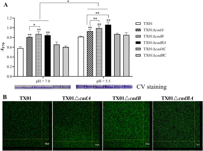
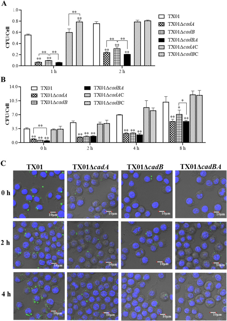
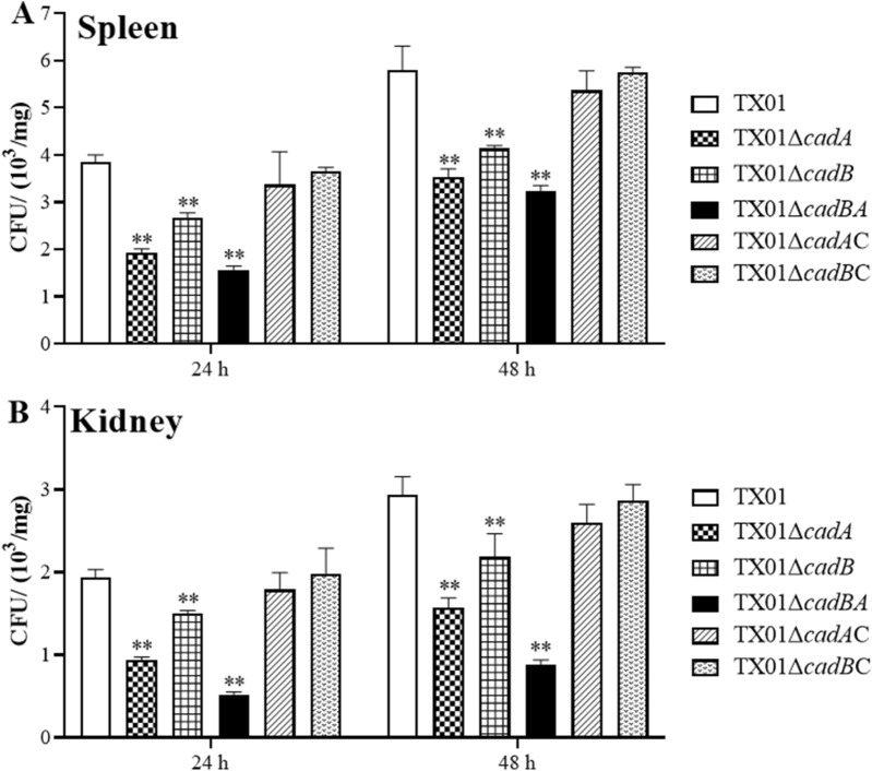
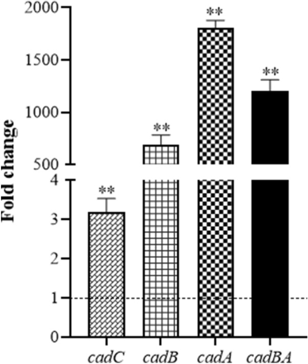
Edwardsiella tarda is a facultative intracellular pathogen in humans and animals. The Gram-negative bacterium is widely considered a potentially important bacterial pathogen. Adaptation to acid stress is important for the transmission of intestinal microbes, so the acid-resistance (AR) system is essential. However, the AR systems of E. tarda are totally unknown. In this study, a lysine-dependent acid resistance (LDAR) system in E. tarda, CadBA, was characterized and identified. CadB is a membrane protein and shares high homology with the lysine/cadaverine antiporter. CadA contains a PLP-binding core domain and a pyridoxal phosphate-binding motif. It shares high homology with lysine decarboxylase. cadB and cadA are co-transcribed under one operon. To study the function of the cadBA operon, isogenic cadA, cadB and cadBA deletion mutant strains TX01ΔcadA, TX01ΔcadB and TX01ΔcadBA were constructed. When cultured under normal conditions, the wild type strain and three mutants exhibited the same growth performance. However, when cultured under acid conditions, the growth of three mutants, especially TX01ΔcadA, were obviously retarded, compared to the wild strain TX01, which indicates the important involvement of the cadBA operon in acid resistance. The deletion of cadB or cadA, especially cadBA, significantly attenuated bacterial activity of lysine decarboxylase, suggesting the vital participation of cadBA operon in lysine metabolism, which is closely related to acid resistance. The mutations of cadBA operon enhanced bacterial biofilm formation, especially under acid conditions. The deletions of the cadBA operon reduced bacterial adhesion and invasion to Hela cells. Consistently, the deficiency of cadBA operon abated bacterial survival and replication in macrophages, and decreased bacterial dissemination in fish tissues. Our results also show that the expression of cadBA operon and regulator cadC were up-regulated upon acid stress, and CadC rigorously regulated the expression of cadBA operon, especially under acid conditions. These findings demonstrate that the AR CadBA system was a requisite for the resistance of E. tarda against acid stress, and played a critical role in bacterial infection of host cells and in host tissues. This is the first study about the acid resistance system of E. tarda and provides new insights into the acid-resistance mechanism and pathogenesis of E. tarda.

Keywords: Edwardsiella tarda; acid resistance; biofilm; cadBA; pathogenicity; regulation.


Conflict of interest statement

The authors have no conflicting commercial or financial interest in publishing this paper.

Figures

Figure 1
Figure 1
Schematic organization and co-transcriptional verification of the cadBA operon in E. tarda.A Schematic organization of the cadBA operon in E. tarda. The cadBA operon consists of cadB and cadA. The cadBA operon is under the control of the predictive promoter Pcad. The upstream gene of the cadBA operon is cadC, which encodes transcriptional regulator. Therefore, the operon was named cadBA. B Verification of cadB and cadA co-transcription. Genomic DNA and total RNA were isolated from overnight cultures of E. tarda. RNA was treated with DNase I and cDNA was synthesized. PCR were conducted with specific primer pair CadBAF/CadBAR using genomic DNA, cDNA, and RNA as the template. PCR products were analyzed by agarose gel electrophoresis.
Figure 2
Figure 2
Growth analysis of E. tarda in different conditions. The wild-type TX01, its isogenic cadA, cadB and cadBA deletion mutant strains TX01ΔcadA, TX01ΔcadB, and TX01ΔcadBA were cultured to the exponential phase. After diluting serially, bacteria were cultured in fresh LB broth with pH adjusted to 7.0, 5.5, 5.0, and 4.5 (AD). Bacteria were added into fresh medium with 500 μM of diamide (E) and 50 μM of 2,2ʹ-dipyridyl (Dp) (F), respectively. Cell density was monitored at 2-h intervals by measuring the OD600. Experiment was performed three times, data are presented as the means ± SEM (N = 3). N, the number of times the experiment was performed.
Figure 3
Figure 3
The activities of lysine decarboxylase in E. tarda.A TX01, TX01ΔcadA, TX01ΔcadB, TX01ΔcadBA, TX01ΔcadAC, and TX01ΔcadBC were cultured in lysine decarboxylase broth with lysine in static condition at 28 ℃ for 10, 20 and 30 h, respectively. A light purple indicates a weak positive result. Purple and yellow indicate the presence and absence of LDC, respectively. B The same strains were cultured in LB broth to an OD600 of 0.5 and normalized to an OD600 of 1.0, buffered at pH 6.8. The activities of lysine decarboxylase were determined as described in the text. The experiment was performed three times, data are presented as the means ± SEM (N = 3). N, the number of times the experiment was performed. **, P < 0.01.
Figure 4
Figure 4
Effects of cadBA mutations on biofilm formation.A Biofilm-forming capacity of E. tarda in normal and acid conditions. TX01, TX01ΔcadA, TX01ΔcadB, TX01ΔcadBA, TX01ΔcadAC, and TX01ΔcadBC were inoculated into LB broth at pH = 7.0 and 5.5, then incubated in polystyrene plates for 24 h. Biofilm formations were determined by measuring the A570 of the final eluates of crystal violet staining. Data are presented as the means ± SEM (N = 3). N, the number of times the experiment was performed. **, P < 0.01; *, P < 0.05. B The viability of biofilm growth of E.tarda was determined by confocal laser scanning microscopy (CLSM). Cells in the biofilms were stained with a BacLight LIVE/DEAD kit to reveal viable (green fluorescence) and non-viable (red fluorescence) bacteria.
Figure 5
Figure 5
Effects of cadBA mutations on motility of E. tarda. TX01, TX01ΔcadA, TX01ΔcadB, and TX01ΔcadBA were cultured in LB medium to an OD600 of 0.5, then aliquots of cell suspensions (1 μL) were inoculated into the center of swimming plates including 0.3% (W/V) agar with pH = 7.0 (A) or pH = 5.5 (B) at 28 °C for 18 h. C, The diameter of the swimming zone from the swimming plate at pH = 5.5. Data are presented as the means ± SEM (N = 3). N, the number of times the experiment was performed. **, P < 0.01; *, P < 0.05.
Figure 6
Figure 6
Effects of cadBA mutations on cellular infection and replication.A Invasion of HeLa cells by E. tarda. HeLa cells were infected with the same dose of E. tarda TX01, TX01ΔcadA, TX01ΔcadB, TX01ΔcadBA, TX01ΔcadAC and TX01ΔcadBC strains for 1 and 2 h, and washed with PBS. Then, HeLa cells were lysed and the CFU were counted. B Replication of E. tarda in macrophages. The murine macrophage cell line RAW264.7 was infected with E. tarda and mutants above mentioned for 2 h, followed by treatment with gentamicin for 2 h to kill extracellular bacteria. After being washed with PBS, the cells were incubated for the time intervals indicated. Then, the cells were lysed and the CFU were counted. Data are the means of three independent experiments and presented as means ± SEM (N = 3). N, the number of times the experiment was performed. **, P < 0.01; *, P < 0.05. C, TX01, TX01ΔcadA, TX01ΔcadB and TX01ΔcadBA containing pGFPuv plasmid were used to infect RAW264.7 cells for 0, 2 and 4 h. DNA was stained blue by DAPI, the cells were observed by confocal microscopy.
Figure 7
Figure 7
Bacterial dissemination in the fish tissues. Tilapia were infected with the same dose of TX01, TX01ΔcadA, TX01ΔcadB, TX01ΔcadBA, TX01ΔcadAC, and TX01ΔcadBC. The recoveries of bacteria in spleen (A) and kidney (B) were determined by plate counting at 24 and 48 h post-infection. Data are presented as means ± SEM (N = 3). N, the number of times the experiment was performed. **, P < 0.01.
Figure 8
Figure 8
The expression of acid resistance genes from TX01 under acidic conditions. The exponential phase of overnight cultures of TX01 were grown in normal LB medium at pH 7.0 and acidic medium at pH 5.5 for 1 h. The relative expression of cadB, cadA, cadBA, and cadC were determined by RT-qPCR. The fold difference derived from the values under acidic conditions compared with the values under normal conditions. Data are presented as the means ± SEM (N = 3). N, the number of times the experiment was performed. **, P < 0.01; *, P < 0.05.
Figure 9
Figure 9
Transcriptional regulation of cadBA by CadC in vitro or in vivo.A Expression profiles of cadBA operon in normal and acidic conditions. E. tarda TX01 and TX01ΔcadC in logarithmic growth phase was culture in normal LB (pH = 7.0) or in acidic LB (pH = 5.5) for 1 h, then the expression of cadB, cadA, and cadBA were examined by RT-qPCR. For convenience of comparison, the expression level of cadBA operon in wild type TX01 set as 1. B The promoter activity of cadBA operon was regulated by CadC in vitro. DH5α/pSC418/pJR21 and DH5α/pSC418/pJR21C were streaked and cultured on an X-gal plate at pH 5.5 in the presence of 5 mM L-lysin. The depth of blue indicates the strength of promoter activity. C The promoter activity of cadBA operon was regulated by CadC in vivo. The wild-type TX01 and TX01ΔcadC mutant carrying the reporter plasmid pJR21-418-lx were cultured to the exponential phase. Then strains were transferred acid LB media (pH = 5.5) supplemented with 5 mM l-lysine and grown for 1 h. The transcriptional regulation of promoter of cadBA was assessed by measuring luciferase activity. Data are the means of three independent experiments and presented as means ± SEM (N = 3). N, the number of times the experiment was performed. **, P < 0.01; *, P < 0.05.

References

    1. Ewing WH, Mcwhorter AC, Escobar MR, Lubin AH. Edwardsiella, a new genus of Enterobacteriaceae based on a new species, E. tarda. Int J Syst Evol Microbiol. 1965;15:33–38.
    1. Abayneh T, Colquhoun DJ, Sørum H. Edwardsiella piscicida sp. nov., a novel species pathogenic to fish. J Appl Microbiol. 2013;114:644–654. doi: 10.1111/jam.12080. - DOI - PubMed
    1. Leung KY, Wang Q, Yang Z, Siame BA. Edwardsiella piscicida: a versatile emerging pathogen of fish. Virulence. 2019;10:555–567. doi: 10.1080/21505594.2019.1621648. - DOI - PMC - PubMed
    1. Griffin MJ, Quiniou SM, Cody T, Tabuchi M, Ware C, Cipriano RC, Mauel MJ, Soto E. Comparative analysis of Edwardsiella isolates from fish in the eastern United States identifies two distinct genetic taxa amongst organisms phenotypically classified as E. tarda. Vet Microbiol. 2013;165:358–372. doi: 10.1016/j.vetmic.2013.03.027. - DOI - PubMed
    1. Wang C, Dong XS, Yang YY, Xu GJ, Wu MM, Yan FJ, Zhang LG, An L, Fu PS, Wang XR, Su YB, Meng QL. Metabolites in the TCA cycle promote resistance to chloramphenicol of Edwardsiella tarda. J Proteome Res. 2021;20:972–981. doi: 10.1021/acs.jproteome.0c00725. - DOI - PubMed

MeSH terms