. 2021 Sep 14;11(1):18194.
doi: 10.1038/s41598-021-97507-w.

The role of DNA-binding and ARNT dimerization on the nucleo-cytoplasmic translocation of the aryl hydrocarbon receptor

Affiliations

The role of DNA-binding and ARNT dimerization on the nucleo-cytoplasmic translocation of the aryl hydrocarbon receptor

Rashad Haidar et al. Sci Rep. .

Abstract

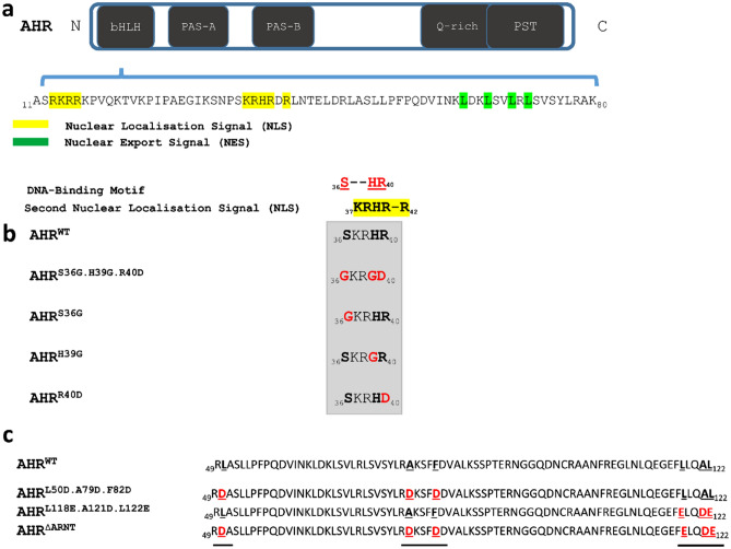
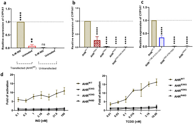
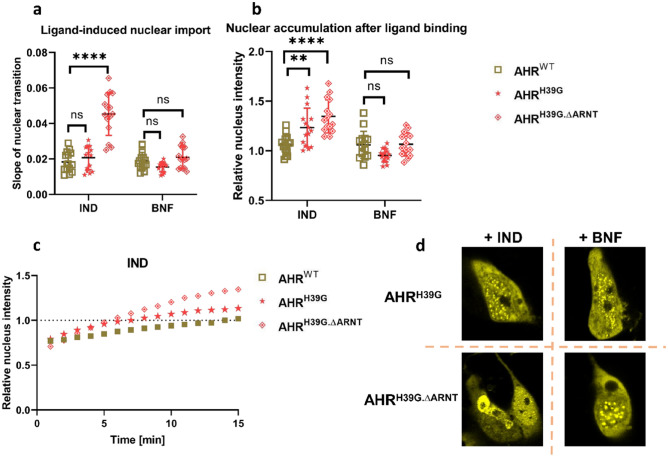
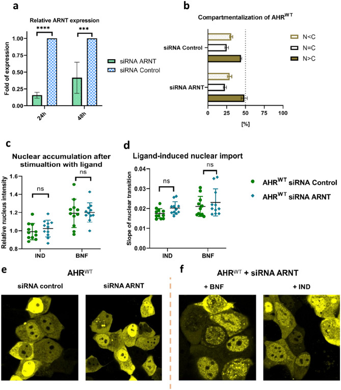
The human aryl hydrocarbon receptor (AHR) is predominantly located in the cytoplasm, while activation depends on its nuclear translocation. Binding to endogenous or xenobiotic ligands terminates the basal nucleo-cytoplasmic shuttling and stabilizes an exclusive nuclear population. The precise mechanisms that facilitate such stable nuclear accumulation remain to be clarified as essential step in the activation cascade. In this study, we have tested whether the sustained nuclear compartmentalization of ligand-bound or basal AHR might further require heterodimerization with the AHR-nuclear translocator (ARNT) and binding to the cognate XRE-motif. Mutagenesis of the DNA-binding motif or of selected individual residues in the ARNT-binding motif did not lead to any variation in AHR's nucleo-cytoplasmic distribution. In response to ligands, all mutants were retained in the nucleus demonstrating that the stable compartmentalization of activated AHR in the nucleus is neither dependent on interactions with DNA, nor ARNT. Knocking down the ARNT gene using small interfering RNA confirmed that ARNT does not play any role in the intracellular trafficking of AHR.


Conflict of interest statement

The authors declare no competing interests.

Figures

Figure 1
Figure 1
Design of AHR mutants to study DNA-binding and ARNT dimerization. (a) Domain architecture of the AHR. The bipartite nuclear localization signal (NLS) and a nuclear export signal (NES) are indicated as amino acid sequence. Graphical representation of mutated amino acids in the DNA-binding motif (b) and ARNT-binding domain (c) of the AHR.
Figure 2
Figure 2
Transcriptional activity of the AHR and AHR mutants. Relative CYP1A1 mRNA level determined by qPCR in MCF-7ΔAHR cells. Values were standardized against HPRT and normalized against AHRWT transfected cells treated with 5 µM indirubin (IND) for three hours. Depicted are the results for a comparison of treatment and AHRWT transfection (a), AHR DNA-binding deficient mutants (b) and AHR mutants deficient for ARNT dimerization (c). (d) Luciferase activity driven by an XRE-promoter after stimulation with increasing concentrations of IND and TCDD for twenty-four hours. (ac) Each bar represents the mean of three biological replicates ± S.D., One way Anova (ns: not significant, ** P < 0.01, **** P < 0.0001). (d) Presented are the means of three biological replicates + /- S.D.
Figure 3
Figure 3
The DNA-binding motif determines the intracellular distribution of the AHR through the overlap with the second nuclear localization signal (NLS). (a) Distribution of the EYFP-tagged AHR and AHR mutants in HepG2 cells are shown. More than 300 cells were randomly selected and classified according to the given distribution pattern. Data represents mean ± S.D from three biological replicates. (b,e) Slopes of nuclear transition the AHRWT and AHRR40D (Arg → Asp) after treatment with 10 µM indirubin (IND), 10 µM β-naphthoflavone (BNF) or 200 nM leptomycin B (LMB). (c,f) Nuclear accumulation of the AHRWT and AHRR40D 15 min or 30 min after treatment with IND, BNF or LMB, respectively. Individual values and the mean ± S.D. of 15 (b,c) or 10 (e,f) cells are presented (two way ANOVA, Dunnett's multiple comparisons test, *** p < 0.001). (d,g) Representative measurements of time-lapse experiments after stimulation with IND (d) or LMB (g) of indicated AHR variants in HepG2 cells. Relative nuclear intensity denotes the intensity of the nucleus against the intensity of the entire cell. (h) Representative images of HepG2 cells transfected with AHRWT or AHRR40D before and after 15 min of treatment with IND, BNF, and LMB.
Figure 4
Figure 4
Forming the AHR-ARNT heterodimer and the active binding to the XRE is not decisive for stable ligand-dependent nuclear retention. (a) Slopes of nuclear transition of the AHRWT, AHRH39G and AHRH39G.∆ARNT after treatment with10µM indirubin (IND) or 10 µM β-naphthoflavone (BNF). (b) Nuclear accumulation of the AHRWT, AHRH39G and AHRH39G.∆ARNT 15 min after treatment with IND or BNF, respectively. Data display the individual values and the mean + /- S.D. of 15 cells (two way ANOVA, Dunnett's multiple comparisons test, ** p < 0.01, *** p < 0.001). (c) Representative measurements of time-lapse experiments after stimulation with IND of indicated AHR variants in HepG2 cells. Relative nucleus intensity denotes the intensity of the nucleus against the intensity of the entire cell. (d) Snapshots of HepG2 cells 15 min after stimulating with IND or BNF.
Figure 5
Figure 5
Down-regulation of ARNT is not critical for the AHR nucleo-cytoplasmic translocation in MCF-7 cells. (a) Relative ARNT mRNA level determined by qPCR in MCF-7ΔAHR cells after transfection with siRNA against ARNT and control siRNA, respectively. Values were standardized against HPRT and normalized against control siRNA transfected cells. Each bar depicts the mean of three biological replicates ± S.D. (two way ANOVA, Dunnett's multiple comparisons test, *** p < 0.001,**** P < 0.0001). (b) Intracellular distribution pattern of EYFP-tagged AHRWT in MCF-7 ΔAHR cells are shown. At least 300 transfected cells are randomly selected and sorted according to the following classification: N > C predominantly nuclear; N = C equal distribution; N < C predominantly cytoplasmic. Each bar represents the mean + /- S.D. of three biological experiments. Depicted are relative nuclear intensity (c) and slope (d) of time-lapse measurements after stimulation with 10 µM indirubin (IND) or 10 µM β-naphthoflavone (BNF) in control siRNA and ARNT siRNA transfected MCF-7ΔAHR cells. The presented data contains individual values and the mean + /- S.D. of 11 cells. (e) Snapshots of MCF-7 ΔAHR cells before and after knocking down the ARNT gene. (f) Snapshots of the MCF-7 cells after stimulation with BNF and IND.

References

    1. Beischlag TV. The aryl hydrocarbon receptor complex and the control of gene expression. Crit. Rev. Eukaryot. Gene Exp. 2008;18(3):207–250. doi: 10.1615/CritRevEukarGeneExpr.v18.i3.20. - DOI - PMC - PubMed
    1. Chen G, Bunce NJ. Interaction between halogenated aromatic compounds in the Ah receptor signal transduction pathway. Environ. Toxicol. 2004;19(5):480–489. doi: 10.1002/tox.20053. - DOI - PubMed
    1. Luch A. Nature and nurture—lessons from chemical carcinogenesis. Nat. Rev. Cancer. 2005;5(2):113–125. doi: 10.1038/nrc1546. - DOI - PubMed
    1. Tkachenko A, et al. Nuclear transport of the human aryl hydrocarbon receptor and subsequent gene induction relies on its residue histidine 291. Arch. Toxicol. 2018;92(3):1151–1160. doi: 10.1007/s00204-017-2129-0. - DOI - PubMed
    1. Puga A, Ma C, Marlowe JL. The aryl hydrocarbon receptor cross-talks with multiple signal transduction pathways. Biochem. Pharmacol. 2009;77(4):713–722. doi: 10.1016/j.bcp.2008.08.031. - DOI - PMC - PubMed

Publication types

MeSH terms

Substances