. 2021 Sep 14;11(1):18227.
doi: 10.1038/s41598-021-97771-w.

Longitudinal choriocapillaris changes in the presence of reticular pseudodrusen

Affiliations

Longitudinal choriocapillaris changes in the presence of reticular pseudodrusen

Christoph R Clemens et al. Sci Rep. .

Abstract

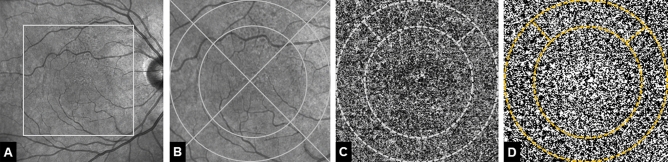
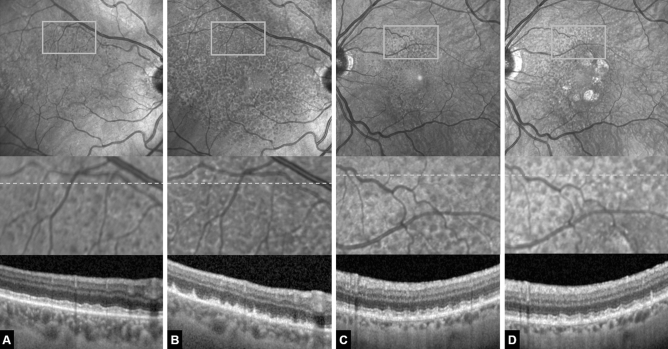
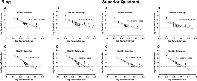
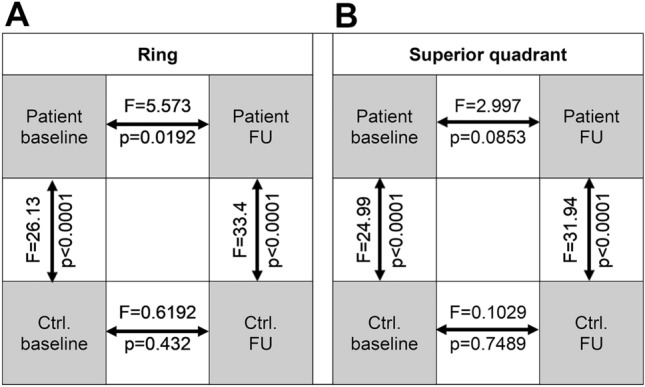
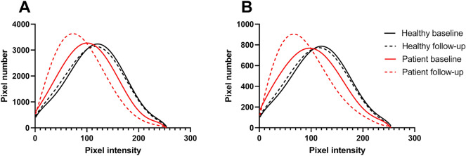
To determine longitudinal changes in choriocapillaris (CC) measures in eyes with reticular pseudodrusen (RPD) using optical coherence tomography angiography (OCTA). In this observational prospective study, 20 patients with exclusively RPD and no other alteration due to age-related macular degeneration were included. Eight RPD patients were re-examined at 5-year follow-up. Multimodal imaging was performed at baseline and at 5-year follow-up. OCTA CC images were analyzed for number, size and total area of flow deficits (FD), mean signal intensity, signal intensity standard deviation and kurtosis of signal intensity distribution in the ring area between a circle of 4 mm diameter and a circle of 6 mm diameter and in the superior ring quadrant. Area affected by RPD increased from 19.36 ± 8.39 mm2 at baseline to 37.77 ± 9.03 mm2 at 5-year follow-up. At baseline, percent of CC FD area was greater in RPD eyes (quadrant: p < 0.001; ring: p < 0.001) compared to controls. Besides, RPD eyes revealed a lower mean intensity signal (quadrant: p < 0.001; ring: p < 0.001). Evaluation of CC parameters suggested significant group × time interaction effects for CC FD (p = 0.04) and mean intensity signal (p = 0.004), in that RPD eyes presented increased CC FD and decreased mean intensity signal at follow-up. OCTA CC decorrelation signal further decreases in RPD patients over 5 years in both RPD-affected and RPD-unaffected macular areas.


Conflict of interest statement

Christoph Clemens and Florian Alten are consultants to Bayer, Nicole Eter is a consultant to Heidelberg Engineering, Novartis, Bayer, Sanofi Aventis, Allergan, Bausch and Lomb. Jost Lauermann and Boris Schmitz: None.

Figures

Figure 1
Figure 1
Representative patient with reticular pseudodrusen (RPD) at baseline. (A) Near-infrared confocal scanning laser opththalmoscopy (cSLO) image showing hyporeflective dots located superiorly to the fovea. Rectangle marks the area of 6 × 6 mm magnified in (B) Overlaid grid with two circles of 4 mm and 6 mm diameter as well as two lines dividing the area in four quadrants. (C) 6 × 6 mm optical coherence tomography angiography choriocapillaris image. Choriocapillaris layer segmentation extending from 10 μm above to 30 μm beneath the Bruch’s membrane. Dotted line marks the 4-mm-diameter, 6-mm-diameter circles centered on the fovea and the superior quadrant. (D) Image after automatic local thresholding done with the Phansalkar method, flow deficits are shown in white.
Figure 2
Figure 2
(A, B) Example of a 70-year-old female reticular pseudodrusen (RPD) patient that developed a significant increase in RPD-affected area over the 5-year follow-up yet no late-stage form of age-related macular degeneration. (A) Baseline. Above near-infrared confocal scanning laser ophthalmoscopy (cSLO) image showing hyporeflective dots superiorly located to the fovea. Rectangle marks the area magnified below. Grey dotted line marks the location of the optical coherence tomography (OCT) scan below. (B) Same eye after 5 years. Note the increase in RPD-affected area and the increase in lesion density visible both in the cSLO en-face image above as well as in the magnified image. The OCT scans reveal early RPD stages at baseline and predominantly stage-three RPD lesions at 5-year follow-up. Also note signs of incomplete outer retinal atrophy in (B). There is detritus on the intact RPE monolayer with remnants of the ellipsoid zone but no hypertransmission. Individual RPD lesions are harder to discern. (C, D) Example of a 71-year-old female RPD patient that developed geographic atrophy over the 5-year follow-up. (C) Baseline. (D) Same eye after 5 years. Note the perifoveal patches of geographic atrophy and a moderate increase in RPD-affected area and lesion density. The OCT scan appears rather similar to the baseline image without any signs of outer retinal atrophy.
Figure 3
Figure 3
Baseline to follow-up changes of flow deficits shown by log–log plot with logarithmic binning in the ring area (above) and in the superior ring quadrant (below). Individual data points are shown with linear regression and 95% confidence Interval. The respective equations are given with slope (m-values) and Y-intercept (c-value).
Figure 4
Figure 4
Comparison matrix of flow deficit linear regression slopes for (A) the ring area and (B) the superior ring quadrant. Arrows indicate respective comparisons with p and F values (derived from F tests). Ctrl control, FU follow-up.
Figure 5
Figure 5
Plots illustrating the distribution of frequencies of grey level intensities in (A) the ring area and (B) the superior ring quadrant of patients and controls at baseline and 5-year follow-up. Mean pixel intensity distribution in healthy controls (n = 8) was identical at baseline and follow-up. Mean pixel intensity distribution in patients (n = 8) was non-identical at baseline and follow-up and different to patients’ distribution at both time points. Individual data points were fitted by sixth order polynomial nonlinear regression.

References

    1. Finger RP, Wu Z, Luu CD, et al. Reticular pseudodrusen: A risk factor for geographic atrophy in fellow eyes of individuals with unilateral choroidal neovascularization. Ophthalmology. 2014;121:1252–1256. doi: 10.1016/j.ophtha.2013.12.034. - DOI - PMC - PubMed
    1. Dutheil C, Le Goff M, Cougnard-Grégoire A, et al. Incidence and risk factors of reticular pseudodrusen using multimodal imaging. JAMA Ophthalmol. 2020;138:467–477. doi: 10.1001/jamaophthalmol.2020.0266. - DOI - PMC - PubMed
    1. Zweifel SA, Spaide RF, Curcio CA, Malek G, Imamura Y. Reticular pseudodrusen are subretinal drusenoid deposits. Ophthalmology. 2010;117:303–312. doi: 10.1016/j.ophtha.2009.07.014. - DOI - PubMed
    1. Greferath U, Guymer RH, Vessey KA, Brassington K, Fletcher EL. Correlation of histologic features with in vivo imaging of reticular pseudodrusen. Ophthalmology. 2016;123:1320–1331. doi: 10.1016/j.ophtha.2016.02.009. - DOI - PubMed
    1. Chen L, Messinger JD, Kar D, Duncan JL, Curcio CA. Biometrics, impact, and significance of basal linear deposit and subretinal drusenoid deposit in age-related macular degeneration. Invest. Ophthalmol. Vis. Sci. 2021;62:33. doi: 10.1167/iovs.62.1.33. - DOI - PMC - PubMed

Publication types