. 2021 Sep 7:15:3783-3808.
doi: 10.2147/DDDT.S328837. eCollection 2021.

An Integrated Analysis of Network Pharmacology and Experimental Validation to Reveal the Mechanism of Chinese Medicine Formula Naotaifang in Treating Cerebral Ischemia-Reperfusion Injury

Affiliations

An Integrated Analysis of Network Pharmacology and Experimental Validation to Reveal the Mechanism of Chinese Medicine Formula Naotaifang in Treating Cerebral Ischemia-Reperfusion Injury

Tong Yang et al. Drug Des Devel Ther. .

Abstract

Background: Cerebral ischemia-reperfusion injury (CIRI) is a crucial factor leading to a poor prognosis for ischemic stroke patients. As a novel Chinese medicine formula, Naotaifang (NTF) was proven to exhibit a neuroprotective effect against ischemic stroke, clinically, and to alleviate CIRI in animals. However, the mechanisms underlying the beneficial effect have not been fully elucidated.

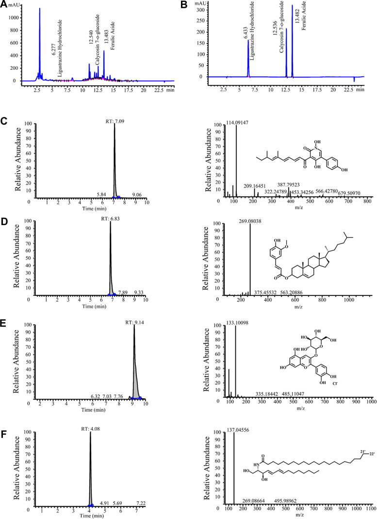
Methods: In this study, we combined a network pharmacology approach and an in vivo experiment to explore the specific effects and underlying mechanisms of NTF in the treatment of ischemia-reperfusion injury. A research strategy based on network pharmacology, combining target prediction, network construction, gene ontology (GO), Kyoto Encyclopedia of Genes and Genomes (KEGG) pathway enrichment analysis, and molecular docking was used to predict the targets of NTF in treating the ischemic stroke and CIRI. On the other hand, we used HPLC and HRMS to identify biologically active components of NTF. Middle cerebral artery occlusion models in rats were utilized to evaluate the effect and the underlying mechanisms of NTF against CIRI after ischemic stroke.

Results: Network pharmacology analysis revealed 43 potential targets and 14 signaling pathways for the treatment of NTF against CIRI after ischemic stroke. Functional enrichment analysis showed that a STAT3/PI3K/AKT signaling pathway serves as the target for in vivo experimental study validation. The results of animal experiments showed that NTF significantly alleviated CIRI by decreasing neurological score, infarct volume, numbers of apoptotic neuronal cells, increasing density of dendritic spines and survival of neurons. Furthermore, NTF could increase the expression of p-STAT3, PI3K, p-AKT. In addition, the detection of apoptosis-related factors showed that the NTF could raise the expression of Bcl-2 and reduce the expression of Bax.

Conclusion: This network pharmacological and experimental study indicated that NTF, as a therapeutic candidate for the management of CIRI following ischemic stroke, may exert a protective effect through the STAT3/PI3K/AKT signaling pathway.

Keywords: STAT3/PI3K/AKT signaling pathway; cerebral ischemia-reperfusion injury; molecular docking; network pharmacology; stroke.


Conflict of interest statement

The authors declare that there are no conflicts of interest for this work.

Figures

Figure 1
Figure 1
The flowchart of network pharmacology and molecular docking-based strategy for deciphering the underlying mechanisms of NTF on the treatment of IS/CIRI.
Figure 2
Figure 2
Chemical ingredients analysis of NTF. Representative ingredients of NTF (A) and standards (B). The chromatogram, mass spectrum and structural formula of four compounds: (C) Compound 1; (D) Compound 2; (E) Compound 3; (F) Compound 4.
Figure 3
Figure 3
Prediction results of network pharmacology of NTF on ischemic stroke and CIRI. (A) The herb-compound network of NTF. Green nodes represent the herbs of NTF, orange nodes represent the central compounds of NTF, and blue nodes represent the other active compounds of NTF. (B) The venn diagram of the targets both in ischemic stroke targets whose color is yellow and NTF targets whose color is blue. (C) The results of topological screening for the PPI network (p>0.99). (D) The graphical interactions network of the 43 key targets. The node color changes from yellow to red reflect the degree centrality changes from low to high. (E) The herb-compound-target network.
Figure 4
Figure 4
Functional analysis of NTF. GO enrichment of related genes of (A) biological process, (B) molecular functions, (C) cellular components and (D) KEGG pathway enrichment analysis for 43 key targets.
Figure 5
Figure 5
3D and 2D interaction diagrams of hyrcanoside (Aa), bassianin (Bb) and cholesteryl ferulate (Cc) in the active site of PIK3CA (PDB ID 4TUU).
Figure 6
Figure 6
Pretreatment of NTF 7 days prior to CIRI reduced infarct volume and ameliorated neurological deficit in rats 24 h after reperfusion. (A) Representative TTC-stained photos of the cerebral infarct coronal sections. (B) Percentage of infarct volume. (C) Neurological scores. All data were presented as mean ± SD. ##p<0.01 versus sham group; **p<0.01, *p<0.05 versus model group, respectively.
Figure 7
Figure 7
Pretreatment of NTF 7 days prior to CIRI increased the spines density in rats 24 h after reperfusion. (A) Representative Golgi-stained photos of the dendritic spines in ischemic cortex sections magnified 400 times. (B) Spines density in ischemic cortex. (C) Representative Golgi-stained photos of the dendritic spines in ischemic hippocampus magnified 400 times. (D) Spines density in ischemic hippocampus. All data were presented as mean ± SD. ##p<0.01 versus sham group; *p<0.05 versus model group, respectively.
Figure 8
Figure 8
Pretreatment of NTF 7 days prior to CIRI increased the neurons activity of the ischemic cortex and hippocampus in rats 24 h after reperfusion. (A) Representative images magnified 200 times and 400 times in ischemic cortex sections. (B) Quantitatively analyzed Nissl bodies in each group after 24 h of reperfusion in ischemic cortex. (C) Representative images magnified 200 times and 400 times in ischemic hippocampus sections. (D) Quantitatively analyzed Nissl bodies in each group after 24 h of reperfusion in ischemic hippocampus. All data were presented as mean ± SD (n = 18 rats in each group). ##p<0.01 versus sham group; **p<0.01, *p<0.05 versus model group, respectively.
Figure 9
Figure 9
Pretreatment of NTF 7 days prior to CIRI reduced the neuronal apoptosis of the ischemic cortex and hippocampus in rats 24 h after reperfusion. (A) Representative images magnified 200 times and 400 times in ischemic cortex sections. (B) The apoptotic index indicates the percentage of TUNEL-positive cells in each group after 24 h of reperfusion in ischemic cortex. (C) Representative images magnified 200 times and 400 times in ischemic hippocampus sections. (D) The apoptotic index indicates the percentage of TUNEL-positive cells in each group after 24 h of reperfusion in ischemic hippocampus. All data were presented as mean ± SD. ##p<0.01 versus sham group; **p<0.01 versus model group, respectively.
Figure 10
Figure 10
Effects of NTF 7 days prior to CIRI on expression of p-STAT3 of the ischemic cortex and hippocampus in rats 24 h after reperfusion. (A) Representative images magnified 400 times in ischemic cortex sections examined with specific antibody against p-STAT3 (red); nuclei were stained with DAPI (blue). (B) The numbers of p-STAT3 positive cells in each group after 24 h of reperfusion in ischemic cortex. (C) Representative images magnified 400 times in ischemic hippocampus sections. (D) The numbers of p-STAT3 positive cells in each group after 24 h of reperfusion in ischemic hippocampus. All data were presented as mean ± SD. ##p<0.01 versus sham group; **p<0.01 versus model group, respectively.
Figure 11
Figure 11
Effects of NTF 7 days prior to CIRI on expression of PI3K of the ischemic cortex and hippocampus in rats 24 h after reperfusion. (A) Representative images magnified 400 times in ischemic cortex sections examined with specific antibody against PI3K (red); nuclei were stained with DAPI (blue). (B) The numbers of PI3K positive cells in each group after 24 h of reperfusion in ischemic cortex. (C) Representative images magnified 400 times in ischemic hippocampus sections. (D) The numbers of PI3K positive cells in each group after 24 h of reperfusion in ischemic hippocampus. All data were presented as mean ± SD. ##p<0.01 versus sham group; *p<0.05 versus model group, respectively.
Figure 12
Figure 12
Effects of NTF extracts 7 days prior to CIRI on expression of p-AKT of the ischemic cortex and hippocampus in rats 24 h after reperfusion. (A) Representative images magnified 400 times in ischemic cortex sections examined with specific antibody against p-AKT (red); nuclei were stained with DAPI (blue). (B) The numbers of p-AKT positive cells in each group after 24 h of reperfusion in ischemic cortex. (C) Representative images magnified 400 times in ischemic hippocampus sections. (D) The numbers of p-AKT positive cells in each group after 24 h of reperfusion in ischemic hippocampus. All data were presented as mean ± SD. ##p<0.01 versus sham group; **p<0.01, *p<0.05 versus model group, respectively.
Figure 13
Figure 13
Effects of NTF 7 days prior to CIRI on expression of Bcl-2 of the ischemic cortex and hippocampus in rats 24 h after reperfusion. (A) Representative images magnified 400 times in ischemic cortex sections examined with specific antibody against Bcl-2 (red); nuclei were stained with DAPI (blue). (B) The numbers of Bcl-2 positive cells in each group after 24 h of reperfusion in ischemic cortex. (C) Representative images magnified 400 times in ischemic hippocampus sections. (D) The numbers of Bcl-2 positive cells in each group after 24 h of reperfusion in ischemic hippocampus. All data were presented as mean ± SD. ##p<0.01 versus sham group; *p<0.05 versus model group, respectively.
Figure 14
Figure 14
Effects of NTF 7 days prior to CIRI on expression of Bax of the ischemic cortex and hippocampus in rats 24 h after reperfusion. (A) Representative images magnified 400 times in ischemic cortex sections examined with specific antibody against Bax (red); nuclei were stained with DAPI (blue). (B) The numbers of Bax positive cells in each group after 24 h of reperfusion in ischemic cortex. (C) Representative images magnified 400 times in ischemic hippocampus sections. (D) The numbers of Bax positive cells in each group after 24 h of reperfusion in ischemic hippocampus. All data were presented as mean ± SD. ##p<0.01 versus sham group; **p<0.01, *p<0.05 versus model group, respectively.
Figure 15
Figure 15
Effects of NTF 7 days prior to CIRI on expression of Bcl-2/Bax, p-STAT3, PI3K and p-AKT/AKT of the ischemic cortex and hippocampus in rats 24 h after reperfusion by Western blotting. (A) The protein band of Bcl-2/Bax, p-STAT3, PI3K and p-AKT/AKT and corresponding GAPDH in ischemic cortex and corresponding bar graphs. (B) The protein band of Bcl-2/Bax, p-STAT3, PI3K and p-AKT/AKT and corresponding GAPDH in ischemic hippocampus and corresponding bar graphs. All data were presented as mean ± SD. ##p<0.01, #p<0.05 versus sham group; *p<0.05 versus model group, respectively.
Figure 16
Figure 16
Proposed scheme of NTF suppressed cerebral ischemia-reperfusion induced apoptosis via STAT3/PI3K/AKT signaling pathway. Our results suggest that NTF pretreatment upregulated p-STAT3, PI3K, p-AKT proteins expression, increased the Bcl-2/Bax ratio, alleviated neurological evaluation, reduced infarct volume and apoptosis-positive cells, increased the density of dendritic spines and survival of neurons in cortex and hippocampus sections.

References

    1. Wu SM, Wu B, Liu M, et al. Stroke in China: advances and challenges in epidemiology, prevention, and management. Lancet Neurol. 2019;18(4):394–405. - PubMed
    1. Benjamin EJ, Muntner P, Alonso A, et al. Heart disease and stroke statistics-2019 update: a report from the American Heart Association. Circulation. 2019;139(10):e56–e528. - PubMed
    1. Liu S, Levine SR, Winn HR. Targeting ischemic penumbra part I: from pathophysiology to therapeutic strategy. J Exp Stroke Transl Med. 2010;3(1):47–55. - PMC - PubMed
    1. Rabinstein AA. Treatment of acute ischemic stroke. Contin Lifelong Learn Neurol. 2017;23(1):62–81. - PubMed
    1. Lahr MMH, Luijckx GJ, Vroomen PCAJ, Van Der Zee DJ, Buskens E. Proportion of patients treated with thrombolysis in a centralized versus a decentralized acute stroke care setting. Stroke. 2012;43(5):1336–1340. - PubMed

Publication types

MeSH terms