Review
. 2021 Aug;11(8):2585-2604.
doi: 10.1016/j.apsb.2021.04.010. Epub 2021 Apr 29.

Delivery strategies of amphotericin B for invasive fungal infections

Affiliations
Review

Delivery strategies of amphotericin B for invasive fungal infections

Xiaochun Wang et al. Acta Pharm Sin B. 2021 Aug.

Abstract

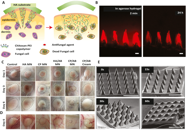
Invasive fungal infections (IFIs) represent a growing public concern for clinicians to manage in many medical settings, with substantial associated morbidities and mortalities. Among many current therapeutic options for the treatment of IFIs, amphotericin B (AmB) is the most frequently used drug. AmB is considered as a first-line drug in the clinic that has strong antifungal activity and less resistance. In this review, we summarized the most promising research efforts on nanocarriers for AmB delivery and highlighted their efficacy and safety for treating IFIs. We have also discussed the mechanism of actions of AmB, rationale for treating IFIs, and recent advances in formulating AmB for clinical use. Finally, this review discusses some practical considerations and provides recommendations for future studies in applying AmB for combating IFIs.

Keywords: ABCD, AmB colloidal dispersion; AIDS, acquired immunodeficiency syndrome; AP, antisolvent precipitation; ARDS, acute respiratory distress syndrome; AmB, amphotericin B; AmB-GCPQ, AmB-encapsulated N-palmitoyl-N-methyl-N,N-dimethyl-N,N,N-trimethyl-6-O-glycol-chitosan nanoparticles; AmB-IONP, AmB-loaded iron oxide nanoparticles; AmB-PM, AmB-polymeric micelles; AmB-SD, AmB sodium deoxycholate; AmBd, AmB deoxycholate; Amphotericin B; Aspergillus fumigatus, A. fumigatus; BBB, blood‒brain barrier; BCS, biopharmaceutics classification system; BDDE, butanediol diglycidyl ether; BSA, bovine serum albumin; BUN, blood urea nitrogen; C. Albicans, Candida Albicans; CFU, colony-forming unit; CLSM, confocal laser scanning microscope; CMC, carboxymethylated l-carrageenan; CP, chitosan-polyethylenimine; CS, chitosan; Conjugates; DDS, drug delivery systems; DMPC, dimyristoyl phosphatidyl choline; DMPG, dimyristoyl phosphatidylglycerole; DMSA, dimercaptosuccinic acid; Drug delivery; GNPs, gelatin nanoparticles; HPH, high-pressure homogenization; HPMC, hydroxypropyl methylcellulose; ICV, intensive care unit; IFIs, invasive fungal infections; Invasive fungal infections; L-AmB, liposomal AmB; LNA, linolenic acid; MAA, methacrylic acid; MFC, minimum fungicidal concentrations; MIC, minimum inhibitory concentration; MN, microneedles; MOP, microneedle ocular patch; MPEG-PCL, monomethoxy poly(ethylene glycol)-poly(epsilon-caprolactone); NEs, nanoemulsions; NLC, nanostructured lipid carriers; NPs, nanoparticles; Nanoparticles; P-407, poloxamer-407; PAM, polyacrylamide; PCL, polycaprolactone; PDA, poly(glycolic acid); PDLLA, poly(d,l-lactic acid); PDLLGA, poly(d,l-lactic-co-glycolic acid); PEG, poly(ethylene glycol); PEG-DSPE, PEG-lipid poly(ethylene glycol)-distearoylphosphatidylethanolamine; PEG-PBC, phenylboronic acid-functionalized polycarbonate/PEG; PEG-PUC, urea-functionalized polycarbonate/PEG; PGA-PPA, poly(l-lysine-b-l-phenylalanine) and poly(l-glutamic acid-b-l-phenylalanine); PLA, poly(lactic acid); PLGA, polyvinyl alcohol poly(lactic-co-glycolic acid); PLGA-PLH-PEG, PLGA-b-poly(l-histidine)-b-poly(ethylene glycol); PMMA, poly(methyl methacrylate); POR, porphyran; PVA, poly(vinyl alcohol); PVP, polyvinylpyrrolidone; Poor water-solubility; RBCs, red blood cells; RES, reticuloendothelial system; ROS, reactive oxygen species; SEM, scanning electron microscope; SL-AmB, sophorolipid-AmB; SLNs, solid lipid nanoparticles; Topical administration; Toxicity; γ-CD, γ-cyclodextrin; γ-PGA, γ-poly(gamma-glutamic acid.


Conflict of interest statement

All the authors declare that this article content has no conflict of interest.

Figures

Image 1
Graphical abstract
Figure 1
Figure 1
Toxicity, spectrum, and available marketed AmB formulations.
Figure 2
Figure 2
Chemical structure (A), and mechanism of AmB on fungal cell (B). AmB induces its action by various means. On cell membrane: (1) It binds with membrane ergosterol and prompt ergosterol sequestration and (2) disrupt membrane stability. Inside the cell: It induces an oxidative burst as a production of prooxidant and (3) increases reactive oxygen species inside the cell. (4) The production of oxygen species (ROS) is produced during respiratory chain reaction, suggested that AmB influences the mitochondrial function, and (5) encourages oxidative burst. Also, (5) the AmB can alter the cellular ionic homeostasis and permit leakage of monovalent Na+, K+, H+, and Cl ions. Thus, the increased level of these free-radicals and imbalance of ions produces multiple deleterious effects on the vital components of cell (membrane, mitochondria, proteins, and DNA) resulting in the cell death.
Figure 3
Figure 3
(A) In vitro hemolytic activity of AmB, Fungizone, Ambisome, CS blank, CS-AmB, and various CS-POR based NPs. The data is represented as mean ± SD (n = 3). Adapted with permission from Ref. . Copyright © 2014 Saurabh Bhatia et al. (B) Inhibition zone of AmB, Fungizone and NPs III against A. fumigatus and AmB, NPs III and Fungizone against C. albicans. Adapted with permission from Ref. . Copyright © 2015 Elsevier. (C) Biodistribution and liver, lung, spleen targeting of orally administered AmB-NPs. Adapted with permission from Ref. . Copyright © 2015 American Chemical Society. (D) Preparation characterization, biocompatibility and antifungal efficacy of Levan-based hydrogels for dermal delivery. Adapted with permission from Ref. . Copyright © 2020 Elsevier.
Figure 4
Figure 4
(A) LIVE/DEAD staining of HEK293 and IMR-90 cells after exposure to AmB-PEG and free AmB for 24 h (Live cells stained green and dead cells stained red). The AmB-PEG did not induce cell death at 139 and 277 μmol/L concentration in HEK293 and IMR-90 cells, respectively. While the molar ratio of AmB to PEG did not have any visible effect on cell toxicity. The experiment was performed twice with three independently prepared AmB-PEG formulations. Adapted with permission from Ref. . Copyright © 2016 Tan et al. (B) Preparation of AmB-loaded micelles using PEG-PBC and PEG-PUC. Adapted with permission from Ref. . Copyright © 2016 Elsevier. (C) Schematic presentation of preparation and action of AmB loaded linolenic acid-modified methoxy poly (ethylene glycol)–oligochitosan conjugate micelles on C. albicans membrane. Adapted with permission from Ref. . Copyright © 2019 Elsevier. (D) Reformulation of AmB-SD with PEG-DSPE micelles in saline and a proposed fungus–membrane interaction of monomeric AmB. Adapted with permission from Ref. . Copyright © 2016 Springer Nature. (E) Preparation of AmB loaded micelle-hydrogel composite by genipin crosslinking method and photograph of AmB loaded micelle-hydrogel composite before and after gelation and as a self-standing gel. (F) AmB release kinetics from micelle-hydrogel at different zeta-potentials. Adapted with permission from Ref. . Copyright © 2017 Royal Society of Chemistry.
Figure 5
Figure 5
(A) Synthesis of triazole based nonionic surfactant (TBNIS). (B) AFM image of AB-loaded TBNIS based vesicles. (C) Plasma drug concentration of AmB-loaded vesicles and plan AmB solution after oral administration at the dose of 6 mg/kg body weight (n = 6, mean ± SE). Adapted with permission from Ref. . Copyright © 2020 Elsevier. (D) Photographic examination of AmB-PEG-NLC prepared by using different molecular weight PEG (from left to right: 1 K, 2 K, 5 K, 10 K, 20 K and the first row shows Day 1 and second row indicates Day 30) and (E) Concentration of AmB (μg/g) in rabbit ocular tissues after hourly instillation of 50 μL (150 μg AmB) AmB-PEG2K-NLC and AmBisome over 6 h, indicates that the AmB concentration for AmBisome and PEGylated NLC were statistically insignificant (P < 0.05). Adapted with permission from Ref. . Copyright © 2019 Elsevier. (F) Schematic presentation of dual alginate-lipid nanocarriers for oral delivery of AmB. Adapted with permission from Ref. . Copyright © 2020 Elsevier. (G) Formulation of ionic-liquid-in-water nanoemulsions for systemic delivery of AmB. (H) Microscope image of zebrafish exposed to the solution of ionic liquid ([DC-7][2NTf2]) indicate complete biocompatibility, and (I) in vitro AmB release from ionic liquid nanoemulsion over 201 h. Adapted with permission from Ref. . Copyright © 2020 American Chemical Society.
Figure 6
Figure 6
(A) The AmB-loaded nanosuspension prepared by high-pressure homogenization method and an antisolvent precipitation method. The relative bioavailability of AmB-HPH was higher than AmB-AP in male SD rats. Adapted with permission from Ref. . Copyright © 2018 Elsevier. (B) Preparation of AmB nanosuspension loaded thermogel for vaginal delivery. The AmB was released in a sustained manner, leading to improved antifungal activity. Adapted with permission from Ref. . Copyright © 2018 Taylor & Francis. (C) CLSM images of cellular uptake of BSA coated AmB loaded iron oxide NPs (AmB-IONP) in C. glabrata. (D) CLSM images of cellular uptake of BSA coated AmB loaded iron oxide NPs (AmB-IONP) in C. albicans. Both (C) and (D) indicated the endolysosomal localization with colocalization of AMB-IONP (green) and lysotracker in red and time-dependent uptake was maximum at 4 h (X = 60 and scale bar was 1 μm for 0.5 h and 2 μm for 4 h. Adapted with permission from Ref. . Copyright © 2020 MDPI.
Figure 7
Figure 7
(A) Schematic description of AB loaded antimicrobial microneedle patch (CP/AB MN) prepared by chitosan-polyethylenimine copolymer (CP) for subcutaneous fungal infection. HA, hyaluronic acid. (B) CLSM images of Cy5 released from CP microneedles in agarose gel at 2 min and after 24 h, respectively (scale bar = 200 μm). (C) Photograph of fungal infection sites after applying different microneedle patches on Days 1, 3, 6, and (D) images taken to observe skin underneath infection on Day 6. Adapted with permission from Ref. . Copyright © 2019 John Wiley & Sons. (E) The SEM images showed dissolution of AmB loaded microneedle ocular patch prepared by dissolvable polymeric matrix (polyvinyl alcohol and polyvinyl pyrrolidone) after insertion in excised cornea at 0, 15, 30 60 s (scale bar = 1 mm). Adapted with permission from Ref. . Copyright © 2019 Elsevier.

References

    1. Mellinghoff S.C., Panse J., Alakel N., Behre G., Buchheidt D., Christopeit M. Primary prophylaxis of invasive fungal infections in patients with haematological malignancies: 2017 update of the recommendations of the Infectious Diseases Working Party (AGIHO) of the German Society for Haematology and Medical Oncology (DGHO) Ann Hematol. 2018;97:197–207. - PMC - PubMed
    1. Kriengkauykiat J., Ito J.I., Dadwal S.S. Epidemiology and treatment approaches in management of invasive fungal infections. Clin Epidemiol. 2011;3:175–191. - PMC - PubMed
    1. Pappas P.G., Lionakis M.S., Arendrup M.C., Ostrosky-Zeichner L., Kullberg B.J. Invasive candidiasis. Nat Rev Dis Primers. 2018;4:18026. - PubMed
    1. Denning D.W., Kneale M., Sobel J.D., Rautemaa-Richardson R. Global burden of recurrent vulvovaginal candidiasis: a systematic review. Lancet Infect Dis. 2018;18:e339–e347. - PubMed
    1. Enoch D., Ludlam H., Brown N. Invasive fungal infections: a review of epidemiology and management options. J Med Microbiol. 2006;55:809–818. - PubMed

LinkOut - more resources