. 2021 Dec;36(12):2317-2328.
doi: 10.1002/jbmr.4443. Epub 2021 Oct 4.

Conductive Hearing Loss in the Hyp Mouse Model of X-Linked Hypophosphatemia Is Accompanied by Hypomineralization of the Auditory Ossicles

Affiliations

Conductive Hearing Loss in the Hyp Mouse Model of X-Linked Hypophosphatemia Is Accompanied by Hypomineralization of the Auditory Ossicles

Maximilian M Delsmann et al. J Bone Miner Res. 2021 Dec.

Abstract

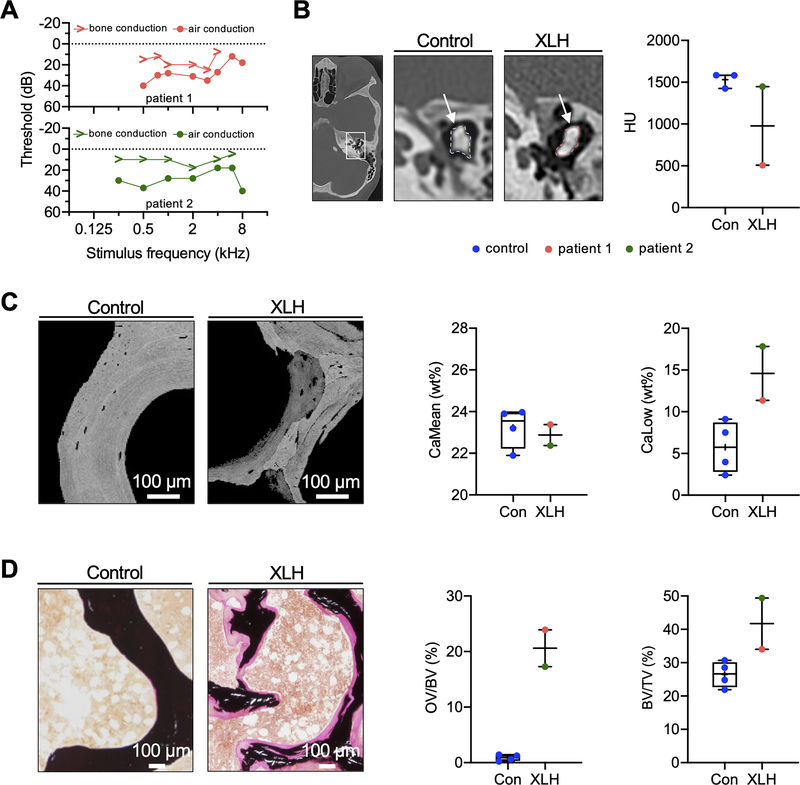
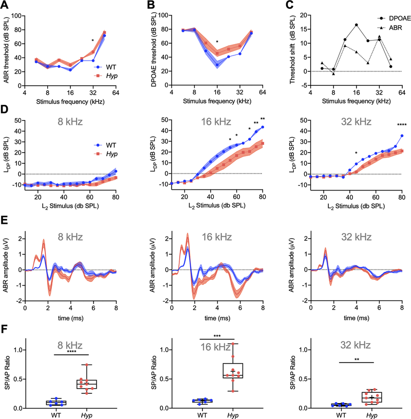
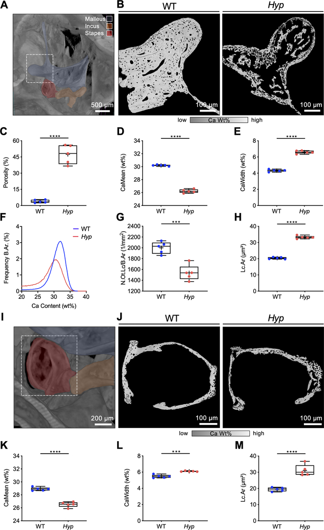
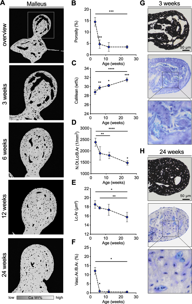
X-linked hypophosphatemia (XLH) is a hereditary musculoskeletal disorder caused by loss-of-function mutations in the PHEX gene. In XLH, increased circulating fibroblast growth factor 23 (FGF23) levels cause renal phosphate wasting and low concentrations of 1,25-dihydroxyvitamin D, leading to an early clinical manifestation of rickets. Importantly, hearing loss is commonly observed in XLH patients. We present here data from two XLH patients with marked conductive hearing loss. To decipher the underlying pathophysiology of hearing loss in XLH, we utilized the Hyp mouse model of XLH and measured auditory brain stem responses (ABRs) and distortion product otoacoustic emissions (DPOAEs) to functionally assess hearing. As evidenced by the increased ABR/DPOAE threshold shifts in the mid-frequency range, these measurements indicated a predominantly conductive hearing loss in Hyp mice compared to wild-type (WT) mice. Therefore, we carried out an in-depth histomorphometric and scanning electron microscopic analysis of the auditory ossicles. Quantitative backscattered electron imaging (qBEI) indicated a severe hypomineralization of the ossicles in Hyp mice, evidenced by lower calcium content (CaMean) and higher void volume (ie, porosity) compared to WT mice. Histologically, voids correlated with unmineralized bone (ie, osteoid), and the osteoid volume per bone volume (OV/BV) was markedly higher in Hyp mice than WT mice. The density of osteocyte lacunae was lower in Hyp mice than in WT mice, whereas osteocyte lacunae were enlarged. Taken together, our findings highlight the importance of ossicular mineralization for hearing conduction and point toward the potential benefit of improving mineralization to prevent hearing loss in XLH. © 2021 The Authors. Journal of Bone and Mineral Research published by Wiley Periodicals LLC on behalf of American Society for Bone and Mineral Research (ASBMR).

Keywords: ANIMAL MODELS; BONE MATRIX; CELL/TISSUE SIGNALING - ENDOCRINE PATHWAYS; GENETIC ANIMAL MODELS; MATRIX MINERALIZATION; PTH/Vit D/FGF23.


Conflict of interest statement

Disclosures: All authors state that they have no conflict of interest and nothing to disclose.

Figures

Fig. 1.
Fig. 1.. Conductive hearing loss and severe osteomalacia in patients with XLH.
(A) Pure-tone threshold audiometry of a 27-year-old (patient 1) and a 32-year-old (patient 2) woman with XLH. While both sides were equally affected, the representative measurements of the right ears are shown. The sound frequency (in kHz) is shown on the horizontal axis of the audiogram, while the sound intensity (in dB) is recorded on the vertical axis. The thresholds of bone and air conduction are displayed as arrowheads and circles, respectively. Results revealed higher air conduction thresholds indicating conductive hearing loss. (B) Left: CCT images showing representative auditory ossicles of a control and XLH patient. White arrows point to the measured region of interest. Right: Quantification of average hounsfield units (HU) in two XLH patients and controls. (C) Representative qBEI images of a transiliac crest biopsy of the XLH patients and of healthy age-matched controls. Bone mineral density distribution analysis showed reduced CaMean and increased fraction of CaLow in XLH. (D) Representative von Kossa-van Gieson stained histological images, showing increased OV/BV and BV/TV of the XLH patient compared to age-matched controls.
Fig. 2.
Fig. 2.. Hyp mice exhibit increased ABR and DPOAE thresholds, shifts in DPOAE I/O functions consistent with conductive hearing loss.
(A) ABR and (B) DPOAEs were measured in 6-week-old male WT (blue curve) and Hyp (red curve) mice from mixed litters. (C) Threshold shifts between WT and Hyp mice were similar in ABRs and DPOAEs, except for the mid-frequency region spanning 16.0 – 22.65 kHz where DPOAE shifts (solid line) were about twice as large as ABR shifts (dashed line). (D) DPOAE I/O-functions at 8, 16 and 32 kHz. (E, F) SP/AP ratios at 8, 16 and 32 kHz are increased in Hyp mice (red curve/column) compared to WT mice (blue curve/column). *p<0.05, **p< 0.01, ***p<0.001, ****p<0.0001 (2-way ANOVA with Sidak’s test for multiple comparisons and two-tailed unpaired t-test).
Fig. 3.
Fig. 3.. Irregular and reduced mineralization of the malleus and stapes evaluated by qBEI in WT and Hyp mice.
(A) A representative three-dimensional reconstruction of μCT-imaging of a murine WT middle ear (24-week-old male mice) with virtual fenestration of the temporal bone and pseudocolors highlighting the anatomical localization of the ossicles. (B-H) qBEI analysis of the malleus. (B) Representative qBEI images of the malleus of WT (left panel) and Hyp mice (right panel). (C) Analysis of the porosity, (D) mean calcium content (CaMean) and (E) width of the distribution of mineralization levels (CaWidth) in WT and Hyp mice. (F) Bone mineral density distribution (BMDD) histograms of the mineralization of the mallei of WT (blue curve) and Hyp mice (red curve). (G, H) Quantification of the number (N.Ot.Lc/B.Ar) and mean area of osteocyte lacunae (Lc.Ar) in Hyp mice compared to WT. (I) μCT-image of the middle ear with focus on the stapes marked in red. (J) Representative qBEI images of the stapes in WT (left panel) and Hyp mice (right panel). (K) Quantification of the mean calcium content (CaMean) in Hyp mice compared to WT, (L) mineralization heterogeneity (CaWidth) and (M) the mean osteocyte lacunar area (Lc.Ar). ***p<0.001, ****p<0.0001 (two-tailed unpaired t-test).
Fig. 4.
Fig. 4.. Undecalcified histology identifies osteoid accumulation in the malleus and signs of mild endolymphatic hydrops in the cochlea in Hyp mice.
(A) Representative histological images of toluidine blue stained orbicular apophysis of the malleus of 24-week-old Hyp and WT mice (upper panel) and von Kossa-van Gieson staining (lower panel). (B) Evaluation of bone volume fraction (BV/TV; upper panel), osteoid volume per bone volume (OV/BV; lower panel) in Hyp mice compared to WT. (C) Representative cochlear cross-sections from 6-week-old male WT (left panel) and Hyp mice (right panel) in hematoxylin and eosin (H&E) staining. The otic capsule in Hyp mice shows osteoidosis (green arrows) and a bulging Reissner’s membrane (RM, red arrow). (D) Endolymphatic hydrops (ELH) in the basal turn classified into the severity grades (23). ELH was not observed in WT animals whereas grade 1 ELH was detected in five out of seven analyzed Hyp animals. **p<0.01 (two-tailed unpaired t-test and Mann-Whitney-U-test).
Fig. 5.
Fig. 5.. Postnatal development of the ossicles in WT mice.
(A) qBEI images of the orbicular apophysis of the malleus at selected timepoints. (B, C) Quantification of the osseous porosity, CaMean and CaPeak. (D, E) Analysis of the number of osteocyte lacunae per bone (N.Ot.Lc/B.Ar) and the mean osteocyte lacunar area (Lc.Ar). (F) Quantification of the vascular area per bone area (Vasc.Ar/B.Ar). (G, H) Histological images (von Kossa-van Gieson and toluidine blue staining) of the malleus at the age of 3 weeks and 24 weeks indicating the reduction of porosity with age in WT mice. *p<0.05, **p<0.01, ***p<0.001, ****p<0.0001 (two-tailed paired t-test).

References

    1. Beck-Nielsen SS, Brock-Jacobsen B, Gram J, Brixen K, Jensen TK. Incidence and prevalence of nutritional and hereditary rickets in southern Denmark. Eur J Endocrinol. 2009;160(3):491–7. - PubMed
    1. Liu S, Guo R, Simpson LG, Xiao ZS, Burnham CE, Quarles LD. Regulation of fibroblastic growth factor 23 expression but not degradation by PHEX. J Biol Chem. 2003;278(39):37419–26. - PubMed
    1. Murali SK, Andrukhova O, Clinkenbeard EL, White KE, Erben RG. Excessive Osteocytic Fgf23 Secretion Contributes to Pyrophosphate Accumulation and Mineralization Defect in Hyp Mice. PLoS Biol. 2016;14(4):e1002427. - PMC - PubMed
    1. Haffner D, Emma F, Eastwood DM, Duplan MB, Bacchetta J, Schnabel D, et al. Clinical practice recommendations for the diagnosis and management of X-linked hypophosphataemia. Nat Rev Nephrol. 2019;15(7):435–55. - PMC - PubMed
    1. Lane NE, Parimi N, Corr M, Yao W, Cauley JA, Nielson CM, et al. Association of serum fibroblast growth factor 23 (FGF23) and incident fractures in older men: the Osteoporotic Fractures in Men (MrOS) study. J Bone Miner Res. 2013;28(11):2325–32. - PMC - PubMed

Publication types

MeSH terms

Substances