Review
. 2021 Dec 15;85(4):e0009121.
doi: 10.1128/MMBR.00091-21. Epub 2021 Sep 15.

DNA Repair in Staphylococcus aureus

Affiliations
Review

DNA Repair in Staphylococcus aureus

Kam Pou Ha et al. Microbiol Mol Biol Rev. .

Abstract

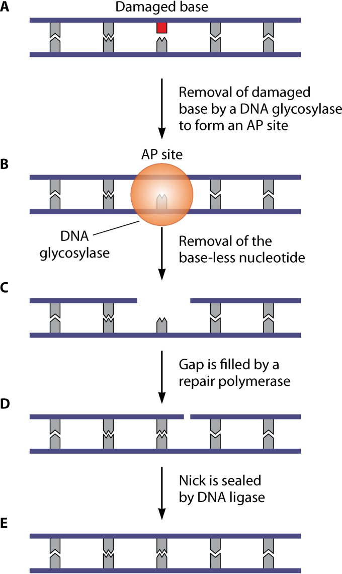
Staphylococcus aureus is a common cause of both superficial and invasive infections of humans and animals. Despite a potent host response and apparently appropriate antibiotic therapy, staphylococcal infections frequently become chronic or recurrent, demonstrating a remarkable ability of S. aureus to withstand the hostile host environment. There is growing evidence that staphylococcal DNA repair makes important contributions to the survival of the pathogen in host tissues, as well as promoting the emergence of mutants that resist host defenses and antibiotics. While much of what we know about DNA repair in S. aureus is inferred from studies with model organisms, the roles of specific repair mechanisms in infection are becoming clear and differences with Bacillus subtilis and Escherichia coli have been identified. Furthermore, there is growing interest in staphylococcal DNA repair as a target for novel therapeutics that sensitize the pathogen to host defenses and antibiotics. In this review, we discuss what is known about staphylococcal DNA repair and its role in infection, examine how repair in S. aureus is similar to, or differs from, repair in well-characterized model organisms, and assess the potential of staphylococcal DNA repair as a novel therapeutic target.

Keywords: DNA damage; DNA repair; SOS; Staphylococcus aureus; antibiotic resistance; bacteriophage; neutrophil.


Figures

FIG 1
FIG 1
Model for activation of the SOS response in S. aureus. (A) In this model, the SOS response is induced by a double-stranded DNA break (DSB), but SOS induction can occur via other mechanisms that result in a ssDNA being formed during DNA repair or replication of the damaged DNA template. (B) The DSB undergoes end processing to produce ssDNA, carried out by the RecBCD or AddAB (also known as RexAB) complexes. (C) The RecA protein forms filaments on the ssDNA, leading to RecA activation. (D) Activated RecA interacts with the LexA repressor, activating its latent protease activity. (E) This results in autocleavage of LexA, inactivating the LexA repressor and leading to derepression of the SOS genes.
FIG 2
FIG 2
Model for base excision repair (BER) in S. aureus. (A and B) BER is initiated by specific DNA glycosylases that recognize and remove damaged bases (shown in red) (A), generating abasic or apurinic/apyrimidinic sites (AP sites) (B). (C) Next, the 5′ and 3′ ends of the AP sites are nicked by AP endonucleases and AP lyases, followed by processing by an exonuclease or deoxyribophosphodiesterase (dRpase) to remove the baseless nucleotide. (D) This leaves a gap, which is filled by a repair polymerase (such as Pol I). (E) The remaining nick is sealed by DNA ligase.
FIG 3
FIG 3
Model for the nucleotide excision repair (NER) pathway in S. aureus. (A) The NER pathway repairs bulky helix-distorting DNA lesions, such as thymine dimers and DNA cross-links. (B) In this pathway, damaged DNA is recognized by the UvrAB complex. (C and D) This leads to UvrA dissociation to enable formation of the UvrBC complex (C), which removes approximately 10 to 15 nucleotides surrounding the lesion (D). This removal is facilitated by PcrA, which enables release of the nucleotide segment. (E and F) The resultant gap is filled by Pol I (E), with the nick sealed by DNA ligase (F).
FIG 4
FIG 4
Model for DSB repair by homologous recombination in S. aureus. (A and B) When an active replication fork encounters a single-strand nick (A), this produces a double-strand break (DSB) and the replication fork collapses (B). (C) The DSB is processed by the RexAB complex, generating a 3′-ssDNA overhang. (D) RecOR is recruited to load the RecA recombinase onto the ssDNA region, with RecF increasing the loading efficiency. (E) RecA pairs with the homologous DNA sequence and mediates strand invasion, producing a D-loop structure. This is aided by the accessory proteins RecX and RarA. (F) DNA polymerase extends the 3′ end of the filament to form a stable four-stranded DNA structure called a Holliday junction, which is moved along the DNA by RuvAB or RecG. (G) In the final step, this junction is cleaved by RecU, and replication restarts.
FIG 5
FIG 5
Model for processing of DNA ends by S. aureus RexAB or E. coli RecBCD. (A and B) DNA damage can lead to a double-strand break (DSB) (A), which can be lethal if not repaired. RexAB or RecBCD binds to the broken end and unwinds the DNA (B). A Chi (crossover hot spot instigator) site is denoted in red. (C) The enzyme translocates along the DNA, degrading both DNA strands. Degradation is performed by the RecB and RecD subunits in RecBCD, or both subunits in RexAB. (D and E) When a Chi site is encountered in the 3′ strand, this induces changes that attenuate degradation of the 3′–5′ nuclease (D), resulting in a 3′ overhang (E). (F) The RecA protein is loaded onto the 3′ overhang to form a filament (green circles) for the next step of the recombinational repair pathway.
FIG 6
FIG 6
Structural homology of S. aureus RexAB to B. subtilis AddAB. Predicted structural models of S. aureus RexA and RexB superimposed onto AddA and AddB subunits of the B. subtilis AddAB crystal structure. Ribbon representation of S. aureus and B. subtilis proteins individually and superimposed. Individual protein structures are colored from blue to red from the N terminus to the C terminus. When superimposed, S. aureus proteins are shown in blue, and B. subtilis proteins are shown in orange. The predicted three-dimensional models were generated by Phyre2 and protein structures visualized using PyMOL. PDB 3U4Q was used for the B. subtilis AddAB structure.

References

    1. Friedberg EC, Wood RD, Walker GC, Schultz RA, Siede W, Ellenberger T. 2006. DNA repair and mutagenesis. American Society for Microbiology Press, Washington, DC.
    1. Rosen R, Buchinger S, Pfänder R, Pedhazur R, Reifferscheid G, Belkin S. 2016. SOS gene induction and possible mutagenic effects of freeze-drying in Escherichia coli and Salmonella Typhimurium. Appl Microbiol Biotechnol 100:9255–9264. doi: 10.1007/s00253-016-7751-x. - DOI - PubMed
    1. Maeda T, Horinouchi T, Sakata N, Sakai A, Furusawa C. 2019. High-throughput identification of the sensitivities of an Escherichia coli ΔrecA mutant strain to various chemical compounds. J Antibiot (Tokyo) 72:566–573. doi: 10.1038/s41429-019-0160-5. - DOI - PubMed
    1. Imlay JA. 2003. Pathways of oxidative damage. Annu Rev Microbiol 57:395–418. doi: 10.1146/annurev.micro.57.030502.090938. - DOI - PubMed
    1. Chapman JR, Taylor MRG, Boulton SJ. 2012. Playing the end game: DNA double-strand break repair pathway choice. Mol Cell 47:497–510. doi: 10.1016/j.molcel.2012.07.029. - DOI - PubMed

Publication types

Substances

LinkOut - more resources