. 2021 Oct 31;9(2):e0100021.
doi: 10.1128/Spectrum.01000-21. Epub 2021 Sep 15.

Grass Carp Reovirus VP56 Allies VP4, Recruits, Blocks, and Degrades RIG-I to More Effectively Attenuate IFN Responses and Facilitate Viral Evasion

Affiliations

Grass Carp Reovirus VP56 Allies VP4, Recruits, Blocks, and Degrades RIG-I to More Effectively Attenuate IFN Responses and Facilitate Viral Evasion

Hang Su et al. Microbiol Spectr. .

Abstract

Grass carp reovirus (GCRV), the most virulent aquareovirus, causes epidemic hemorrhagic disease and tremendous economic loss in freshwater aquaculture industry. VP56, a putative fibrin inlaying the outer surface of GCRV-II and GCRV-III, is involved in cell attachment. In the present study, we found that VP56 localizes at the early endosome, lysosome, and endoplasmic reticulum, recruits the cytoplasmic viral RNA sensor retinoic acid-inducible gene I (RIG-I) and binds to it. The interaction between VP56 and RIG-I was detected by endogenous coimmunoprecipitation (co-IP), glutathione S-transferase (GST) pulldown, and subsequent liquid chromatography-tandem mass spectrometry (LC-MS/MS) and was then confirmed by traditional co-IPs and a novel far-red mNeptune-based bimolecular fluorescence complementation system. VP56 binds to the helicase domain of RIG-I. VP56 enhances K48-linked ubiquitination of RIG-I to degrade it by the proteasomal pathway. Thus, VP56 impedes the initial immune function of RIG-I by dual mechanisms (blockade and degradation) and attenuates signaling from RIG-I recognizing viral RNA, subsequently weakening downstream signaling transduction and interferon (IFN) responses. Accordingly, host antiviral effectors are reduced, and cytopathic effects are increased. These findings were corroborated by RNA sequencing (RNA-seq) and VP56 knockdown. Finally, we found that VP56 and the major outer capsid protein VP4 bind together in the cytosol to enhance the degradation of RIG-I and more efficiently facilitate viral replication. Collectively, the results indicated that VP56 allies VP4, recruits, blocks, and degrades RIG-I, thereby attenuating IFNs and antiviral effectors to facilitate viral evasion more effectively. This study reveals a virus attacking target and an escaping strategy from host antiviral immunity for GCRV and will help understand mechanisms of infection of reoviruses. IMPORTANCE Grass carp reovirus (GCRV) fibrin VP56 and major outer capsid protein VP4 inlay and locate on the outer surface of GCRV-II and GCRV-III, which causes tremendous loss in grass carp and black carp industries. Fibrin is involved in cell attachment and plays an important role in reovirus infection. The present study identified the interaction proteins of VP56 and found that VP56 and VP4 bind to the different domains of the viral RNA sensor retinoic acid-inducible gene I (RIG-I) in grass carp to block RIG-I sensing of viral RNA and induce RIG-I degradation by the proteasomal pathway to attenuate signaling transduction, thereby suppressing interferons (IFNs) and antiviral effectors, facilitating viral replication. VP56 and VP4 bind together in the cytosol to more efficiently facilitate viral evasion. This study reveals a virus attacking a target and an escaping strategy from host antiviral immunity for GCRV and will be helpful in understanding the mechanisms of infection of reoviruses.

Keywords: RIG-I; fibrin VP56; grass carp reovirus; host-virus protein interaction; interferon; major outer capsid protein VP4; viral evasion.


Figures

FIG 1
FIG 1
Schematic diagram of endogenous co-IP and GST pulldown. (A) Purified recombinant protein GST-VP56 was checked by SDS-PAGE. (B) Workflow on exploring interaction proteins with VP56 by endogenous co-IP and GST pulldown. Left, detecting VP56 interacting proteins via co-IP and LC-MS/MS. CIK cells were infected with GCRV for 24 h, and co-IP with GST monoclonal Ab, GST-VP56 purified polyclonal Ab, or negative serum (NS) was performed. LC-MS/MS analysis was performed on a Q Exactive mass spectrometer. Right, detecting VP56 interacting proteins via GST pulldown and LC-MS/MS. CIK proteins were exacted from CIK cells and incubated with purified recombinant GST or GST-VP56 protein. Pulldown was performed with GST resin, and the elution was examined by LC-MS/MS on a Q Exactive mass spectrometer. Candidate interaction proteins were obtained by analyzing these LC-MS/MS results. Triplicate independent experiments were performed with three biological replicates.
FIG 2
FIG 2
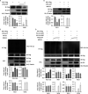
Verification of the interaction between VP56 and the helicase domain of RIG-I. (A) Verification of the interaction between VP56 and RIG-I by traditional co-IPs. Top, FHM cells were cotransfected with VP56-GFP/vector and RIG-I-HA for 48 h. Co-IP was performed with anti-HA monoclonal Ab and mouse IgG (control) and immunoblotting with the corresponding Abs. Bottom, FHM cells were cotransfected with RIG-I-HA/vector and VP56-Flag for 48 h. Co-IP was performed with anti-Flag monoclonal Ab and mouse IgG (control) and immunoblotting with the corresponding Abs. IB, immunoblot; WCL, whole-cell lysate. (B) Imaging of the VP56-RIG-I interaction by far-red mNeptune-based BiFC. Corresponding vectors were transfected alone or cotransfected into CIK cells under normal conditions. In the BiFC system, the fluorescence of the mNeptune channel was red, and the nucleus was stained with DAPI. Images were acquired using confocal microscopy under a 40× lens objective. Appearance of red fluorescence represents the positive observation. BiFC experiments were repeated in triplicate, and the images were selected and cropped to show the positive results. (C) VP56 interacts with the RIG-I-DExD/H helicase domain. FHM cells were cotransfected with 4 μg of VP56-GFP and 4 μg of RIG-I-CARD-HA or RIG-I-helicase-HA or RIG-I-RD-HA for 24 h in 10-cm2 dishes. Co-IP was performed using HA Ab, and mouse IgG was used as a control. IPs were analyzed by immunoblotting with anti-HA and anti-GFP, respectively. Expression of VP56-GFP (input) was examined with GFP Ab. All the co-IP and BiFC assays were repeated independently at least three times.
FIG 3
FIG 3
VP56 promotes RIG-I degradation via K48-linked ubiquitination. (A) VP56 degrades RIG-I protein. FHM cells were seeded into 6-well plates overnight and transfected with pRIG-I-Flag (1 μg each), pVP5-GFP (0, 1, 2 μg), and empty vector (2, 1, 0 μg). After 24 h, immunoblotting was performed with lysates and indicated Abs. (B) MG132 rescues VP56-induced RIG-I degradation. FHM cells were seeded into 6-well plates overnight and transfected with 1 μg of RIG-I-Flag, VP56-GFP, or empty vector. Eighteen hours posttransfection, dimethyl sulfoxide (DMSO) or MG132 (25 μM) was added for 6 h. Cells were harvested for immunoblotting with the indicated Abs. (C) VP56 promotes RIG-I ubiquitination. FHM cells were seeded in 10-cm2 dishes for 24 h and transfected with 2 μg of HA-Ub, VP56-GFP (0, 1.5, and 3 μg) together with empty vector (3, 1.5, and 0 μg), and 4 μg of RIG-I-Flag. At 42 h posttransfection, the cells were treated with MG132 for 6 h. The cells were then harvested for IP with Flag Ab and immunoblotting with the indicated Abs. (D) VP56 enhances K48-linked ubiquitination of RIG-I. FHM cells were seeded in 10-cm2 dishes for 24 h and transfected with 1 μg of HA-Ub-K63O or HA-Ub-K48O, VP56 (0, 1.5, and 3 μg) together with empty vector (3, 1.5, and 0 μg), and 4 μg of RIG-I-Flag. At 42 h posttransfection, the cells were treated with MG132 for 6 h. The cells were then harvested for IP with Flag Ab and immunoblotting with the indicated Abs. All experiments were repeated at least three times. The histograms below the immunoblotting results show the relative expression levels, which were quantified using ImageJ software.
FIG 4
FIG 4
VP56 inhibits RLR signaling pathway key gene expression. (A) VP56 inhibits RLR signaling pathway key gene promoter activities. CIK cells seeded in 24-well plates overnight were cotransfected with 380 ng of VP56/empty vector, 380 ng of each target plasmid (pRIG-Ipro-Luc, pIPS-1pro-Luc, pSTINGpro-Luc, pTBK1pro-Luc, and pIRF3pro-Luc), and 38 ng of pRL-TK. Twenty-four hours later, the cells were uninfected (top) or infected with GCRV (bottom). The luciferase activities were examined at 24 h postchallenge. Rel. Luc. Act., relative luciferase activity. (B) VP56 decreases RLR-related gene mRNA expression in uninfected (top) or GCRV-infected (bottom) CIK cells. CIK cells transiently transfected with VP56/empty vector were seeded in 12-well plates. After 24 h, CIK cells were uninfected or infected with GCRV. Twenty-four hours postinfection, total RNAs were extracted, and mRNA expression was examined for RIG-I, IPS-1, STING, TBK1, and IRF3 genes. Data of reporter assays and qRT-PCR are shown as mean ± standard deviation (SD) of 4 wells of cells per group with three independent experiments. Significance was calculated in relation to the control group; *, P < 0.05; **, P < 0.01. The relative transcription levels were normalized to mRNA expression of the EF1α gene and are represented as fold change relative to the transcription level in control cells, which was set to 1. (C) VP56 suppresses IRF3 protein and phosphorylation levels. CIK cells transiently transfected with VP56/empty vector were seeded in 6-well plates. After 24 h, cell lysate was used for Western blotting using IRF3 polyclonal antiserum. β-Tubulin was used to normalize the protein concentration. All the experiments were repeated independently at least three times. The histograms beside the Western blotting results show the IRF3 and phospho-IRF3 (IRF3-P) expression levels, which were quantified using ImageJ software.
FIG 5
FIG 5
VP56 suppresses IFNs and inflammatory responses. (A) VP56 decreases IFN1, IFN3, IFN-γ2, and NF-κB1 promoter activities in uninfected (top) or GCRV-infected (bottom) CIK cells. (B) VP56 decreases IFN1, IFN3, IFN-γ2, and NF-κB1 mRNA expression levels in uninfected (top) or GCRV-infected (bottom) CIK cells. (C) VP56 reduces IFN1 and NF-κB1 protein expression. CIK cells transiently transfected with VP56/empty vector were seeded in 6-well plates. After 24 h, cell lysate was used for Western blotting using IFN1 or NF-κB1 polyclonal antiserum. The histogram exhibits the relative protein expression levels, which were quantified using ImageJ software. (D) VP56 inhibits IFNs and NF-κB1 promoter activities induced by RIG-I in uninfected (top) or GCRV-infected (bottom) CIK cells. (E) VP56 inhibits IFNs, NF-κB1, and antiviral effector Mx2 mRNA expression levels induced by RIG-I in uninfected (top) or GCRV-infected (bottom) CIK cells. All the experiments were repeated at least three times. Other figure captions are the same as Fig. 4A and B.
FIG 6
FIG 6
VP56 represses antiviral effectors and facilitates GCRV replication. (A) VP56 reduces antiviral effector mRNA expression in uninfected (left) or GCRV-infected (right) CIK cells. (B) VP56 facilitates mRNA expression of viral segments. Other figure captions are the same as Fig. 4B (C) VP56 promotes GCRV infection. CIK cells transiently transfected with empty vector/VP56 (trans. vector/trans. VP56) as well as stably transfected with empty vector/VP56 (stab. vector/stab. VP56) were seeded in 6-well plates overnight and infected with GCRV, and the supernatants were collected at 24 h postinfection for viral titer assays by TCID50. (D) VP56 facilitates GCRV-induced cell death. CIK cells transiently transfected with empty vector/VP56 were seeded in 24-well plates for 24 h, treated with PBS or infected with GCRV for 24 h, and fixed and stained with crystal violet. All the experiments were repeated at least three times. The histogram exhibits the relative crystal violet staining levels, which were quantified using ImageJ software.
FIG 7
FIG 7
RNA-seq analyses and qRT-PCR verification with CIK cells stably expressing VP56. (A) Bubble chart of functional annotation of 4-fold differentially expressed genes based on KEGG categorization. The y axis indicates the signaling pathway category, and the x axis indicates the enrichment factor. (B) Heat map of raw gene expression of specific genes in the transcriptome database. Values on the right indicate FPKM (fragments per kilobase million). (C to E) Verification of gene expression. Transcriptome sample CIK cells that were stably expressed with empty vector (stab. vector) or VP56 (stab. VP56) were seeded into 12-well plates for 24 h and examined for the expression of RLR-related genes (C), IFNs (D), and NF-κB-related genes (E). Other figure captions are the same as Fig. 4B.
FIG 8
FIG 8
siRNA-mediated knockdown of VP56 in CIK cells stably expressing VP56 enhances antiviral immunity and restrains viral infection. (A) Screening the highly efficient siRNA by qRT-PCR. CIK cells stably expressing VP56 were seeded into 6-well plates overnight and transfected with 50 nM si-NC, si-VP56-1, si-VP56-2, or si-VP56-3 for 24 h, respectively. The total RNAs were extracted to examine mRNA expression of VP56. The experiment was repeated at least three times. (B to E) Effects of siRNA on mRNA expression of IFNs and NF-κB1 (B), protein levels of IFNs and NF-κB1 (C), the viral segment transcripts of GCRV (D), and viral titer (E). (B) CIK cells were seeded into 6-well plates overnight and transfected with 50 nM si-NC/si-VP56-1 for 24 h and infected with GCRV for 24 h. Total RNAs were isolated for qRT-PCR. (C) CIK cells were seeded into 6-well plates overnight and transfected with 50 nM si-NC/si-VP56-1 for 24 h. The cell lysates were collected for immunoblotting with IFN1 and NF-κB1 polyclonal antiserum. β-Tubulin was used as a control. The histogram shows the relative protein expression levels, which were quantified using ImageJ software. (D, E) VP56 knockdown reduces GCRV infection. CIK cells stably expressing VP56 were seeded in 6-well plates overnight, transiently transfected with si-NC or si-VP56-1 for 24h, and infected with GCRV for 24 h. The cells were collected for qRT-PCR assays of viral fragments (D), and the supernatants were gathered for viral titer assays by TCID50 (E). Other figure captions are the same as Fig. 4B.
FIG 9
FIG 9
VP56 and VP4 bind together and more efficiently boost viral evasion. (A) VP56 colocalizes with VP4. FHM cells were cotransfected with VP4-GFP and VP56-RFP for 48 h, fixed, stained, and observed under a confocal microscope. (B) Co-IP between VP56 and VP4. Top, FHM cells were cotransfected with VP4-Flag and VP56-GFP/vector for 48 h. Co-IP was performed with Flag monoclonal Ab and mouse IgG (control) and immunoblotting with the corresponding Abs. Bottom, FHM cells were cotransfected with VP56-Flag and VP4-GFP/vector for 48 h. Co-IP was performed with Flag monoclonal Ab and mouse IgG (control) and immunoblotting with the corresponding Abs. (C) VP56 allies VP4 to more efficiently degrade RIG-I. FHM cells were cotransfected with RIG-I-HA, VP4-Flag, and VP56-GFP as indicated for 24 h, and immunoblotting was performed with lysates and indicated Abs. (D) VP56 and VP4 synergistically inhibit antiviral immunity. FHM cells were cotransfected as in Fig. 9C, and the total RNAs were prepared for qRT-PCR assays of IFN1 and Mx2 genes. (E) VP56 unites VP4 to more efficiently facilitate viral replication. FHM cells were cotransfected, and gene expression (VP1 and VP35) was quantified as in Fig. 9D. (F) VP56 and VP4 promote GCRV infection. CIK cells transfected with empty vector/VP4/VP56/VP4+VP56 were seeded in 6-well plates overnight and infected with GCRV, and the supernatants were collected at 24 h postinfection for viral titer assays by TCID50. All the experiments were performed in triplicate.
FIG 10
FIG 10
Illustration of VP56 in the regulation of the antiviral signaling pathway in grass carp. Following GCRV-II/GCRV-III infection, fibrin VP56 and major outer capsid protein VP4 bind together in the cytosol and disperse at the early endosome, lysosome, and ER, etc., where they recruit the viral RNA sensor RIG-I at different regions (VP56 binds to the helicase domain, VP4 binds to CARDs and RD [29]), form a shield, and obstruct RIG-I sensing of viral RNA. Meanwhile, they synergistically enhance the K48-linked ubiquitination of RIG-I to degrade RIG-I by the proteasomal pathway. Signal transduction from RIG-I to downstream adaptors IPS-1 and STING, TBK1, and IRF3 are then attenuated. Furthermore, VP56 restrains protein expression and phosphorylation of IRF3 and degrades IRF7 (11), thereby inhibiting the subsequent signals of IFNs and NF-κB. Consequently, antiviral effectors are suppressed, and GCRV accomplishes immune evasion and infection.

Similar articles

Cited by

References

    1. Su H, Su JG. 2018. Cyprinid viral diseases and vaccine development. Fish Shellfish Immunol 83:84–95. doi: 10.1016/j.fsi.2018.09.003. - DOI - PMC - PubMed
    1. Mohd Jaafar F, Goodwin AE, Belhouchet M, Merry G, Fang Q, Cantaloube J-F, Biagini P, de Micco P, Mertens PPC, Attoui H. 2008. Complete characterisation of the American grass carp reovirus genome (genus Aquareovirus: family Reoviridae) reveals an evolutionary link between aquareoviruses and coltiviruses. Virology 373:310–321. doi: 10.1016/j.virol.2007.12.006. - DOI - PubMed
    1. Wang Q, Zeng WW, Liu C, Zhang C, Wang YY, Shi CB, Wu SQ. 2012. Complete genome sequence of a reovirus isolated from grass carp, indicating different genotypes of GCRV in China. J Virol 86:12466. doi: 10.1128/JVI.02333-12. - DOI - PMC - PubMed
    1. Yu F, Wang LL, Li WJ, Lu LQ. 2019. Identification of a novel membrane-associated protein from the S7 segment of grass carp reovirus. J Gen Virol 100:369–379. doi: 10.1099/jgv.0.001223. - DOI - PubMed
    1. Attoui H, Fang Q, Jaafar FM, Cantaloube JF, Biagini P, de Micco P, de Lamballerie X. 2002. Common evolutionary origin of aquareoviruses and orthoreoviruses revealed by genome characterization of Golden shiner reovirus, Grass carp reovirus, Striped bass reovirus and golden ide reovirus (genus Aquareovirus, family Reoviridae). J Gen Virol 83:1941–1951. doi: 10.1099/0022-1317-83-8-1941. - DOI - PubMed

Publication types