. 2022 Jan;157(1):27-38.
doi: 10.1007/s00418-021-02030-8. Epub 2021 Sep 15.

Fat causes necrosis and inflammation in parenchymal cells in human steatotic liver

Affiliations

Fat causes necrosis and inflammation in parenchymal cells in human steatotic liver

Eddie Wisse et al. Histochem Cell Biol. 2022 Jan.

Abstract

Adapted fixation methods for electron microscopy allowed us to study liver cell fine structure in 217 biopsies of intact human livers over the course of 10 years. The following novel observations and concepts arose: single fat droplets in parenchymal cells can grow to a volume four times larger than the original cell, thereby extremely marginalizing the cytoplasm with all organelles. Necrosis of single parenchymal cells, still containing one huge fat droplet, suggests death by fat in a process of single-cell steatonecrosis. In a later stage of single-cell steatonecrosis, neutrophils and erythrocytes surround the single fat droplet, forming an inflammatory fat follicle indicating the apparent onset of inflammation. Also, fat droplets frequently incorporate masses of filamentous fragments and other material, most probably representing Mallory substance. No other structure or material was found that could possibly represent Mallory bodies. We regularly observe the extrusion of huge fat droplets, traversing the peripheral cytoplasm of parenchymal cells, the Disse space and the endothelium. These fat droplets fill the sinusoid as a sinusoidal lipid embolus. In conclusion, adapted methods of fixation applied to human liver tissue revealed that single, huge fat droplets cause necrosis and inflammation in single parenchymal cells. Fat droplets also collect Mallory substance and give rise to sinusoidal fat emboli. Therefore, degreasing of the liver seems to be an essential therapeutic first step in the self-repairing of non-alcoholic fatty liver disease. This might directly reduce single-cell steatotic necrosis and inflammation as elements in non-alcoholic steatohepatitis progression.

Keywords: Fat; Human liver; Inflammation; Necrosis; Parenchymal cell; Steatosis.


Figures

Fig. 1
Fig. 1
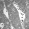
Representative image of perfusion fixation quality of a needle biopsy of diseased human liver. A survey transmission electron microscopy picture of a human liver needle biopsy fixed with jet fixation, of a patient with fibrosis, showing an intralobular region with normal parenchymal cells and sinusoids. Scale bar, 10 µm
Fig. 2
Fig. 2
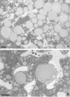
Fat accumulation causes necrosis of liver parenchymal cells at the single cell level. A Low magnification of a jet-fixed needle biopsy of a steatotic human liver. Large fat droplets with a preserved lipid content leave little room for the cytoplasms of the parenchymal cells. Notice that these “clean” fat droplets do not contain other material and have an equal electron density. Four stellate cells with small fat droplets, next to the open sinusoids, are dispersed in the tissue. Scale bar, 20 µm. B Transmission electron microscopy picture of a single-cell steatonecrosis of a steatotic human liver, in an injection-fixed wedge biopsy. To the right, we observe a parenchymal cell in necrosis with a large fat droplet, an electron lucent cytoplasm, dispersed organelles and morphologically intact plasma membrane. To the left, in a neighbouring cell, a fat droplet with Mallory substance. Scale bar, 20 µm
Fig. 3
Fig. 3
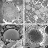
Inflammation at the single cell level. A Transmission electron microscopy picture of an inflammatory fat follicle in a human liver fixed with injection fixation, showing a fat droplet surrounded by inflammatory cells and a few red blood cells. Scale bar, 10 µm. B One of a sequential series of 350 micrographs together composing a three-dimensional dataset, observable as a video through the following link: https://www.dropbox.com/s/ho2l4cju16gj1vn/HM53%20Z-stack.avi?dl=0. The figure contains two SCNs, four inflammatory fat follicles, and large fat droplets; in the top left corner, an inflammatory fat follicle, showing neutrophils and erythrocytes surrounding a large fat droplet in a necrotic parenchymal cell; in the middle of the picture, a single-cell steatonecrosis, showing a large fat droplet not yet surrounded by neutrophils. Three other inflammatory fat follicles are visible at 12, 14 and 17 o’clock. Neutrophils are also seen in the sinusoids. The three-dimensional video is not based on a timeline, but takes you on a trip along the Z-axis down the tissue block. The preparation regards an injection-fixed wedge biopsy of a steatotic human liver with inflammation. Scale bar, 30 µm. C Scanning electron microscopy (SEM) picture of an inflammatory fat follicle, showing the uptake, or release, of filaments, supposedly being Mallory substance. As in Fig. 3A, the fat droplet is surrounded also by red blood cells. Scale bar, 10 µm. D SEM image of an inflammatory fat follicle. During preparation, the fat droplet has dropped out, leaving behind the cells that directly surrounded the fat droplet. The cells involved are neutrophils and red blood cells, apparently forming a monolayer of closely neighbouring cells. Neutrophils show a different contact pattern as compared with the smooth surface of the red blood cells. Scale bar, 10 µm
Fig. 4
Fig. 4
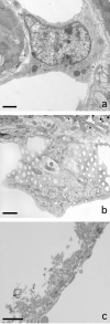
Escape of fat from parenchymal cells. A Transmission electron microscopy picture of a huge fat droplet in a parenchymal cell of a patient with liver steatosis. The fat droplet is free of filaments and Mallory-like substances, except for a little dense structure at the right margin of the droplet. To the left and the top right are the lumina of two sinusoids. At the bottom right we observe a sinusoidal endothelial cell. B Formation of a nozzle pointing into the direction of the sinusoidal membrane of the parenchymal cell. C Extrusion of a fat droplet, breaking out of the parenchymal cell, traversing the space of Disse and the endothelial lining and entering the lumen of the sinusoid. D Sinusoidal fat embolus: a large fat droplet is plugging the lumen of a sinusoid. Scale bars, 5 µm
Fig. 5
Fig. 5
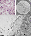
Mallory bodies (MB) developing within fat droplets. A Light microscopical picture of a pathological routine paraffin section showing eosinophilic MB in the lower half of the figure. The darker, irregular structures are the MB. Scale bar, 20 µm. B Transmission electron microscope (TEM) image showing a lipid droplet of a jet-fixed needle biopsy of a patient with non-alcoholic steatohepatitis. The fat droplet is more than half filled with a mass of filamentous and irregular material, shown at higher magnification in Fig. 5C. In the same liver, we find droplets with less Mallory substance. Scale bar, 2 µm. C TEM picture with higher magnification of the material included into the fat droplet in the liver of a non-alcoholic steatohepatitis patient. Pieces of filaments are clearly present, next to irregular material that is difficult to describe. Note that there is no membrane between the lipid droplet and the cytoplasm of the parenchymal cell. Scale bar, 1 µm
Fig. 6
Fig. 6
Liver sinusoidal endothelial cells and fenestrae in NASH. A Transmission electron microscope (TEM) picture of a sinusoidal endothelial cell in a jet-fixed needle biopsy of human liver of a patient with non-alcoholic steatohepatitis (NASH). Note the presence of organelles such as mitochondria, lysosomes (dense bodies), a nucleus with eu- and heterochromatin and a nucleolus, many pinocytotic vesicles and in the right lower corner the connection with a sieve plate. Scale bar, 2 µm. B TEM image of fenestrae arranged in a sieve plate in a sinusoidal endothelial cell in a jet-fixed needle biopsy of an intact human liver of a patient with NASH, illustrating that normal fenestrae are present. This liver and other livers with NASH, contain a normal population of fenestrae as the picture is showing. To image sieve plates, it is necessary that the thin layer of endothelium is within the volume of the ultrathin section and has an orientation parallel to the surface of the section. Scale bar, 0.5 µm. C: TEM picture showing an “endothelial complex” ruptured off from the surface of the parenchymal cell. The complex consists of the endothelial lining, with recognizable fenestrae, processes of stellate cells, microvilli of the parenchymal cell and fibres and fluffy material belonging to the extracellular matrix normally present in the space of Disse. It is supposed that this complex has been ripped off as a coherent unit by the force of the fluid stream during injection fixation. The patient had received chemotherapy for colon carcinoma metastasis, last chemotherapy 132 days before the wedge biopsy was taken. The liver was diagnosed as steatosis scale 3. Scale bar, 1 µm

Similar articles

Cited by

References

    1. Arias IM. The biology of hepatic endothelial cell fenestrae. Prog Liver Dis. 1990;9:11–26. - PubMed
    1. Braet F, De Zanger R, Baekeland M, Crabbe E, Van Der Smissen P, Wisse E. Structure and dynamics of the fenestrae-associated cytoskeleton of rat liver sinusoidal endothelial cells. Hepatology. 1995;21(1):180–189. doi: 10.1002/hep.1840210130. - DOI - PubMed
    1. Braet F, Spector I, De Zanger R, Wisse E. A novel structure involved in the formation of liver endothelial cell fenestrae revealed by using the actin inhibitor misakinolide. Proc Natl Acad Sci U S A. 1998;95(23):13635–13640. doi: 10.1073/pnas.95.23.13635. - DOI - PMC - PubMed
    1. Brenner DA. Molecular pathogenesis of liver fibrosis. Trans Am Clin Climatol Assoc. 2009;120:361–368. - PMC - PubMed
    1. Caldwell S, Ikura Y, Dias D, Isomoto K, Yabu A, Moskaluk C, Pramoonjago P, Simmons W, Scruggs H, Rosenbaum N, Wilkinson T, Toms P, Argo CK, Al-Osaimi AM, Redick JA. Hepatocellular ballooning in NASH. J Hepatol. 2010;53(4):719–723. doi: 10.1016/j.jhep.2010.04.031. - DOI - PMC - PubMed